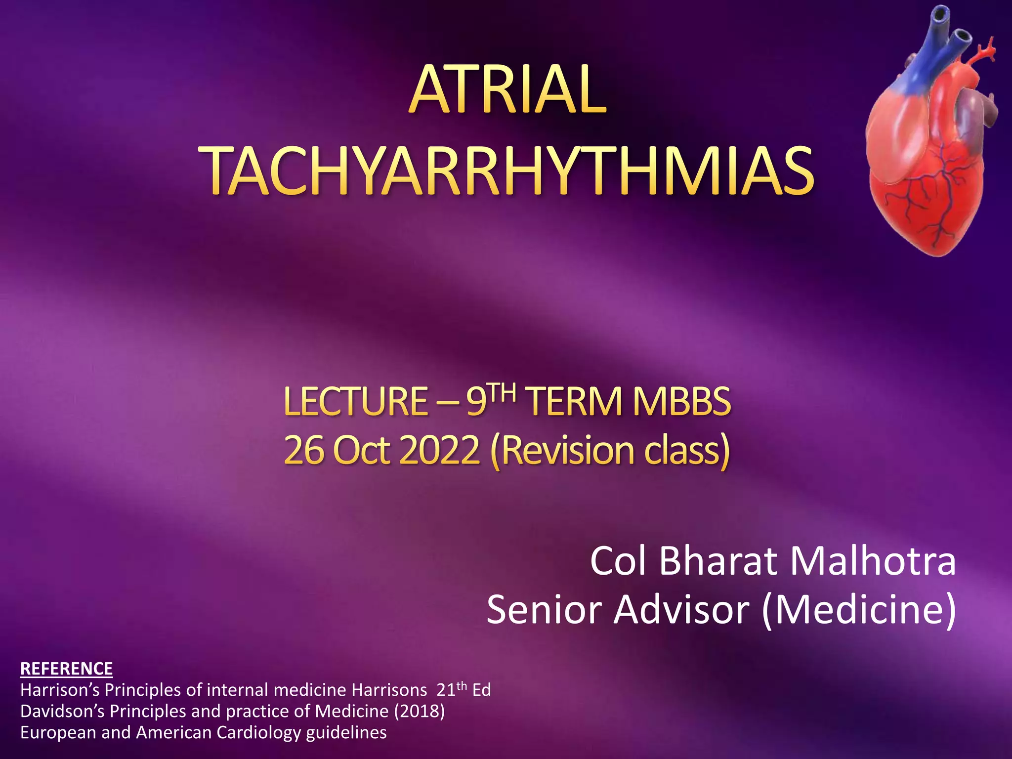
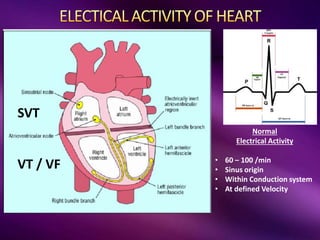
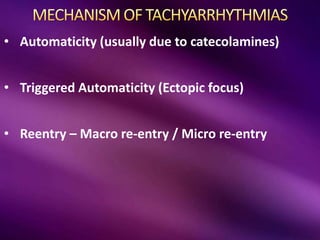
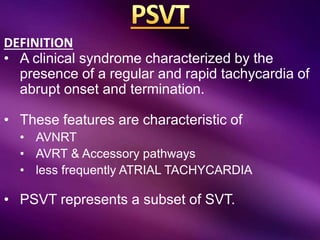
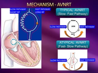
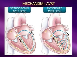
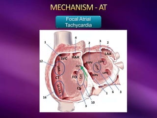
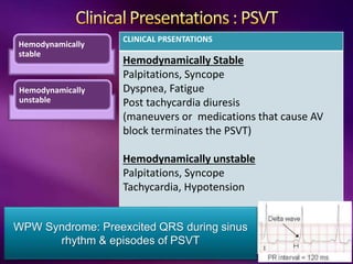
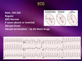
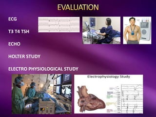
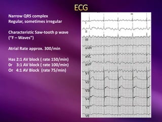
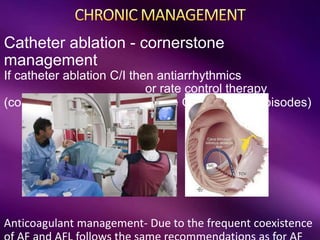
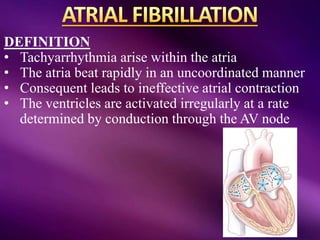
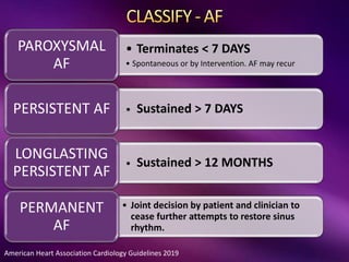
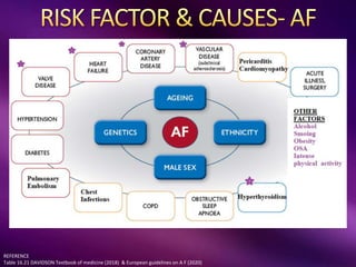
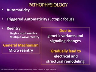
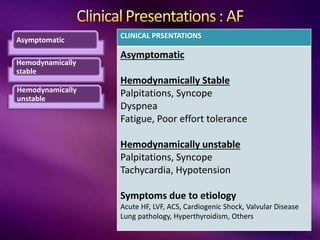
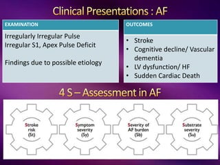
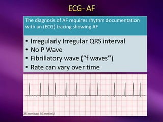
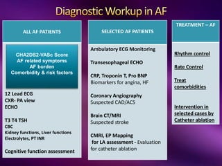
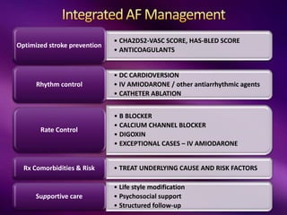
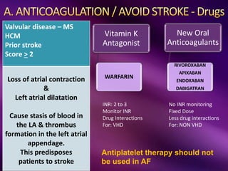
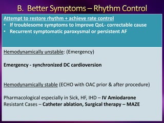
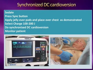
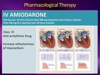
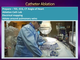
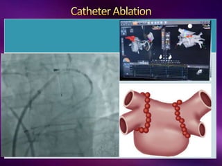
Col Bharat Malhotra
Senior Advisor (Medicine)
REFERENCE
Harrison’s Principles of internal medicine Harrisons 21th Ed
Davidson’s Principles and practice of Medicine (2018)
European and American Cardiology guidelines
LEFT ATRIA
Normal
Electrical Activity
• 60 – 100 /min
• Sinus origin
• Within Conduction system
• At defined Velocity
SVT
VT / VF
LEFT ATRIA
Sinus Tachycardia
Inappropriate sinus tachycardia
Paroxysmal Supraventricular tachycardia
Atrial Flutter, Atrial Fibrillation
Ventricular Tachycardia & Fibrillation
100 /min
60 /min
Sinus Bradycardia
SA Disease or Sick Sinus syndrome
AV Block
First degree block
Second degree block – type 1 & Type 2
Third degree block
Can have
Atrial/ Ventricular Ectopic
LBBB, RBBB
NORMAL
TACHYARRHYTHMIA
S
BRADYARRHYTHMIA
S
Atrial Tachycardia
AVNRT
AVRT & Accessory pathways
• Automaticity (usually due to catecolamines)
• Triggered Automaticity (Ectopic focus)
• Reentry – Macro re-entry / Micro re-entry
DEFINITION
• A clinical syndrome characterized by the
presence of a regular and rapid tachycardia of
abrupt onset and termination.
• These features are characteristic of
• AVNRT
• AVRT & Accessory pathways
• less frequently ATRIAL TACHYCARDIA
• PSVT represents a subset of SVT.
TYPICAL AVNRT
(Slow- Fast Pathway)
ATYPICAL AVNRT
(Fast- Slow Pathway)
FAST PATHWAY
LONG RP
SLOW PATHWAY
SHORT RP
FAST PATHWAY
SLOW PATHWAY
Slow
Pathway
SLOW PATHWAY
FAST PATHWAY
ORTHODROMIC
AVRT (90%)
ANTIDROMIC
AVRT (10%)
Focal Atrial
Tachycardia
Hemodynamically
stable
Hemodynamically
unstable
CLINICAL PRSENTATIONS
Hemodynamically Stable
Palpitations, Syncope
Dyspnea, Fatigue
Post tachycardia diuresis
(maneuvers or medications that cause AV
block terminates the PSVT)
Hemodynamically unstable
Palpitations, Syncope
Tachycardia, Hypotension
WPW Syndrome: Preexcited QRS during sinus
rhythm & episodes of PSVT
Rate: 150-250
Regular
QRS Narrow
P wave absent or inverted
Abrupt Onset
Abrupt termination – by AV block drugs
ECG
T3 T4 TSH
ECHO
HOLTER STUDY
ELECTRO PHYSIOLOGICAL STUDY
STABLE
HAEMODYNAMICALLY UNSTABLE
Hemodynamically Unstable
Sync DC
Cardioversion
Hemodynamically Stable
ECG
Vagal manoeuvre
IV Adenosine 6-12mg fast
bolus
IV Diltiazem or verapamil
IV Betablocker
If Ineffective
Sync DC Cardioversion
STABLE
Only if No preexcitation
HAEMODYNAMICALLY UNSTABLE
Hemodynamically Unstable
Sync DC
Cardioversion
Hemodynamically Stable
ECG
Vagal manoeuvre
IV Adenosine – if no
preexcitation
IV Procainamide or
IV Amiodarone
Not recommended – CCB &
BB
• ORAL B BLOCKER
• OR DILTIAZEM
• OR VERAPAMIL
• EP STUDY &
CATHETER
ABLATION
(Recurrent PSVT)
Macro reentrant circuit around the tricuspid
valve Atypical flutters -> cardiac surgery or
ablation
NON ISTHMUS
DEPENDENT FLUTTER
General Mechanism
Macro reentry
CAVO-TRICUSPID ISTHMUS
DEPENDENT FLUTTER
Narrow QRS complex
Regular, sometimes irregular
Characteristic Saw-tooth p wave
(“F – Waves”)
Atrial Rate approx. 300/min
Has 2:1 AV block ( rate 150/min)
0r 3:1 AV block ( rate 100/min)
Or 4:1 AV Block (rate 75/min)
Hemodynamically unstable
• Synchronized DC cardioversion
Hemodynamically stable
• Synchronized DC cardioversion If trained person present
• Rate Control  oral B Blocker/ CCB
(avoid amiodarone in acute setting)
Catheter ablation - cornerstone
management
If catheter ablation C/I then antiarrhythmics
or rate control therapy
(considered after a first episode OR recurrent episodes)
Anticoagulant management- Due to the frequent coexistence
DEFINITION
• Tachyarrhythmia arise within the atria
• The atria beat rapidly in an uncoordinated manner
• Consequent leads to ineffective atrial contraction
• The ventricles are activated irregularly at a rate
determined by conduction through the AV node
American Heart Association Cardiology Guidelines 2019
• Terminates < 7 DAYS
• Spontaneous or by Intervention. AF may recur
PAROXYSMAL
AF
• Sustained > 7 DAYS
PERSISTENT AF
• Sustained > 12 MONTHS
LONGLASTING
PERSISTENT AF
• Joint decision by patient and clinician to
cease further attempts to restore sinus
rhythm.
PERMANENT
AF
REFERENCE
Table 16.21 DAVIDSON Textbook of medicine (2018) & European guidelines on A F (2020)
Yu-ki Iwasaki. Circulation. Atrial Fibrillation Pathophysiology, Volume: 124, Issue: 20, Pages: 2264-2274
• Automaticity
• Triggered Automaticity (Ectopic focus)
• Reentry
Single circuit reentry
Multiple wave reentry
Due to
genetic variants and
signaling changes
Gradually lead to
electrical and
structural remodeling
General Mechanism
Micro reentry
SVC
IVC
P Artery P Vein
Aorta
Lungs
Lt A
Rt A
Rt V Lt V
Asymptomatic
Hemodynamically
stable
Hemodynamically
unstable
CLINICAL PRSENTATIONS
Asymptomatic
Hemodynamically Stable
Palpitations, Syncope
Dyspnea
Fatigue, Poor effort tolerance
Hemodynamically unstable
Palpitations, Syncope
Tachycardia, Hypotension
Symptoms due to etiology
Acute HF, LVF, ACS, Cardiogenic Shock, Valvular Disease
Lung pathology, Hyperthyroidism, Others
EXAMINATION
Irregularly Irregular Pulse
Irregular S1, Apex Pulse Deficit
Findings due to possible etiology
OUTCOMES
• Stroke
• Cognitive decline/ Vascular
dementia
• LV dysfunction/ HF
• Sudden Cardiac Death
The diagnosis of AF requires rhythm documentation
with an (ECG) tracing showing AF
• Irregularly Irregular QRS interval
• No P Wave
• Fibrillatory wave (“f waves”)
• Rate can vary over time
ALL AF PATIENTS
12 Lead ECG
CXR- PA view
ECHO
T3 T4 TSH
CBC
Kidney functions, Liver functions
Electrolytes, PT INR
Cognitive function assessment
TREATMENT – AF
Rhythm control
Rate Control
Treat
comorbidities
Intervention in
selected cases by
Catheter ablation
SELECTED AF PATIENTS
Ambulatory ECG Monitoring
Transesophageal ECHO
CRP, Troponin T, Pro BNP
Biomarkers for angina, HF
Coronary Angiography
Suspected CAD/ACS
Brain CT/MRI
Suspected stroke
CMRI, EP Mapping
for LA assessment - Evaluation
for catheter ablation
CHA2DS2-VASc Score
AF related symptoms
AF burden
Comorbidity & risk factors
• CHA2DS2-VASC SCORE, HAS-BLED SCORE
• ANTICOAGULANTS
Optimized stroke prevention
• DC CARDIOVERSION
• IV AMIODARONE / other antiarrhythmic agents
• CATHETER ABLATION
Rhythm control
• B BLOCKER
• CALCIUM CHANNEL BLOCKER
• DIGOXIN
• EXCEPTIONAL CASES – IV AMIODARONE
Rate Control
• TREAT UNDERLYING CAUSE AND RISK FACTORS
Rx Comorbidities & Risk
• Life style modification
• Psychosocial support
• Structured follow-up
Supportive care
Valvular disease – MS
HCM
Prior stroke
Score > 2
Loss of atrial contraction
&
Left atrial dilatation
Cause stasis of blood in
the LA & thrombus
formation in the left atrial
appendage.
This predisposes
patients to stroke
Antiplatelet therapy should not
be used in AF
Vitamin K
Antagonist
WARFARIN
New Oral
Anticoagulants
RIVOROXABAN
APIXABAN
ENDOXABAN
DABIGATRAN
No INR monitoring
Fixed Dose
Less drug interactions
For: NON VHD
INR: 2 to 3
Monitor INR
Drug Interactions
For: VHD
Attempt to restore rhythm + achieve rate control
• If troublesome symptoms to improve QoL- correctable cause
• Recurrent symptomatic paroxysmal or persistent AF
Hemodynamically unstable: (Emergency)
Emergency - synchronized DC cardioversion
Hemodynamically stable (ECHO with OAC prior & after procedure)
Pharmacological especially in Sick, HF, IHD – IV Amiodarone
Resistant Cases – Catheter ablation, Surgical therapy – MAZE
Sedate
Press Sync button
Apply jelly over pads and place over chest as demonstrated
Select Charge 100-200 J
Do synchronized DC cardioversion
Monitor patient
IV AMIODARONE
150 mg over 10 mins infusion then 360 mg (1mg/min) over 6 hours infusion
Then 540 mg (0.5 mg/min) over 18 hours infusion
Class III
Anti arrhythmic Drug
Increase refractoriness
of myocardium
Prepare – TEE, ECG, CT Angio of Heart
Ablation Cath Lab
Electrical mapping
Ablate around pulmonary veins
Attempt to restore rhythm + achieve rate control
Accept presence of AF but achieve rate control
B Blocker (IV Metoprolol, IV Esmolol)
(Oral Metoprolol, Bisoprolol, Carvedilol)
Calcium Channel Blocker – (Diltiazem, Verapamil)
Digoxin
Exceptional Cases- Implant a permanent pacemaker
Plus Complete AV node block with catheter ablation
Last Resort - IV Amiodarone
<110/min
< 80/Min
Treat the coexisting illness & Risk Factors

Atrial tachy 26 Oct 22.pptx

  • 1.
    Col Bharat Malhotra SeniorAdvisor (Medicine) REFERENCE Harrison’s Principles of internal medicine Harrisons 21th Ed Davidson’s Principles and practice of Medicine (2018) European and American Cardiology guidelines
  • 2.
    LEFT ATRIA Normal Electrical Activity •60 – 100 /min • Sinus origin • Within Conduction system • At defined Velocity SVT VT / VF
  • 3.
    LEFT ATRIA Sinus Tachycardia Inappropriatesinus tachycardia Paroxysmal Supraventricular tachycardia Atrial Flutter, Atrial Fibrillation Ventricular Tachycardia & Fibrillation 100 /min 60 /min Sinus Bradycardia SA Disease or Sick Sinus syndrome AV Block First degree block Second degree block – type 1 & Type 2 Third degree block Can have Atrial/ Ventricular Ectopic LBBB, RBBB NORMAL TACHYARRHYTHMIA S BRADYARRHYTHMIA S Atrial Tachycardia AVNRT AVRT & Accessory pathways
  • 4.
    • Automaticity (usuallydue to catecolamines) • Triggered Automaticity (Ectopic focus) • Reentry – Macro re-entry / Micro re-entry
  • 5.
    DEFINITION • A clinicalsyndrome characterized by the presence of a regular and rapid tachycardia of abrupt onset and termination. • These features are characteristic of • AVNRT • AVRT & Accessory pathways • less frequently ATRIAL TACHYCARDIA • PSVT represents a subset of SVT.
  • 6.
    TYPICAL AVNRT (Slow- FastPathway) ATYPICAL AVNRT (Fast- Slow Pathway) FAST PATHWAY LONG RP SLOW PATHWAY SHORT RP FAST PATHWAY SLOW PATHWAY Slow Pathway SLOW PATHWAY FAST PATHWAY
  • 7.
  • 8.
  • 9.
    Hemodynamically stable Hemodynamically unstable CLINICAL PRSENTATIONS Hemodynamically Stable Palpitations,Syncope Dyspnea, Fatigue Post tachycardia diuresis (maneuvers or medications that cause AV block terminates the PSVT) Hemodynamically unstable Palpitations, Syncope Tachycardia, Hypotension WPW Syndrome: Preexcited QRS during sinus rhythm & episodes of PSVT
  • 10.
    Rate: 150-250 Regular QRS Narrow Pwave absent or inverted Abrupt Onset Abrupt termination – by AV block drugs
  • 11.
    ECG T3 T4 TSH ECHO HOLTERSTUDY ELECTRO PHYSIOLOGICAL STUDY
  • 12.
    STABLE HAEMODYNAMICALLY UNSTABLE Hemodynamically Unstable SyncDC Cardioversion Hemodynamically Stable ECG Vagal manoeuvre IV Adenosine 6-12mg fast bolus IV Diltiazem or verapamil IV Betablocker If Ineffective Sync DC Cardioversion
  • 13.
    STABLE Only if Nopreexcitation HAEMODYNAMICALLY UNSTABLE Hemodynamically Unstable Sync DC Cardioversion Hemodynamically Stable ECG Vagal manoeuvre IV Adenosine – if no preexcitation IV Procainamide or IV Amiodarone Not recommended – CCB & BB
  • 14.
    • ORAL BBLOCKER • OR DILTIAZEM • OR VERAPAMIL • EP STUDY & CATHETER ABLATION (Recurrent PSVT)
  • 15.
    Macro reentrant circuitaround the tricuspid valve Atypical flutters -> cardiac surgery or ablation NON ISTHMUS DEPENDENT FLUTTER General Mechanism Macro reentry CAVO-TRICUSPID ISTHMUS DEPENDENT FLUTTER
  • 16.
    Narrow QRS complex Regular,sometimes irregular Characteristic Saw-tooth p wave (“F – Waves”) Atrial Rate approx. 300/min Has 2:1 AV block ( rate 150/min) 0r 3:1 AV block ( rate 100/min) Or 4:1 AV Block (rate 75/min)
  • 17.
    Hemodynamically unstable • SynchronizedDC cardioversion Hemodynamically stable • Synchronized DC cardioversion If trained person present • Rate Control  oral B Blocker/ CCB (avoid amiodarone in acute setting)
  • 18.
    Catheter ablation -cornerstone management If catheter ablation C/I then antiarrhythmics or rate control therapy (considered after a first episode OR recurrent episodes) Anticoagulant management- Due to the frequent coexistence
  • 19.
    DEFINITION • Tachyarrhythmia arisewithin the atria • The atria beat rapidly in an uncoordinated manner • Consequent leads to ineffective atrial contraction • The ventricles are activated irregularly at a rate determined by conduction through the AV node
  • 20.
    American Heart AssociationCardiology Guidelines 2019 • Terminates < 7 DAYS • Spontaneous or by Intervention. AF may recur PAROXYSMAL AF • Sustained > 7 DAYS PERSISTENT AF • Sustained > 12 MONTHS LONGLASTING PERSISTENT AF • Joint decision by patient and clinician to cease further attempts to restore sinus rhythm. PERMANENT AF
  • 21.
    REFERENCE Table 16.21 DAVIDSONTextbook of medicine (2018) & European guidelines on A F (2020)
  • 22.
    Yu-ki Iwasaki. Circulation.Atrial Fibrillation Pathophysiology, Volume: 124, Issue: 20, Pages: 2264-2274 • Automaticity • Triggered Automaticity (Ectopic focus) • Reentry Single circuit reentry Multiple wave reentry Due to genetic variants and signaling changes Gradually lead to electrical and structural remodeling General Mechanism Micro reentry
  • 23.
    SVC IVC P Artery PVein Aorta Lungs Lt A Rt A Rt V Lt V
  • 24.
    Asymptomatic Hemodynamically stable Hemodynamically unstable CLINICAL PRSENTATIONS Asymptomatic Hemodynamically Stable Palpitations,Syncope Dyspnea Fatigue, Poor effort tolerance Hemodynamically unstable Palpitations, Syncope Tachycardia, Hypotension Symptoms due to etiology Acute HF, LVF, ACS, Cardiogenic Shock, Valvular Disease Lung pathology, Hyperthyroidism, Others
  • 25.
    EXAMINATION Irregularly Irregular Pulse IrregularS1, Apex Pulse Deficit Findings due to possible etiology OUTCOMES • Stroke • Cognitive decline/ Vascular dementia • LV dysfunction/ HF • Sudden Cardiac Death
  • 26.
    The diagnosis ofAF requires rhythm documentation with an (ECG) tracing showing AF • Irregularly Irregular QRS interval • No P Wave • Fibrillatory wave (“f waves”) • Rate can vary over time
  • 27.
    ALL AF PATIENTS 12Lead ECG CXR- PA view ECHO T3 T4 TSH CBC Kidney functions, Liver functions Electrolytes, PT INR Cognitive function assessment TREATMENT – AF Rhythm control Rate Control Treat comorbidities Intervention in selected cases by Catheter ablation SELECTED AF PATIENTS Ambulatory ECG Monitoring Transesophageal ECHO CRP, Troponin T, Pro BNP Biomarkers for angina, HF Coronary Angiography Suspected CAD/ACS Brain CT/MRI Suspected stroke CMRI, EP Mapping for LA assessment - Evaluation for catheter ablation CHA2DS2-VASc Score AF related symptoms AF burden Comorbidity & risk factors
  • 28.
    • CHA2DS2-VASC SCORE,HAS-BLED SCORE • ANTICOAGULANTS Optimized stroke prevention • DC CARDIOVERSION • IV AMIODARONE / other antiarrhythmic agents • CATHETER ABLATION Rhythm control • B BLOCKER • CALCIUM CHANNEL BLOCKER • DIGOXIN • EXCEPTIONAL CASES – IV AMIODARONE Rate Control • TREAT UNDERLYING CAUSE AND RISK FACTORS Rx Comorbidities & Risk • Life style modification • Psychosocial support • Structured follow-up Supportive care
  • 29.
    Valvular disease –MS HCM Prior stroke Score > 2 Loss of atrial contraction & Left atrial dilatation Cause stasis of blood in the LA & thrombus formation in the left atrial appendage. This predisposes patients to stroke Antiplatelet therapy should not be used in AF Vitamin K Antagonist WARFARIN New Oral Anticoagulants RIVOROXABAN APIXABAN ENDOXABAN DABIGATRAN No INR monitoring Fixed Dose Less drug interactions For: NON VHD INR: 2 to 3 Monitor INR Drug Interactions For: VHD
  • 30.
    Attempt to restorerhythm + achieve rate control • If troublesome symptoms to improve QoL- correctable cause • Recurrent symptomatic paroxysmal or persistent AF Hemodynamically unstable: (Emergency) Emergency - synchronized DC cardioversion Hemodynamically stable (ECHO with OAC prior & after procedure) Pharmacological especially in Sick, HF, IHD – IV Amiodarone Resistant Cases – Catheter ablation, Surgical therapy – MAZE
  • 31.
    Sedate Press Sync button Applyjelly over pads and place over chest as demonstrated Select Charge 100-200 J Do synchronized DC cardioversion Monitor patient
  • 32.
    IV AMIODARONE 150 mgover 10 mins infusion then 360 mg (1mg/min) over 6 hours infusion Then 540 mg (0.5 mg/min) over 18 hours infusion Class III Anti arrhythmic Drug Increase refractoriness of myocardium
  • 33.
    Prepare – TEE,ECG, CT Angio of Heart Ablation Cath Lab Electrical mapping Ablate around pulmonary veins
  • 35.
    Attempt to restorerhythm + achieve rate control Accept presence of AF but achieve rate control B Blocker (IV Metoprolol, IV Esmolol) (Oral Metoprolol, Bisoprolol, Carvedilol) Calcium Channel Blocker – (Diltiazem, Verapamil) Digoxin Exceptional Cases- Implant a permanent pacemaker Plus Complete AV node block with catheter ablation Last Resort - IV Amiodarone <110/min < 80/Min
  • 36.
    Treat the coexistingillness & Risk Factors