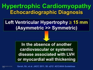
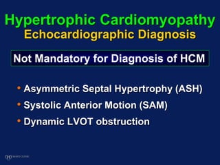
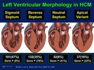
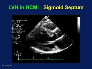
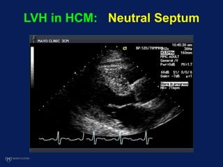
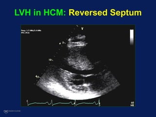
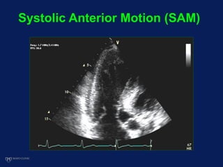
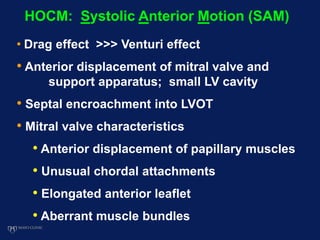
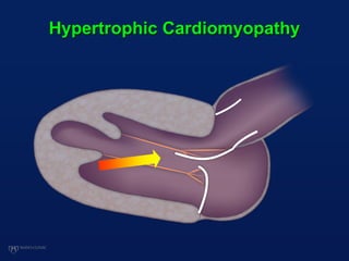
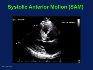
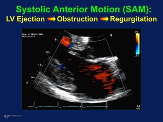
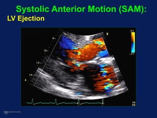
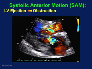
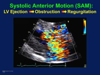
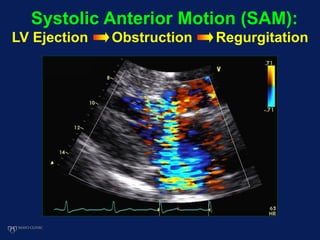
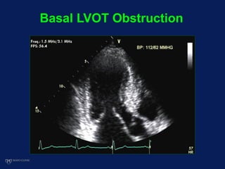
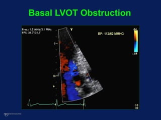
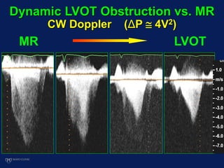
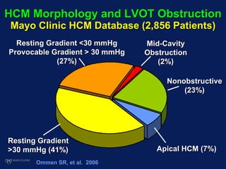
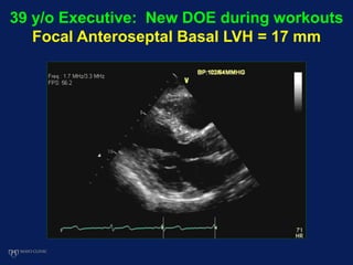
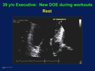
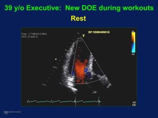
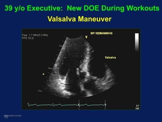
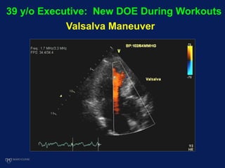
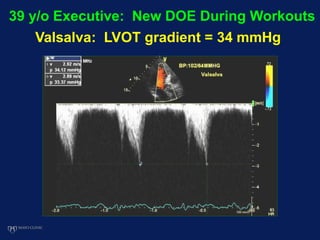
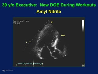
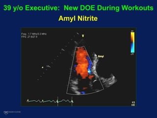
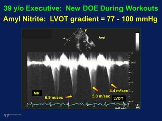
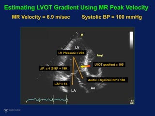
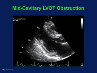
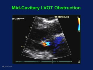
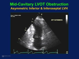
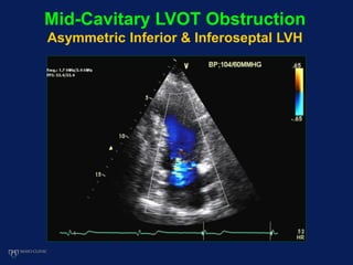
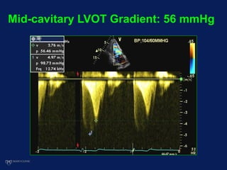
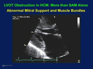
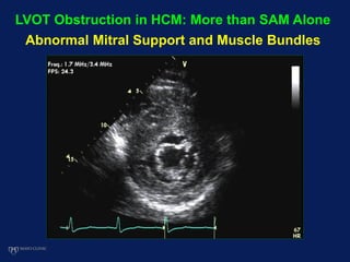
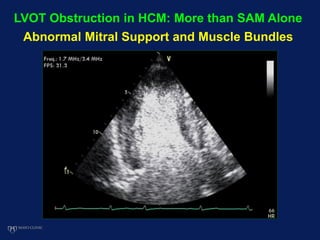
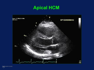
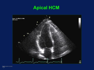
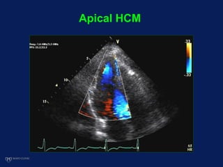
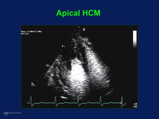
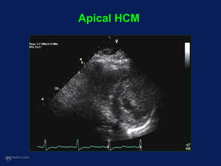
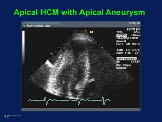
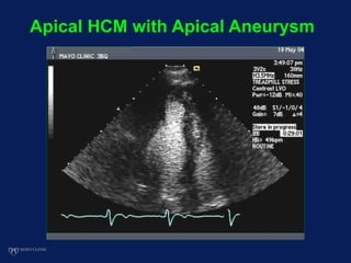
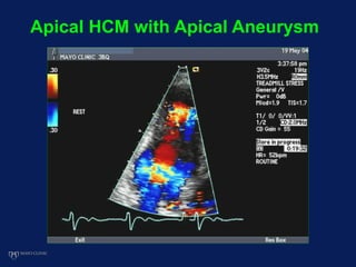
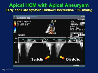
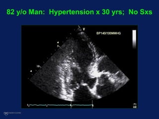
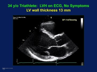
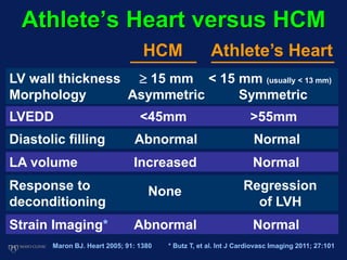
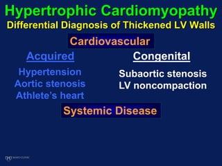
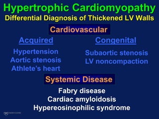
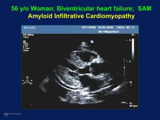
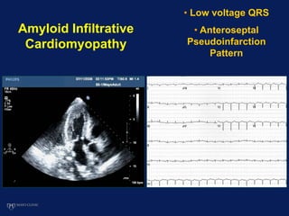
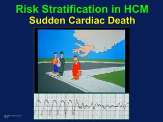
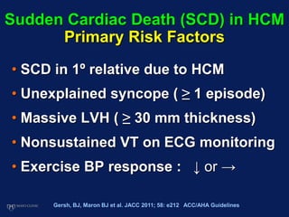
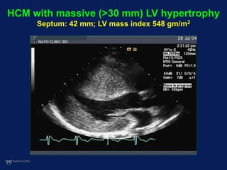
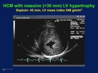
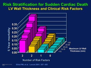
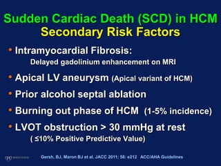
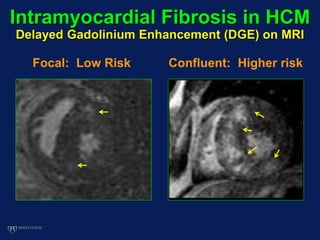
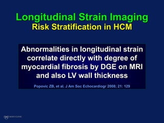
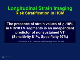
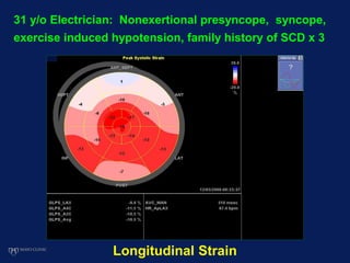
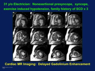
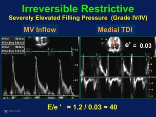
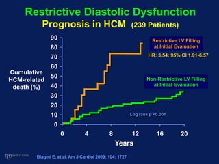
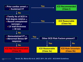
Hypertrophic
Cardiomyopathy (HCM)
William K. Freeman, MD, FACC, FASE
Evaluation and
Differential Diagnosis
Role of Echocardiography
DISCLOSURES
Relevant Financial Relationship(s)
None
Off Label Usage
None
Hypertrophic Cardiomyopathy
Echocardiographic Diagnosis
Left Ventricular Hypertrophy  15 mm
(Asymmetric >> Symmetric)
In the absence of another
cardiovascular or systemic
disease associated with LVH
or myocardial wall thickening
Gersh, BJ, et al. JACC 2011; 58: e212 ACC/AHA Guidelines
Hypertrophic Cardiomyopathy
Echocardiographic Diagnosis
• Asymmetric Septal Hypertrophy (ASH)
• Systolic Anterior Motion (SAM)
• Dynamic LVOT obstruction
Not Mandatory for Diagnosis of HCM
Hypertrophic Cardiomyopathy
Distribution of LVH (600 Patients)
Anterior and
inferior septum
(31%)
Anterior
septum only
(25%)
Klues HG, JACC 1995; 26: 1699
Septum & ant lat freewall (17%)
Septum & all
freewalls (17%)
Anterior septum
& ant lat freewall
(7%)
Lateral
freewall
(1%)
Apex only
(2%)
Left Ventricular Morphology in HCM
Binder J, et al. Mayo Clin Proc 2006; 81: 459.
181(47%)
Gene + (8%)
132(35%)
Gene + (79%)
37(10%)
Gene + (32%)
32(8%)
Gene + (41%)
Sigmoid
Septum
Reverse
Septum
Neutral
Septum
Apical
Variant
Genetic testing for HCM
Mayo Clinic Database (389 Patients)
• Echocardiographic anatomic
phenotypes are not specific
for individual gene mutations
• Specific gene mutations not
predictive of prognosis or
need for myectomy
Van Driest SL, et al. Mayo Clin Proc 2005; 80: 739
LVH in HCM: Sigmoid Septum
LVH in HCM: Neutral Septum
LVH in HCM: Reversed Septum
Systolic Anterior Motion (SAM)
• Drag effect >>> Venturi effect
• Anterior displacement of mitral valve and
support apparatus; small LV cavity
• Septal encroachment into LVOT
• Mitral valve characteristics
• Anterior displacement of papillary muscles
• Unusual chordal attachments
• Elongated anterior leaflet
• Aberrant muscle bundles
HOCM: Systolic Anterior Motion (SAM)
Normal Anatomy of the
LV Outflow Tract
Hypertrophic Cardiomyopathy
Systolic Anterior Motion (SAM)
Systolic Anterior Motion (SAM):
LV Ejection Obstruction Regurgitation
Systolic Anterior Motion (SAM):
LV Ejection
Systolic Anterior Motion (SAM):
LV Ejection Obstruction
Systolic Anterior Motion (SAM):
LV Ejection Obstruction Regurgitation
Systolic Anterior Motion (SAM):
LV Ejection Obstruction Regurgitation
Basal LVOT Obstruction
MR LVOT
Dynamic LVOT Obstruction vs. MR
CW Doppler (ΔP  4V2)
HCM Morphology and LVOT Obstruction
Mayo Clinic HCM Database (2,856 Patients)
Resting Gradient
>30 mmHg (41%)
Resting Gradient <30 mmHg
Provocable Gradient > 30 mmHg
(27%)
Apical HCM (7%)
Nonobstructive
(23%)
Mid-Cavity
Obstruction
(2%)
Ommen SR, et al. 2006
39 y/o Executive: New DOE during workouts
Focal Anteroseptal Basal LVH = 17 mm
39 y/o Executive: New DOE during workouts
Rest
39 y/o Executive: New DOE during workouts
Rest
39 y/o Executive: New DOE During Workouts
Resting LVOT gradient = 12 mmHg
39 y/o Executive: New DOE During Workouts
Valsalva Maneuver
39 y/o Executive: New DOE During Workouts
Valsalva Maneuver
39 y/o Executive: New DOE During Workouts
Valsalva: LVOT gradient = 34 mmHg
39 y/o Executive: New DOE During Workouts
Amyl Nitrite
39 y/o Executive: New DOE During Workouts
Amyl Nitrite
39 y/o Executive: New DOE During Workouts
Amyl Nitrite: LVOT gradient = 77 - 100 mmHg
5.0 m/sec
4.4 m/sec
LVOT6.9 m/sec
MR
LAP  15
P  4 (6.9)2 = 190
LV Pressure  205
Aortic  Systolic BP = 100
LV
Ao
LA
LVOT gradient  105
Estimating LVOT Gradient Using MR Peak Velocity
MR Velocity = 6.9 m/sec Systolic BP = 100 mmHg
Mid-Cavitary LVOT Obstruction
Asymmetric Inferior & Inferoseptal LVH
Mid-Cavitary LVOT Obstruction
Asymmetric Inferior & Inferoseptal LVH
Mid-cavitary LVOT Gradient: 56 mmHg
LVOT Obstruction in HCM: More than SAM Alone
Abnormal Mitral Support and Muscle Bundles
LVOT Obstruction in HCM: More than SAM Alone
Abnormal Mitral Support and Muscle Bundles
LVOT Obstruction in HCM: More than SAM Alone
Abnormal Mitral Support and Muscle Bundles
Apical HCM
Apical HCM
Apical HCM
Apical HCM
Apical HCM
Apical HCM with Apical Aneurysm
Early and Late Systolic Outflow Obstruction ~ 60 mmHg
Systolic Diastolic
• Apical abnormalities in apical HCM:
Pouch: 15%; Aneurysm: 3%
• Adverse events associated with aneurysm
(not apical pouch)
• Progressive heart failure/death (18%)
• SCD or revived cardiac arrest (14%)
• Appropriate ICD discharge (11%)
• Nonfatal embolic stroke (7%)
Hypertrophic Cardiomyopathy
Complicated by Apical Aneurysm
Binder J et al JASE 2011;24:775
Maron MS, et al. Circulation 2008;118:1541
Hypertrophic Cardiomyopathy
Differential Diagnosis of Thickened LV Walls
Cardiovascular
Systemic Disease
Acquired
Hypertension
Aortic stenosis
Athlete’s heart
82 y/o Man: Hypertension x 30 yrs; No Sxs
34 y/o Triathlete: LVH on ECG, No Symptoms
LV wall thickness 13 mm
Athlete’s Heart versus HCM
HCM Athlete’s Heart
LV wall thickness  15 mm < 15 mm (usually < 13 mm)
Morphology Asymmetric Symmetric
LVEDD <45mm >55mm
Diastolic filling Abnormal Normal
LA volume Increased Normal
Response to Regression
deconditioning of LVH
None
Maron BJ. Heart 2005; 91: 1380
Strain Imaging* Abnormal Normal
* Butz T, et al. Int J Cardiovasc Imaging 2011; 27:101
Hypertrophic Cardiomyopathy
Differential Diagnosis of Thickened LV Walls
Cardiovascular
Systemic Disease
Acquired Congenital
Hypertension
Aortic stenosis
Athlete’s heart
Subaortic stenosis
LV noncompaction
71 y/o Woman: Murmur Since Childhood;
Previously Treated as HOCM
Congenital Fibromuscular Subaortic Stenosis
68 y/o Woman: Abnormal ECG; Asymptomatic
Left Ventricular Noncompaction Syndrome
68 y/o Woman: Abnormal ECG; Asymptomatic
Left Ventricular Noncompaction Syndrome
Hypertrophic Cardiomyopathy
Differential Diagnosis of Thickened LV Walls
Cardiovascular
Systemic Disease
Acquired Congenital
Hypertension
Aortic stenosis
Athlete’s heart
Subaortic stenosis
LV noncompaction
Fabry disease
Cardiac amyloidosis
Hypereosinophilic syndrome
70 y/o Man: Dyspnea on exertion
Fabry Disease (Alpha-Galactosidase A Deficiency)
Glycosphingolipid
Accumulation
Pieroni M, et al. JACC 2006; 47: 1663
Hyper-refractile
subendocardial border:
94% Sensitive
100% Specific
56 y/o Woman: Biventricular heart failure; SAM
Amyloid Infiltrative Cardiomyopathy
Amyloid Infiltrative
Cardiomyopathy
• Low voltage QRS
• Anteroseptal
Pseudoinfarction
Pattern
Risk Stratification in HCM
Sudden Cardiac Death
Hypertrophic Cardiomyopathy (HCM)
Arrhythmogenic Myocardial Substrate
Myocyte
Disarray
Coronary
Arteriole
Remodeling
Ischemia
Micro-infarction
Fibrosis
Maron BJ. Circulation 2010; 121: 445
Sudden Cardiac Death (SCD) in HCM
Primary Risk Factors
• SCD in 1º relative due to HCM
• Unexplained syncope ( ≥ 1 episode)
• Massive LVH ( ≥ 30 mm thickness)
• Nonsustained VT on ECG monitoring
• Exercise BP response : ↓ or →
Gersh, BJ, Maron BJ et al. JACC 2011; 58: e212 ACC/AHA Guidelines
HCM with massive (>30 mm) LV hypertrophy
Septum: 42 mm; LV mass index 548 gm/m2
HCM with massive (>30 mm) LV hypertrophy
Septum: 42 mm; LV mass index 548 gm/m2
Risk Stratification for Sudden Cardiac Death
LV Wall Thickness and Clinical Risk Factors
0.00
0.05
0.10
0.15
0.20
0.25
0.30
0.35
5Yearmortality
3 2 1 0
<15
15-19
20-24
25-29
>30
Number of Risk Factors
Elliot PM, et al. Lancet 2001; 357: 420
Maximum LV Wall
Thickness (mm)
Sudden Cardiac Death (SCD) in HCM
Secondary Risk Factors
• Intramyocardial Fibrosis:
Delayed gadolinium enhancement on MRI
• Apical LV aneurysm (Apical variant of HCM)
• Prior alcohol septal ablation
• Burning out phase of HCM (1-5% incidence)
• LVOT obstruction > 30 mmHg at rest
( ≤10% Positive Predictive Value)
Gersh, BJ, Maron BJ et al. JACC 2011; 58: e212 ACC/AHA Guidelines
Intramyocardial Fibrosis in HCM
Delayed Gadolinium Enhancement (DGE) on MRI
Focal: Low Risk Confluent: Higher risk
27
8
0
20
40
60
DGE
Present
DGE
Absent
Nonsustained VT
(4314 Months F/U)
%
(n = 126) (n = 94)
Rubinshtein R, et al. Circ Heart Fail 2010; 3:51
• Reversed septal
morphology
• Septal thickness
> 20 mm
• LV Mass > 150 gm/m2
• LVEF < 50%
Predictors of DGE
Intramyocardial Fibrosis in HCM
Delayed Gadolinium Enhancement (DGE) on MRI
Intramyocardial Fibrosis in HCM:
Detection by Echocardiography ?
Speckle Tracking
Strain Imaging
Apparent normal
global and regional
LV systolic function
Abnormal global
and/or regional LV
systolic function
Fibrosis likely where
LV is dysfunctional
Longitudinal Strain Imaging
Risk Stratification in HCM
Abnormalities in longitudinal strain
correlate directly with degree of
myocardial fibrosis by DGE on MRI
and also LV wall thickness
Popovic ZB, et al. J Am Soc Echocardiogr 2008; 21: 129
The presence of strain values of ≥ -10%
in > 3/18 LV segments is an independent
predictor of nonsustained VT
(Sensitivity 81%, Specificity 97%)
Di Salvo G, et al. J Am Soc Echocardiogr 2010; 23: 581
Longitudinal Strain Imaging
Risk Stratification in HCM
31 y/o Electrician: Nonexertional presyncope, syncope,
exercise induced hypotension, family history of SCD x 3
31 y/o Electrician: Nonexertional presyncope, syncope,
exercise induced hypotension, family history of SCD x 3
Longitudinal Strain
31 y/o Electrician: Nonexertional presyncope, syncope,
exercise induced hypotension, family history of SCD x 3
Cardiac MR Imaging: Delayed Gadolinium Enhancement
31 y/o Electrician: Nonexertional presyncope, syncope,
exercise induced hypotension, family history of SCD x 3
Sudden Cardiac Death (SCD) in HCM
• Gene mutation (>1,000 mutations; 11 genes)
• Atrial fibrillation
• Coronary artery bridging
• Diastolic dysfunction
• Highly competitive sports
• Coronary artery disease
Uncertain Risk Factors
Modifiable Risk Factors
Gersh, BJ, Maron BJ et al. JACC 2011; 58: e212 ACC/AHA Guidelines
Abnormal Relaxation
Mildly Elevated Filling Pressure (Grade Ia/IV)
MV Inflow Medial TDI
E/e' = 0.6 / 0.03 = 20
Irreversible Restrictive
Severely Elevated Filling Pressure (Grade IV/IV)
e' = 0.03
E/e ' = 1.2 / 0.03 = 40
MV Inflow Medial TDI
Years
0
10
20
30
40
50
60
70
80
90
0 4 8 12 16 20
Log rank p <0.001
Cumulative
HCM-related
death (%)
Biagini E, et al. Am J Cardiol 2009; 104: 1727
Restrictive Diastolic Dysfunction
Prognosis in HCM (239 Patients)
Restrictive LV Filling
at Initial Evaluation
Non-Restrictive LV Filling
at Initial Evaluation
HR: 3.54; 95% CI 1.91-6.57
Indications for ICD in
Hypertrophic Cardiomyopathy
 Prior cardiac arrest or
 Sustained VT
Yes ICD Recommended
(Class I)
 Family Hx of SCD in
first degree relative or
 Recent unexplained
syncope or
 LV wall thickness
 30 mm
 Nonsustained VT or
 Abnormal Stress BP
response
ICD Not Recommended
(Class III)
No
No
No
Yes ICD Reasonable
(Class IIa)
Yes
Other SCD Risk Factors present?
Yes No
ICD Reasonable
(Class IIa)
ICD Role Uncertain
(Class IIb)
Gersh, BJ, Maron BJ et al. JACC 2011; 58: e212 ACC/AHA Guidelines
Family Screening for HCM by Echo
< 12 Yrs Old
12 to 18-21 Yrs
Old
>18-21 Yrs Old
Optional unless:
 Malignant Family Hx
 Cardiac symptoms
 Competitive sports
 Other signs of LVH
Every 12 to 18
Months
Every 5 Yrs or as per
clinical suspicion
Gersh, BJ, Maron BJ et al. JACC 2011; 58: e212 ACC/AHA Guidelines
Evaluation of HCM by Echocardiography
Comprehensive echocardiography is
indispensable for the diagnosis and
hemodynamic assessment of HCM
Echocardiography plays an important
role in the clinical risk stratification
and also the interventional
management of the patient with HCM
Freeman hypertrophic-cardiomyopathy

Freeman hypertrophic-cardiomyopathy

  • 1.
    Hypertrophic Cardiomyopathy (HCM) William K.Freeman, MD, FACC, FASE Evaluation and Differential Diagnosis Role of Echocardiography
  • 2.
  • 3.
    Hypertrophic Cardiomyopathy Echocardiographic Diagnosis LeftVentricular Hypertrophy  15 mm (Asymmetric >> Symmetric) In the absence of another cardiovascular or systemic disease associated with LVH or myocardial wall thickening Gersh, BJ, et al. JACC 2011; 58: e212 ACC/AHA Guidelines
  • 4.
    Hypertrophic Cardiomyopathy Echocardiographic Diagnosis •Asymmetric Septal Hypertrophy (ASH) • Systolic Anterior Motion (SAM) • Dynamic LVOT obstruction Not Mandatory for Diagnosis of HCM
  • 5.
    Hypertrophic Cardiomyopathy Distribution ofLVH (600 Patients) Anterior and inferior septum (31%) Anterior septum only (25%) Klues HG, JACC 1995; 26: 1699 Septum & ant lat freewall (17%) Septum & all freewalls (17%) Anterior septum & ant lat freewall (7%) Lateral freewall (1%) Apex only (2%)
  • 6.
    Left Ventricular Morphologyin HCM Binder J, et al. Mayo Clin Proc 2006; 81: 459. 181(47%) Gene + (8%) 132(35%) Gene + (79%) 37(10%) Gene + (32%) 32(8%) Gene + (41%) Sigmoid Septum Reverse Septum Neutral Septum Apical Variant
  • 7.
    Genetic testing forHCM Mayo Clinic Database (389 Patients) • Echocardiographic anatomic phenotypes are not specific for individual gene mutations • Specific gene mutations not predictive of prognosis or need for myectomy Van Driest SL, et al. Mayo Clin Proc 2005; 80: 739
  • 8.
    LVH in HCM:Sigmoid Septum
  • 9.
    LVH in HCM:Neutral Septum
  • 10.
    LVH in HCM:Reversed Septum
  • 11.
  • 12.
    • Drag effect>>> Venturi effect • Anterior displacement of mitral valve and support apparatus; small LV cavity • Septal encroachment into LVOT • Mitral valve characteristics • Anterior displacement of papillary muscles • Unusual chordal attachments • Elongated anterior leaflet • Aberrant muscle bundles HOCM: Systolic Anterior Motion (SAM)
  • 13.
    Normal Anatomy ofthe LV Outflow Tract
  • 14.
  • 15.
  • 16.
    Systolic Anterior Motion(SAM): LV Ejection Obstruction Regurgitation
  • 17.
    Systolic Anterior Motion(SAM): LV Ejection
  • 18.
    Systolic Anterior Motion(SAM): LV Ejection Obstruction
  • 19.
    Systolic Anterior Motion(SAM): LV Ejection Obstruction Regurgitation
  • 20.
    Systolic Anterior Motion(SAM): LV Ejection Obstruction Regurgitation
  • 21.
  • 22.
  • 23.
    MR LVOT Dynamic LVOTObstruction vs. MR CW Doppler (ΔP  4V2)
  • 24.
    HCM Morphology andLVOT Obstruction Mayo Clinic HCM Database (2,856 Patients) Resting Gradient >30 mmHg (41%) Resting Gradient <30 mmHg Provocable Gradient > 30 mmHg (27%) Apical HCM (7%) Nonobstructive (23%) Mid-Cavity Obstruction (2%) Ommen SR, et al. 2006
  • 25.
    39 y/o Executive:New DOE during workouts Focal Anteroseptal Basal LVH = 17 mm
  • 26.
    39 y/o Executive:New DOE during workouts Rest
  • 27.
    39 y/o Executive:New DOE during workouts Rest
  • 28.
    39 y/o Executive:New DOE During Workouts Resting LVOT gradient = 12 mmHg
  • 29.
    39 y/o Executive:New DOE During Workouts Valsalva Maneuver
  • 30.
    39 y/o Executive:New DOE During Workouts Valsalva Maneuver
  • 31.
    39 y/o Executive:New DOE During Workouts Valsalva: LVOT gradient = 34 mmHg
  • 32.
    39 y/o Executive:New DOE During Workouts Amyl Nitrite
  • 33.
    39 y/o Executive:New DOE During Workouts Amyl Nitrite
  • 34.
    39 y/o Executive:New DOE During Workouts Amyl Nitrite: LVOT gradient = 77 - 100 mmHg 5.0 m/sec 4.4 m/sec LVOT6.9 m/sec MR
  • 35.
    LAP  15 P 4 (6.9)2 = 190 LV Pressure  205 Aortic  Systolic BP = 100 LV Ao LA LVOT gradient  105 Estimating LVOT Gradient Using MR Peak Velocity MR Velocity = 6.9 m/sec Systolic BP = 100 mmHg
  • 36.
  • 37.
  • 38.
    Mid-Cavitary LVOT Obstruction AsymmetricInferior & Inferoseptal LVH
  • 39.
    Mid-Cavitary LVOT Obstruction AsymmetricInferior & Inferoseptal LVH
  • 40.
  • 41.
    LVOT Obstruction inHCM: More than SAM Alone Abnormal Mitral Support and Muscle Bundles
  • 42.
    LVOT Obstruction inHCM: More than SAM Alone Abnormal Mitral Support and Muscle Bundles
  • 43.
    LVOT Obstruction inHCM: More than SAM Alone Abnormal Mitral Support and Muscle Bundles
  • 44.
  • 45.
  • 46.
  • 47.
  • 48.
  • 49.
    Apical HCM withApical Aneurysm
  • 50.
    Apical HCM withApical Aneurysm
  • 51.
    Apical HCM withApical Aneurysm
  • 52.
    Apical HCM withApical Aneurysm Early and Late Systolic Outflow Obstruction ~ 60 mmHg Systolic Diastolic
  • 53.
    • Apical abnormalitiesin apical HCM: Pouch: 15%; Aneurysm: 3% • Adverse events associated with aneurysm (not apical pouch) • Progressive heart failure/death (18%) • SCD or revived cardiac arrest (14%) • Appropriate ICD discharge (11%) • Nonfatal embolic stroke (7%) Hypertrophic Cardiomyopathy Complicated by Apical Aneurysm Binder J et al JASE 2011;24:775 Maron MS, et al. Circulation 2008;118:1541
  • 54.
    Hypertrophic Cardiomyopathy Differential Diagnosisof Thickened LV Walls Cardiovascular Systemic Disease Acquired Hypertension Aortic stenosis Athlete’s heart
  • 55.
    82 y/o Man:Hypertension x 30 yrs; No Sxs
  • 56.
    34 y/o Triathlete:LVH on ECG, No Symptoms LV wall thickness 13 mm
  • 57.
    Athlete’s Heart versusHCM HCM Athlete’s Heart LV wall thickness  15 mm < 15 mm (usually < 13 mm) Morphology Asymmetric Symmetric LVEDD <45mm >55mm Diastolic filling Abnormal Normal LA volume Increased Normal Response to Regression deconditioning of LVH None Maron BJ. Heart 2005; 91: 1380 Strain Imaging* Abnormal Normal * Butz T, et al. Int J Cardiovasc Imaging 2011; 27:101
  • 58.
    Hypertrophic Cardiomyopathy Differential Diagnosisof Thickened LV Walls Cardiovascular Systemic Disease Acquired Congenital Hypertension Aortic stenosis Athlete’s heart Subaortic stenosis LV noncompaction
  • 59.
    71 y/o Woman:Murmur Since Childhood; Previously Treated as HOCM Congenital Fibromuscular Subaortic Stenosis
  • 60.
    68 y/o Woman:Abnormal ECG; Asymptomatic Left Ventricular Noncompaction Syndrome
  • 61.
    68 y/o Woman:Abnormal ECG; Asymptomatic Left Ventricular Noncompaction Syndrome
  • 62.
    Hypertrophic Cardiomyopathy Differential Diagnosisof Thickened LV Walls Cardiovascular Systemic Disease Acquired Congenital Hypertension Aortic stenosis Athlete’s heart Subaortic stenosis LV noncompaction Fabry disease Cardiac amyloidosis Hypereosinophilic syndrome
  • 63.
    70 y/o Man:Dyspnea on exertion Fabry Disease (Alpha-Galactosidase A Deficiency) Glycosphingolipid Accumulation Pieroni M, et al. JACC 2006; 47: 1663 Hyper-refractile subendocardial border: 94% Sensitive 100% Specific
  • 64.
    56 y/o Woman:Biventricular heart failure; SAM Amyloid Infiltrative Cardiomyopathy
  • 65.
    Amyloid Infiltrative Cardiomyopathy • Lowvoltage QRS • Anteroseptal Pseudoinfarction Pattern
  • 66.
    Risk Stratification inHCM Sudden Cardiac Death
  • 67.
    Hypertrophic Cardiomyopathy (HCM) ArrhythmogenicMyocardial Substrate Myocyte Disarray Coronary Arteriole Remodeling Ischemia Micro-infarction Fibrosis Maron BJ. Circulation 2010; 121: 445
  • 68.
    Sudden Cardiac Death(SCD) in HCM Primary Risk Factors • SCD in 1º relative due to HCM • Unexplained syncope ( ≥ 1 episode) • Massive LVH ( ≥ 30 mm thickness) • Nonsustained VT on ECG monitoring • Exercise BP response : ↓ or → Gersh, BJ, Maron BJ et al. JACC 2011; 58: e212 ACC/AHA Guidelines
  • 69.
    HCM with massive(>30 mm) LV hypertrophy Septum: 42 mm; LV mass index 548 gm/m2
  • 70.
    HCM with massive(>30 mm) LV hypertrophy Septum: 42 mm; LV mass index 548 gm/m2
  • 71.
    Risk Stratification forSudden Cardiac Death LV Wall Thickness and Clinical Risk Factors 0.00 0.05 0.10 0.15 0.20 0.25 0.30 0.35 5Yearmortality 3 2 1 0 <15 15-19 20-24 25-29 >30 Number of Risk Factors Elliot PM, et al. Lancet 2001; 357: 420 Maximum LV Wall Thickness (mm)
  • 72.
    Sudden Cardiac Death(SCD) in HCM Secondary Risk Factors • Intramyocardial Fibrosis: Delayed gadolinium enhancement on MRI • Apical LV aneurysm (Apical variant of HCM) • Prior alcohol septal ablation • Burning out phase of HCM (1-5% incidence) • LVOT obstruction > 30 mmHg at rest ( ≤10% Positive Predictive Value) Gersh, BJ, Maron BJ et al. JACC 2011; 58: e212 ACC/AHA Guidelines
  • 73.
    Intramyocardial Fibrosis inHCM Delayed Gadolinium Enhancement (DGE) on MRI Focal: Low Risk Confluent: Higher risk
  • 74.
    27 8 0 20 40 60 DGE Present DGE Absent Nonsustained VT (4314 MonthsF/U) % (n = 126) (n = 94) Rubinshtein R, et al. Circ Heart Fail 2010; 3:51 • Reversed septal morphology • Septal thickness > 20 mm • LV Mass > 150 gm/m2 • LVEF < 50% Predictors of DGE Intramyocardial Fibrosis in HCM Delayed Gadolinium Enhancement (DGE) on MRI
  • 75.
    Intramyocardial Fibrosis inHCM: Detection by Echocardiography ? Speckle Tracking Strain Imaging Apparent normal global and regional LV systolic function Abnormal global and/or regional LV systolic function Fibrosis likely where LV is dysfunctional
  • 76.
    Longitudinal Strain Imaging RiskStratification in HCM Abnormalities in longitudinal strain correlate directly with degree of myocardial fibrosis by DGE on MRI and also LV wall thickness Popovic ZB, et al. J Am Soc Echocardiogr 2008; 21: 129
  • 77.
    The presence ofstrain values of ≥ -10% in > 3/18 LV segments is an independent predictor of nonsustained VT (Sensitivity 81%, Specificity 97%) Di Salvo G, et al. J Am Soc Echocardiogr 2010; 23: 581 Longitudinal Strain Imaging Risk Stratification in HCM
  • 78.
    31 y/o Electrician:Nonexertional presyncope, syncope, exercise induced hypotension, family history of SCD x 3
  • 79.
    31 y/o Electrician:Nonexertional presyncope, syncope, exercise induced hypotension, family history of SCD x 3
  • 80.
    Longitudinal Strain 31 y/oElectrician: Nonexertional presyncope, syncope, exercise induced hypotension, family history of SCD x 3
  • 81.
    Cardiac MR Imaging:Delayed Gadolinium Enhancement 31 y/o Electrician: Nonexertional presyncope, syncope, exercise induced hypotension, family history of SCD x 3
  • 82.
    Sudden Cardiac Death(SCD) in HCM • Gene mutation (>1,000 mutations; 11 genes) • Atrial fibrillation • Coronary artery bridging • Diastolic dysfunction • Highly competitive sports • Coronary artery disease Uncertain Risk Factors Modifiable Risk Factors Gersh, BJ, Maron BJ et al. JACC 2011; 58: e212 ACC/AHA Guidelines
  • 83.
    Abnormal Relaxation Mildly ElevatedFilling Pressure (Grade Ia/IV) MV Inflow Medial TDI E/e' = 0.6 / 0.03 = 20
  • 84.
    Irreversible Restrictive Severely ElevatedFilling Pressure (Grade IV/IV) e' = 0.03 E/e ' = 1.2 / 0.03 = 40 MV Inflow Medial TDI
  • 85.
    Years 0 10 20 30 40 50 60 70 80 90 0 4 812 16 20 Log rank p <0.001 Cumulative HCM-related death (%) Biagini E, et al. Am J Cardiol 2009; 104: 1727 Restrictive Diastolic Dysfunction Prognosis in HCM (239 Patients) Restrictive LV Filling at Initial Evaluation Non-Restrictive LV Filling at Initial Evaluation HR: 3.54; 95% CI 1.91-6.57
  • 86.
    Indications for ICDin Hypertrophic Cardiomyopathy
  • 87.
     Prior cardiacarrest or  Sustained VT Yes ICD Recommended (Class I)  Family Hx of SCD in first degree relative or  Recent unexplained syncope or  LV wall thickness  30 mm  Nonsustained VT or  Abnormal Stress BP response ICD Not Recommended (Class III) No No No Yes ICD Reasonable (Class IIa) Yes Other SCD Risk Factors present? Yes No ICD Reasonable (Class IIa) ICD Role Uncertain (Class IIb) Gersh, BJ, Maron BJ et al. JACC 2011; 58: e212 ACC/AHA Guidelines
  • 88.
    Family Screening forHCM by Echo < 12 Yrs Old 12 to 18-21 Yrs Old >18-21 Yrs Old Optional unless:  Malignant Family Hx  Cardiac symptoms  Competitive sports  Other signs of LVH Every 12 to 18 Months Every 5 Yrs or as per clinical suspicion Gersh, BJ, Maron BJ et al. JACC 2011; 58: e212 ACC/AHA Guidelines
  • 89.
    Evaluation of HCMby Echocardiography Comprehensive echocardiography is indispensable for the diagnosis and hemodynamic assessment of HCM Echocardiography plays an important role in the clinical risk stratification and also the interventional management of the patient with HCM