Downloaded 631 times


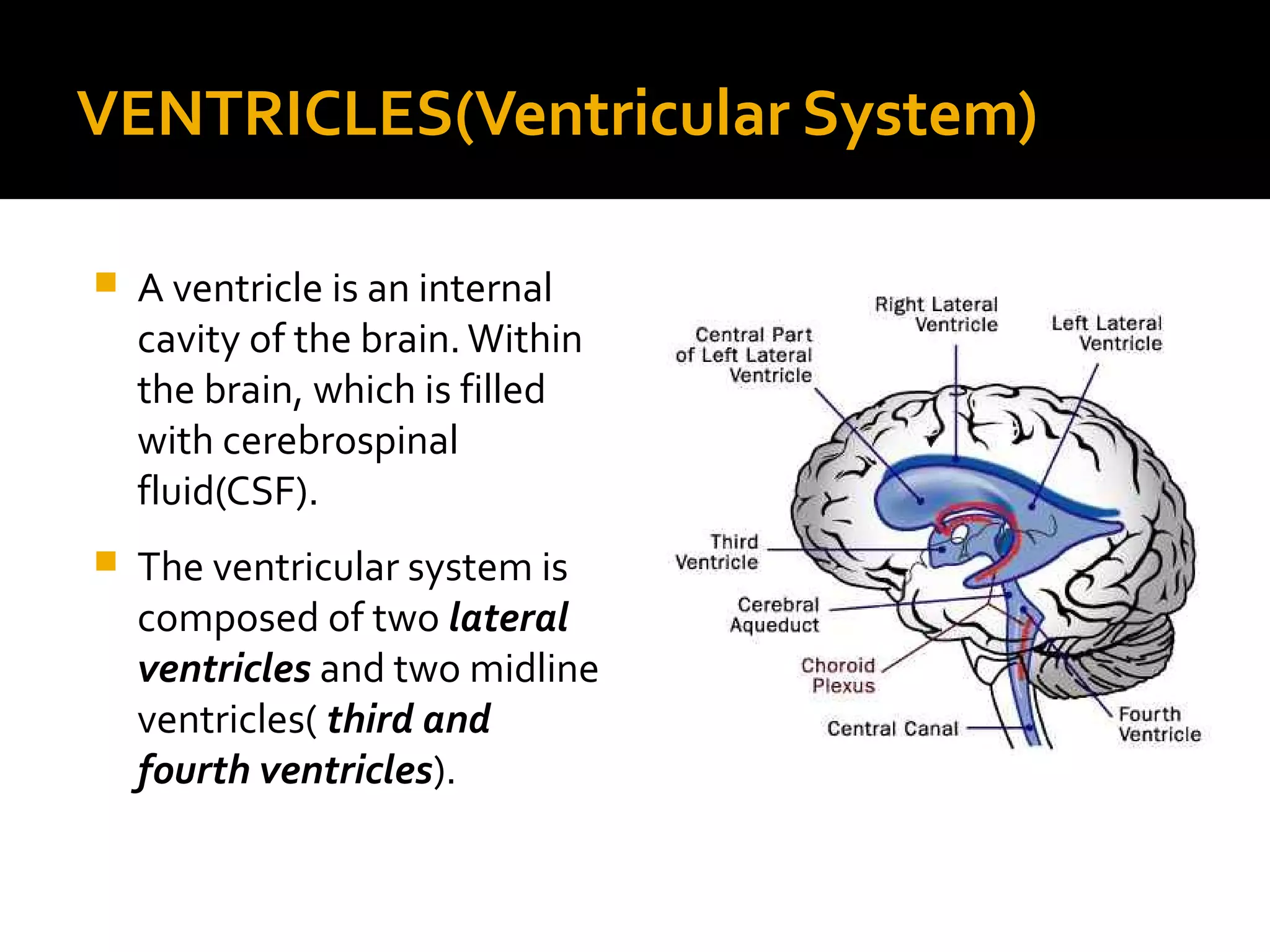

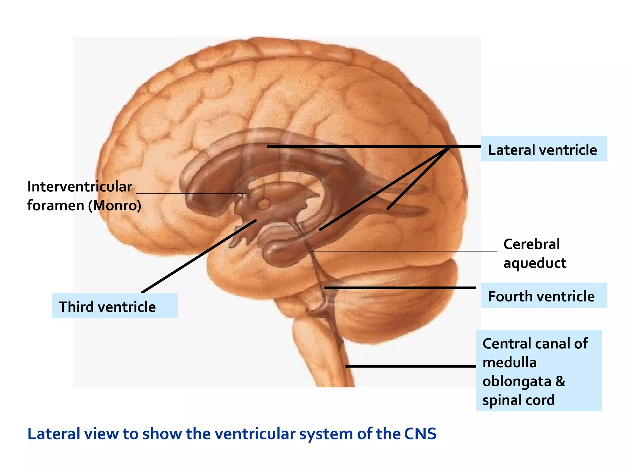
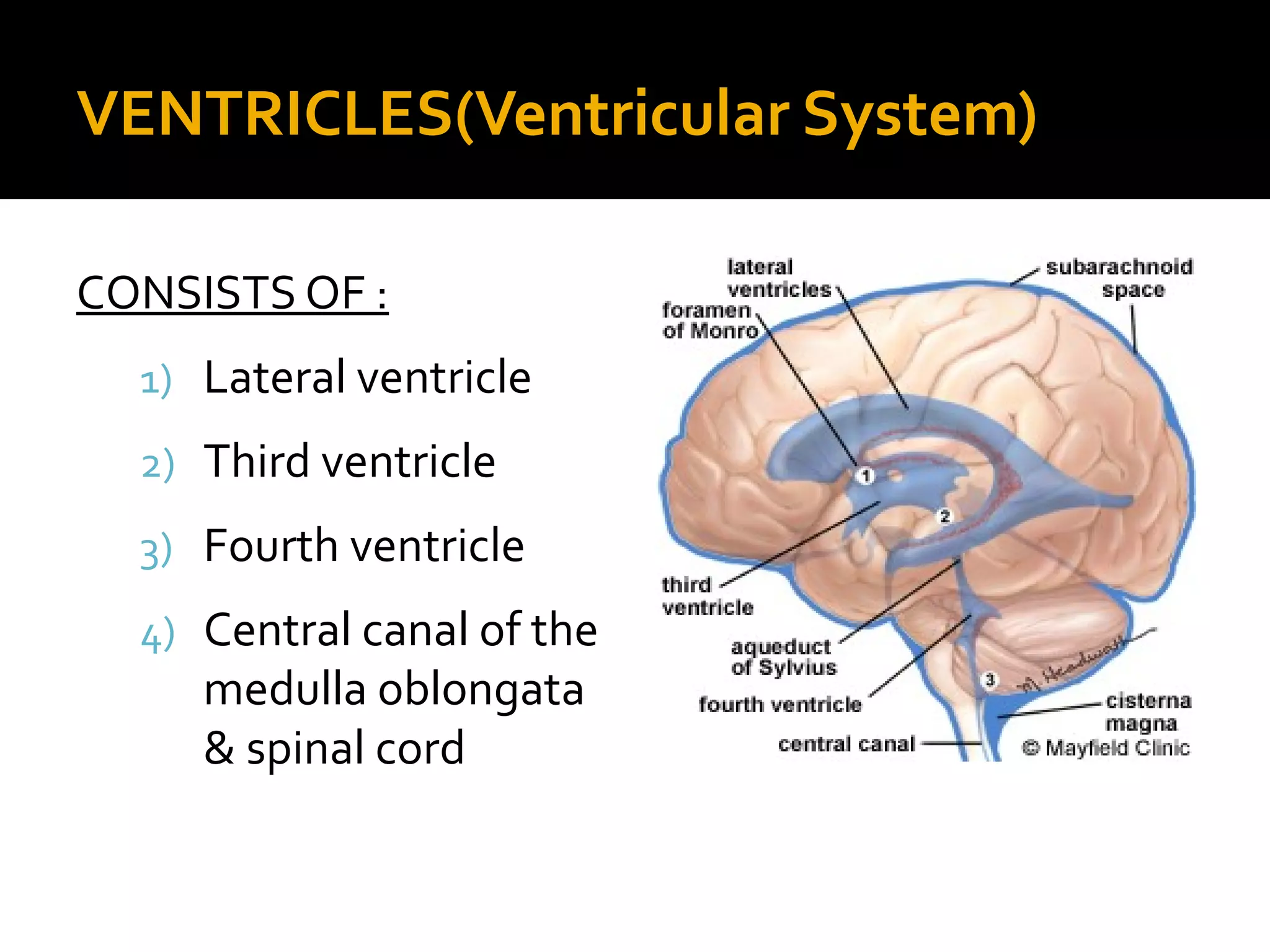
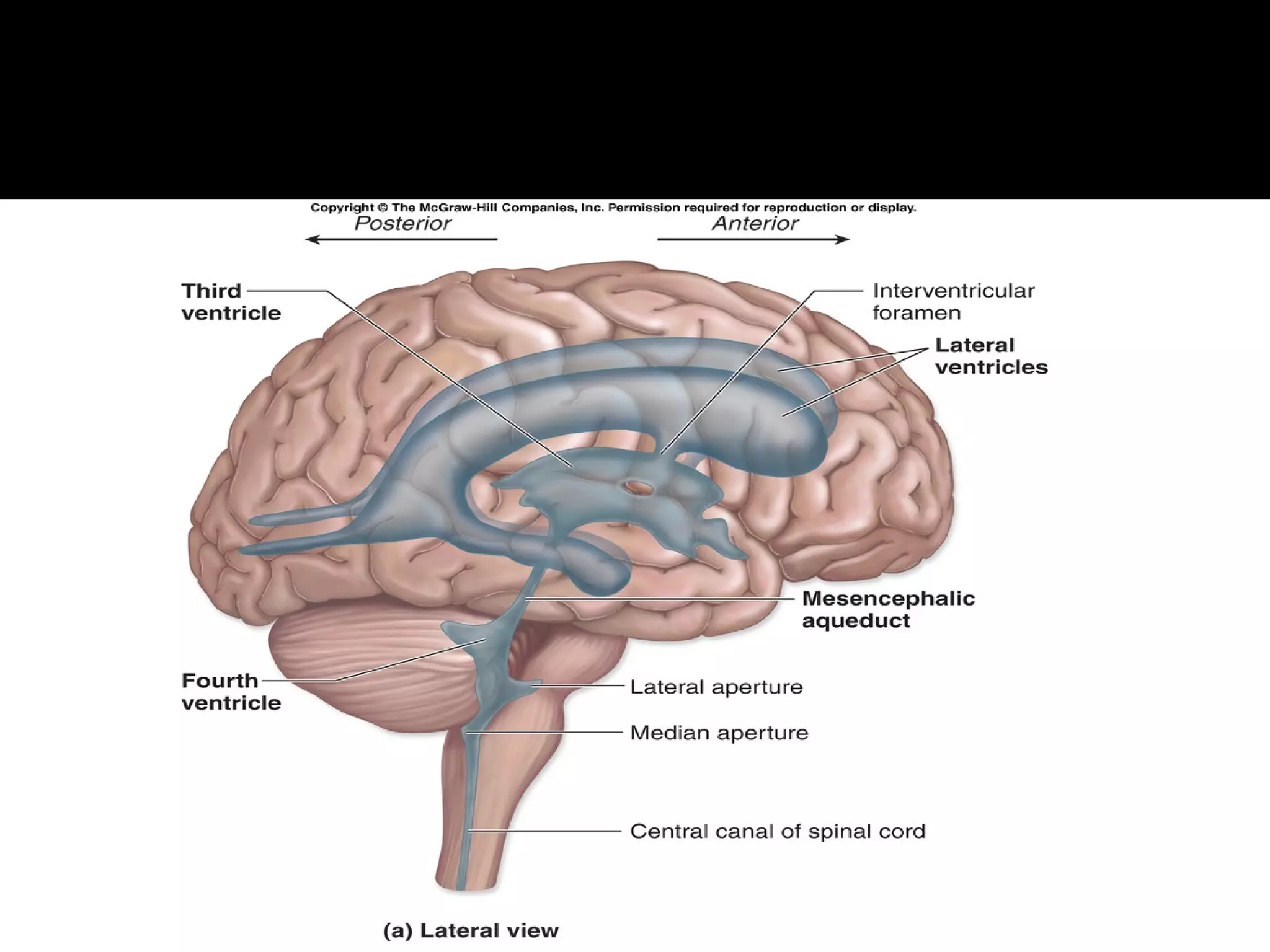
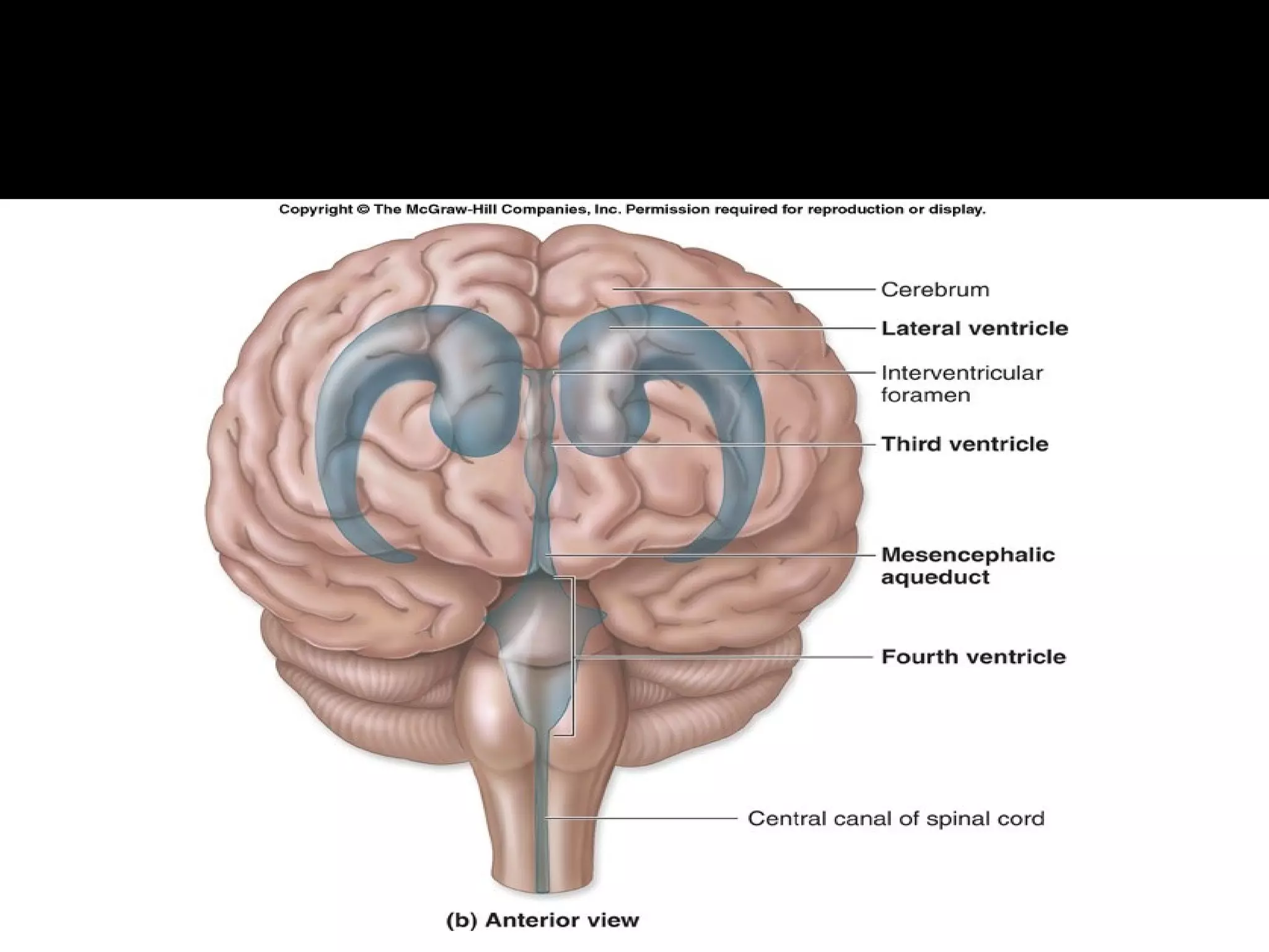
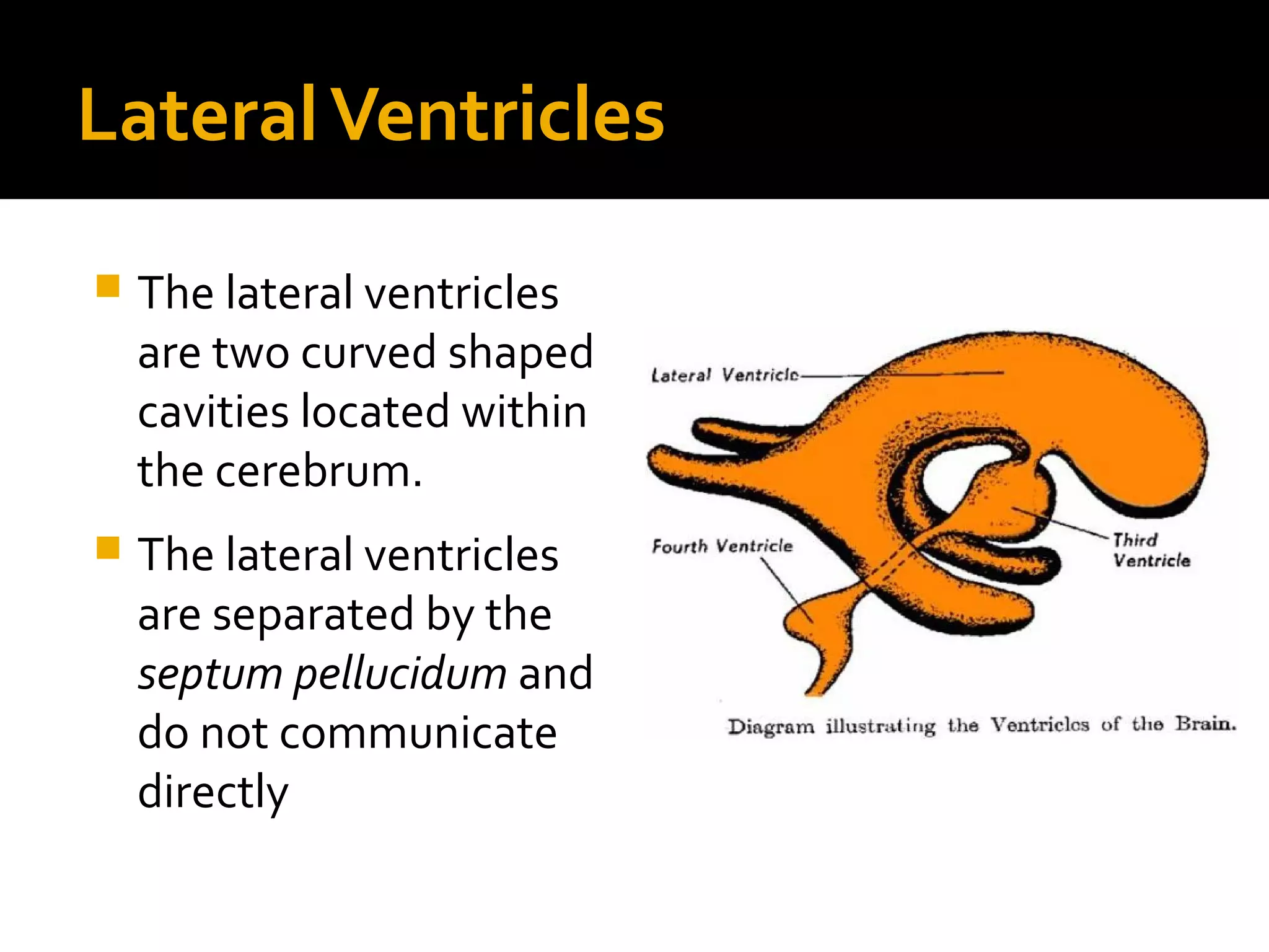
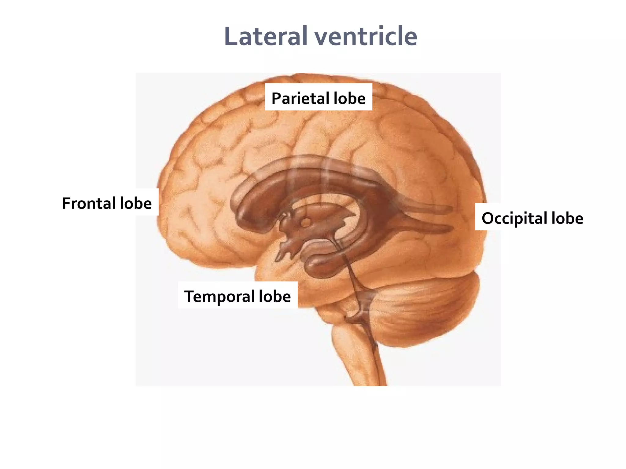
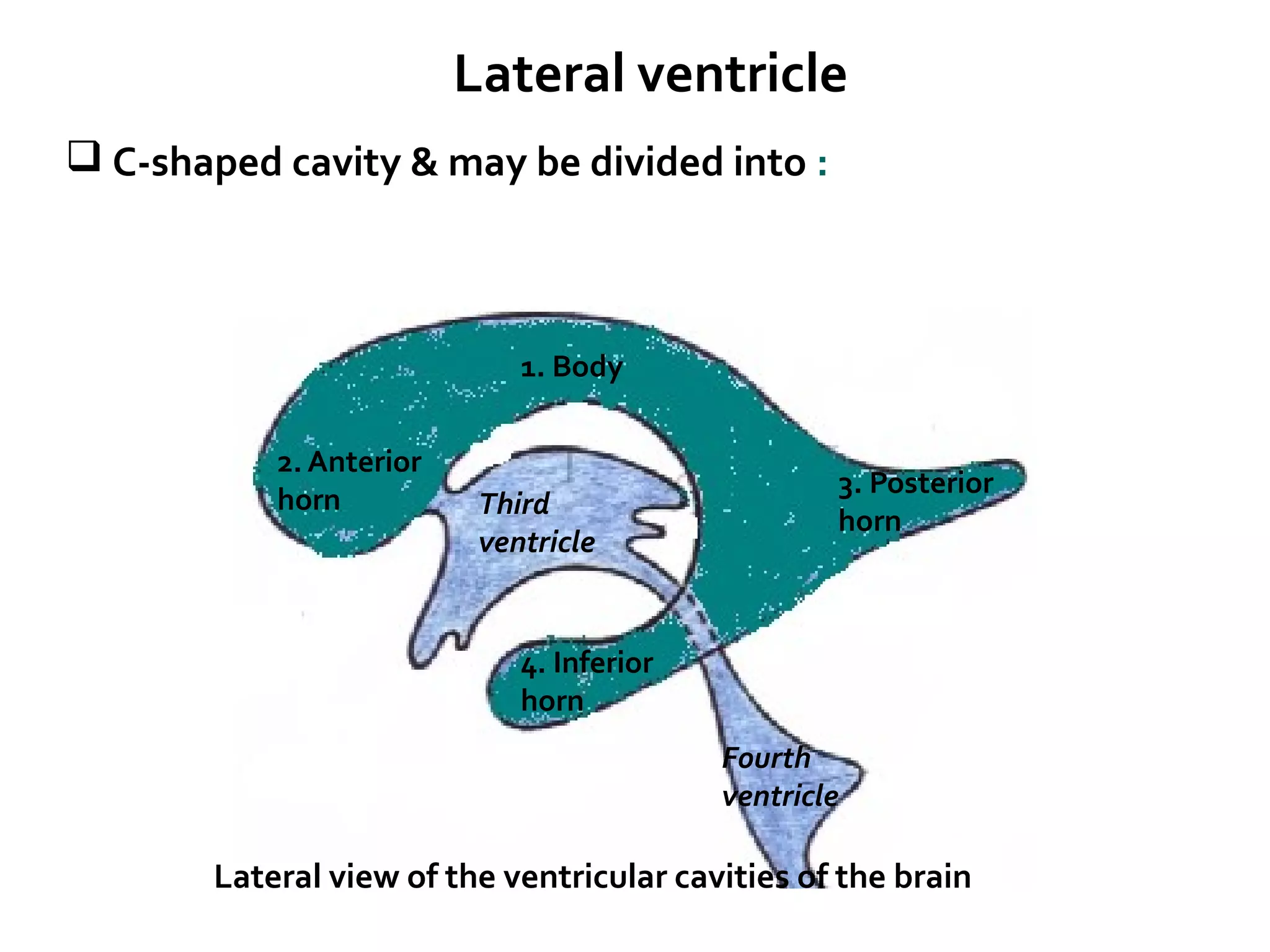
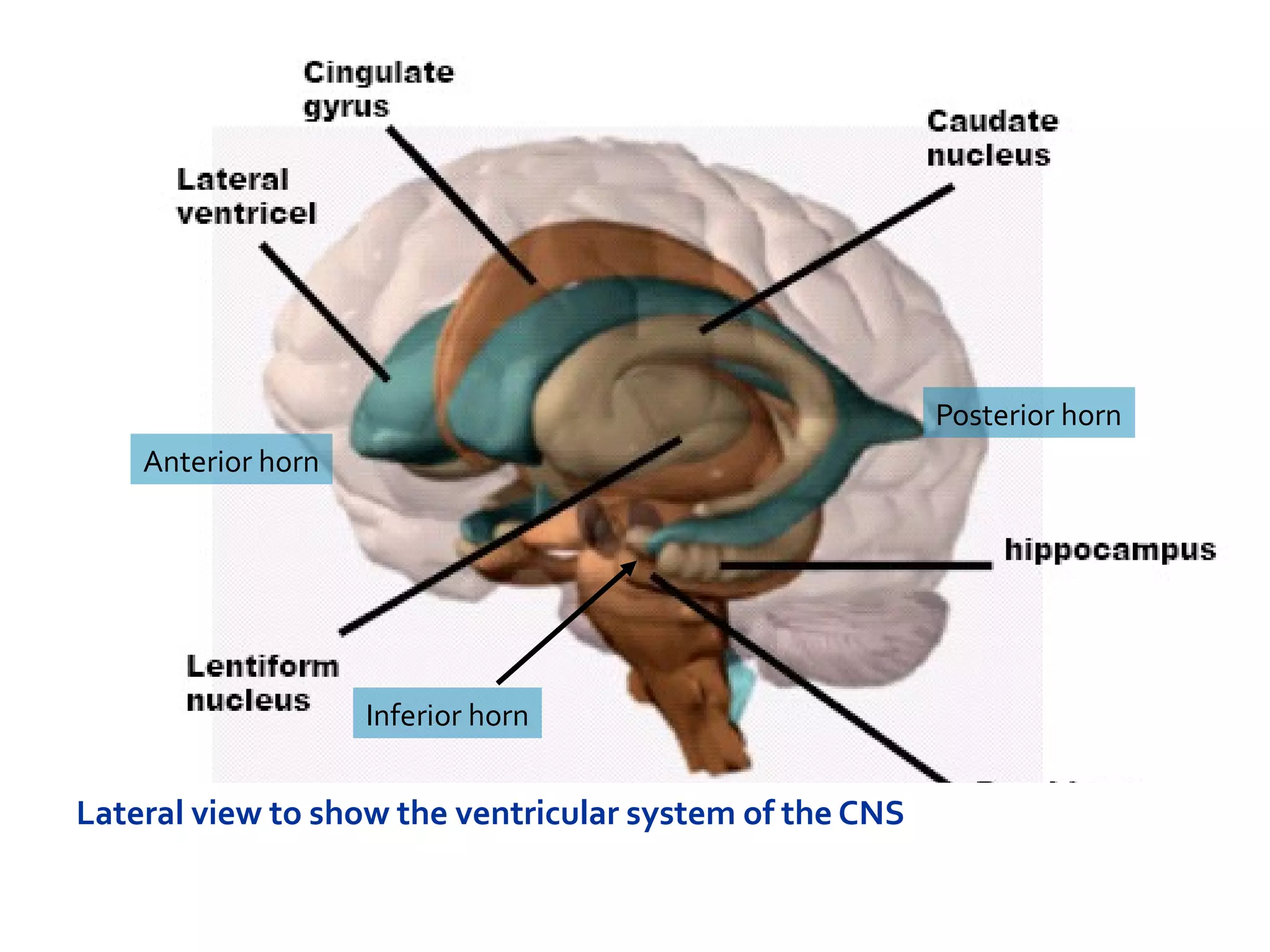
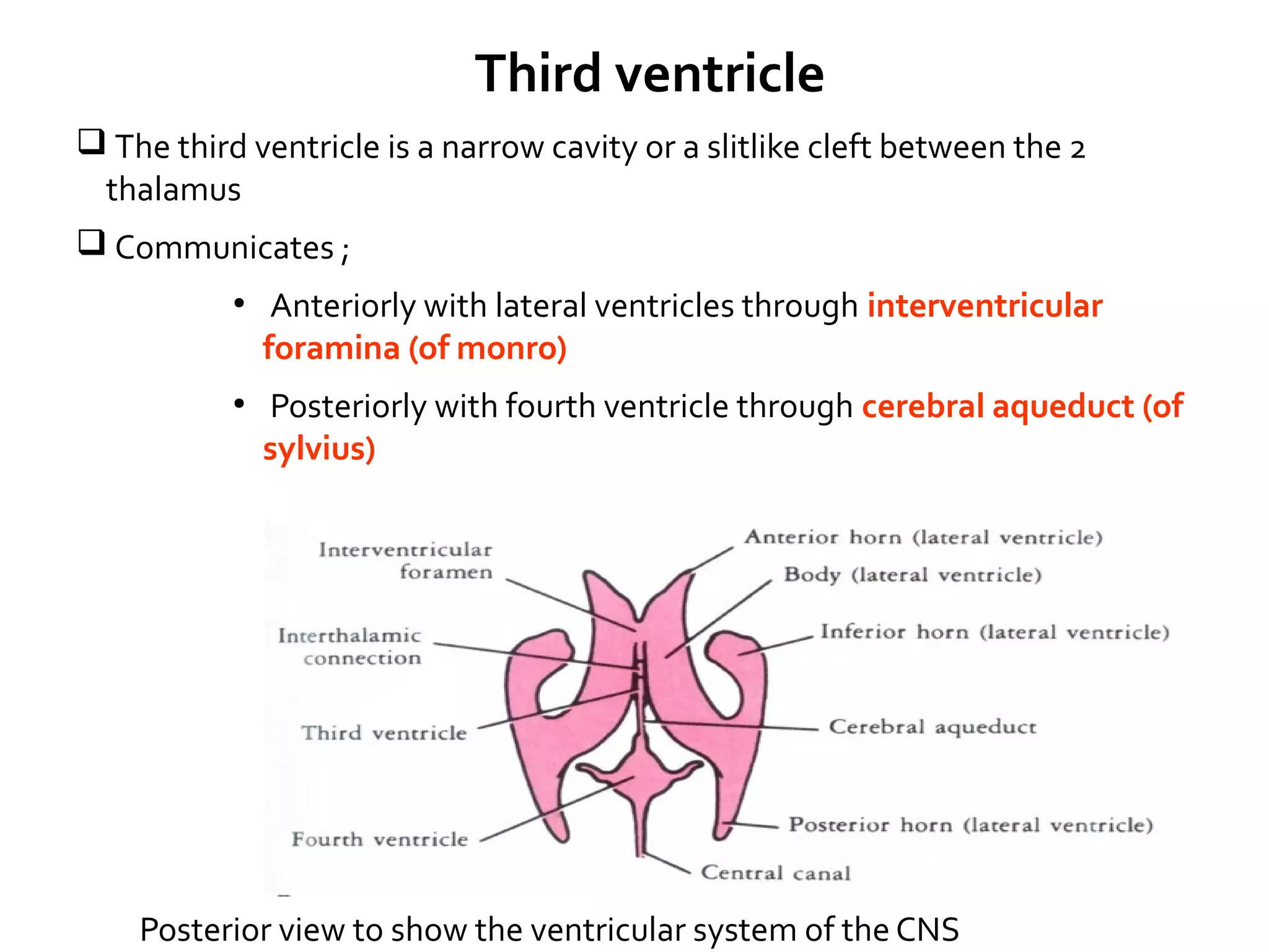

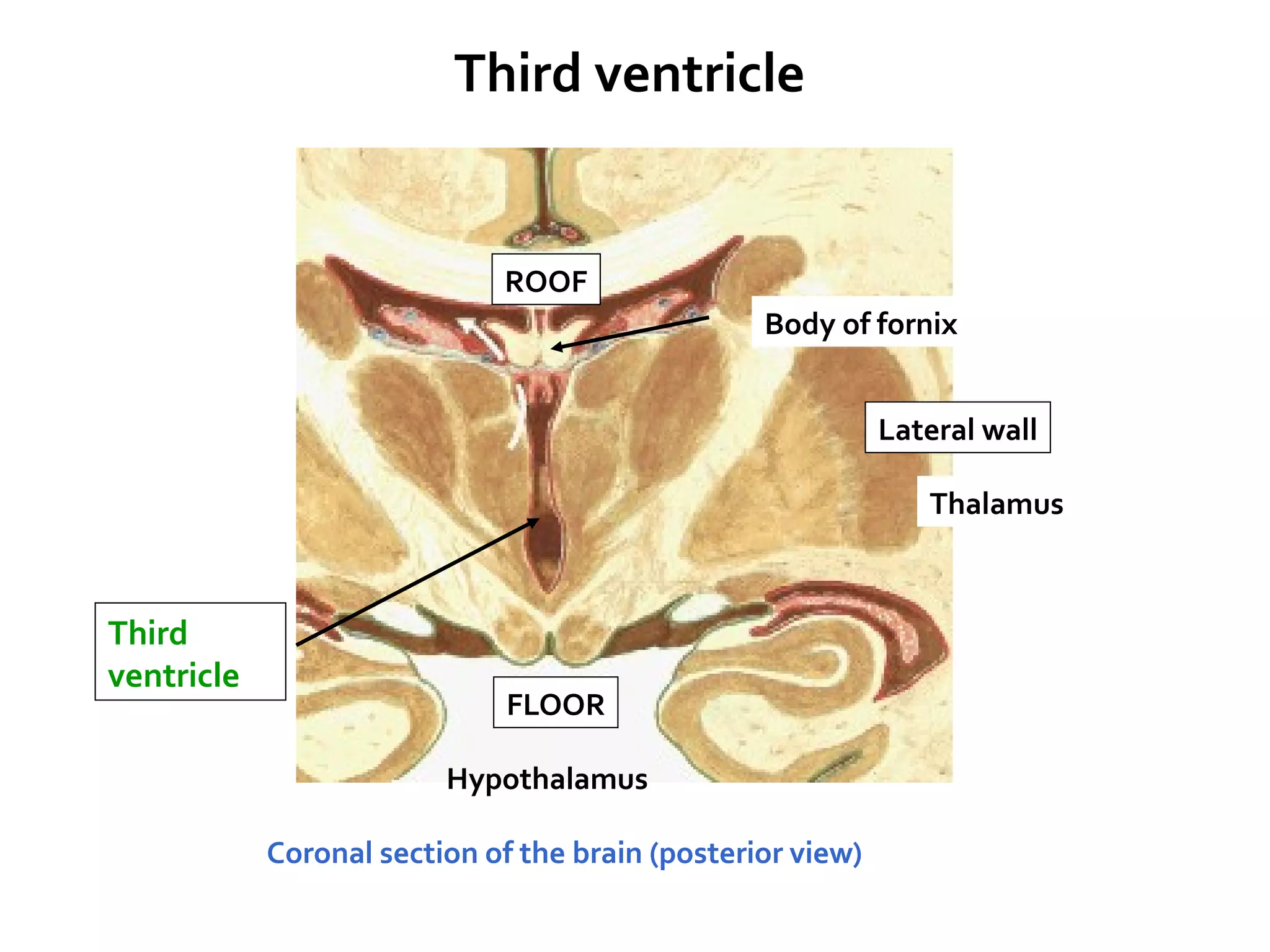
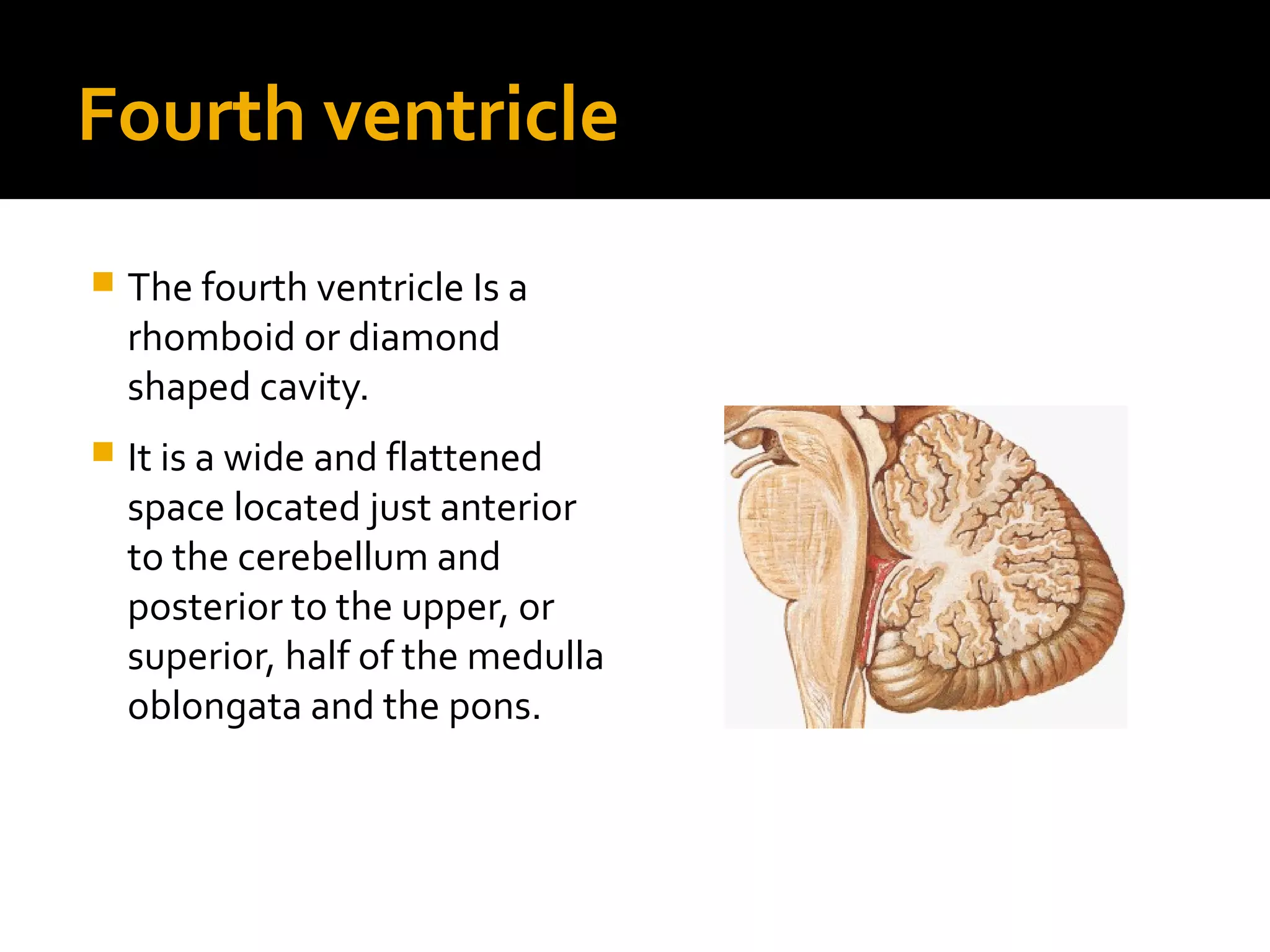
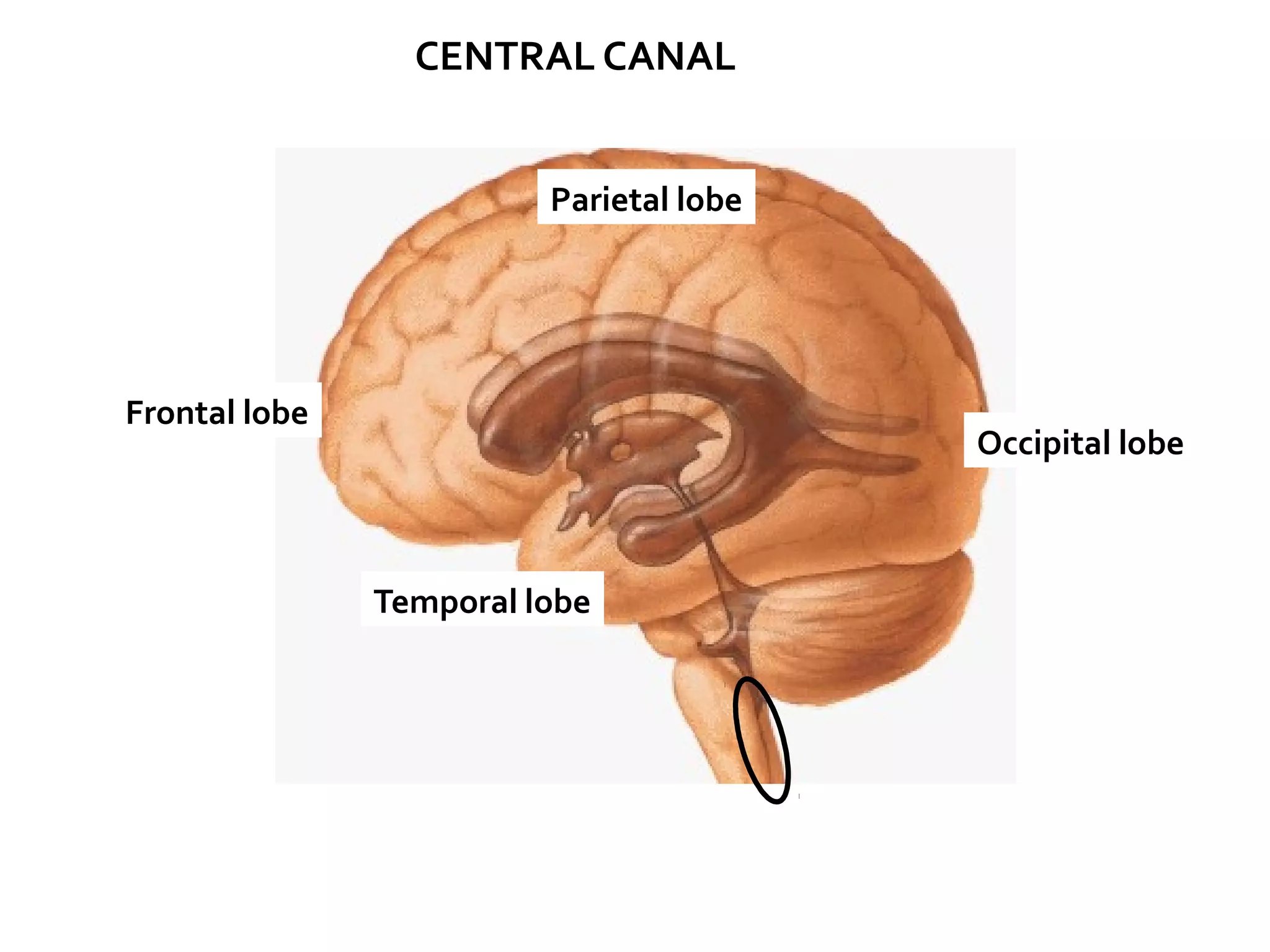
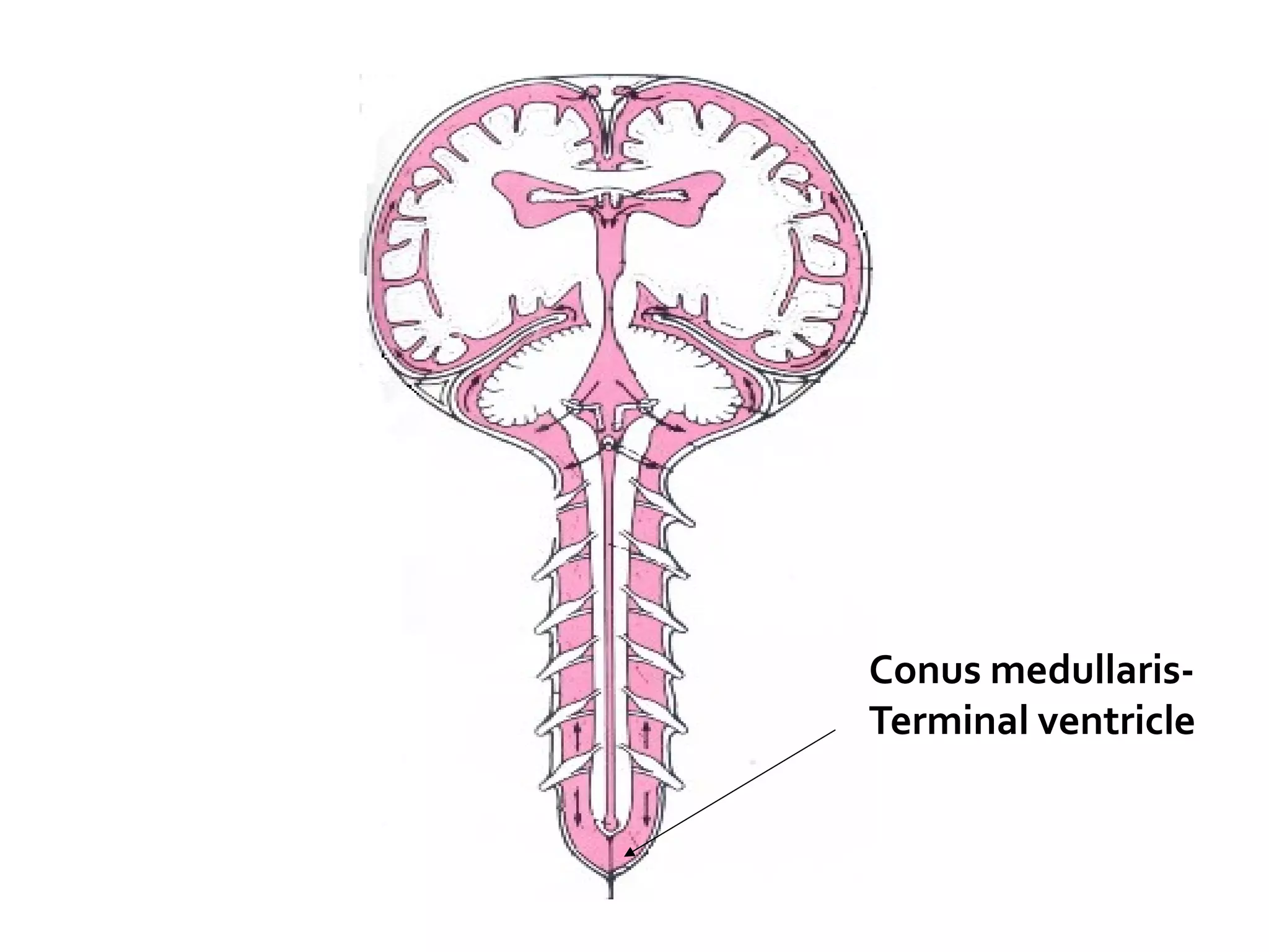

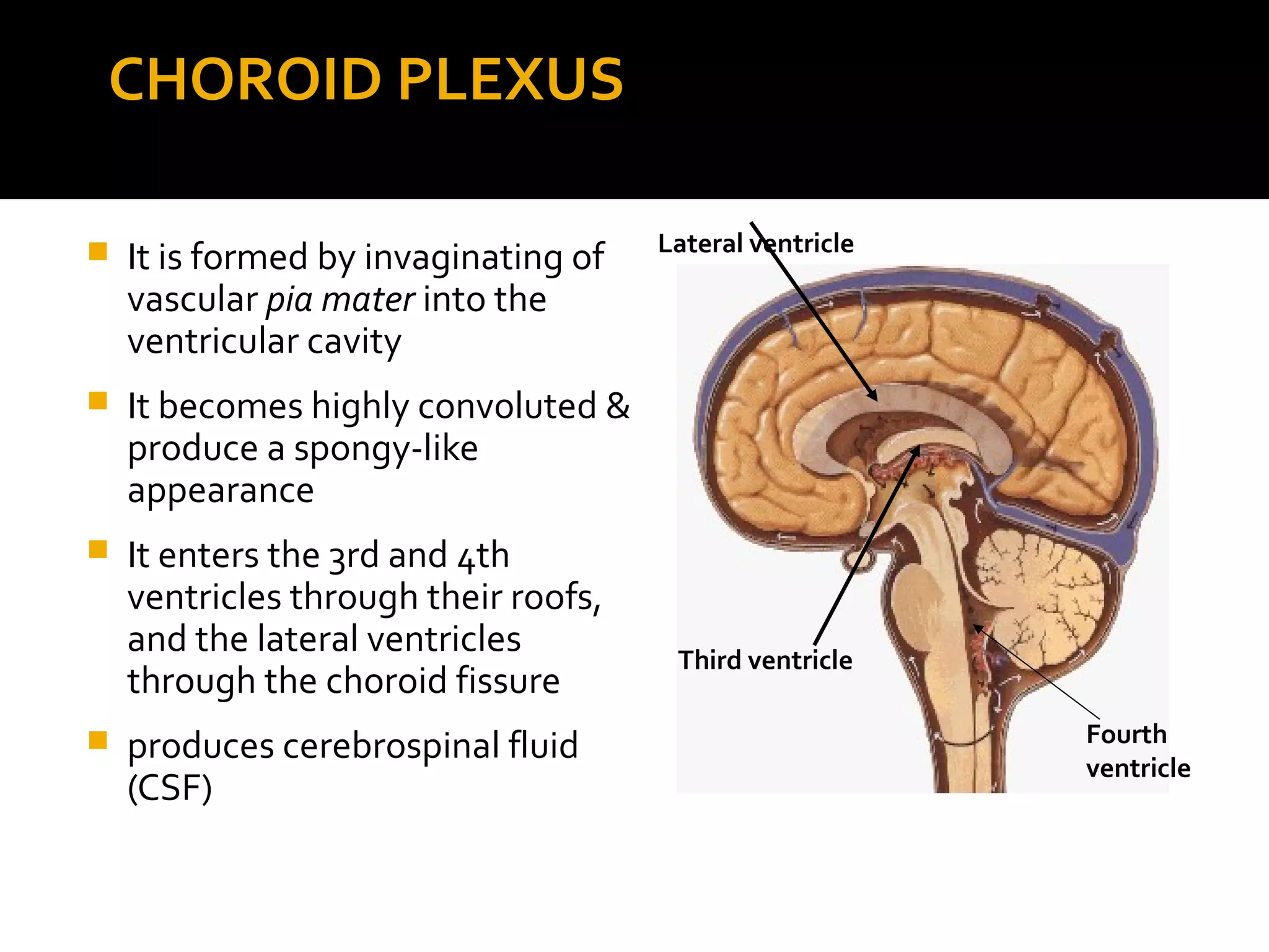

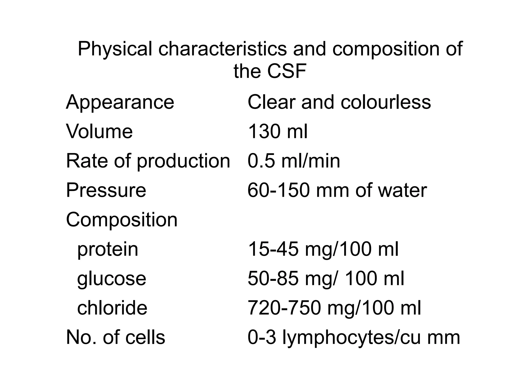


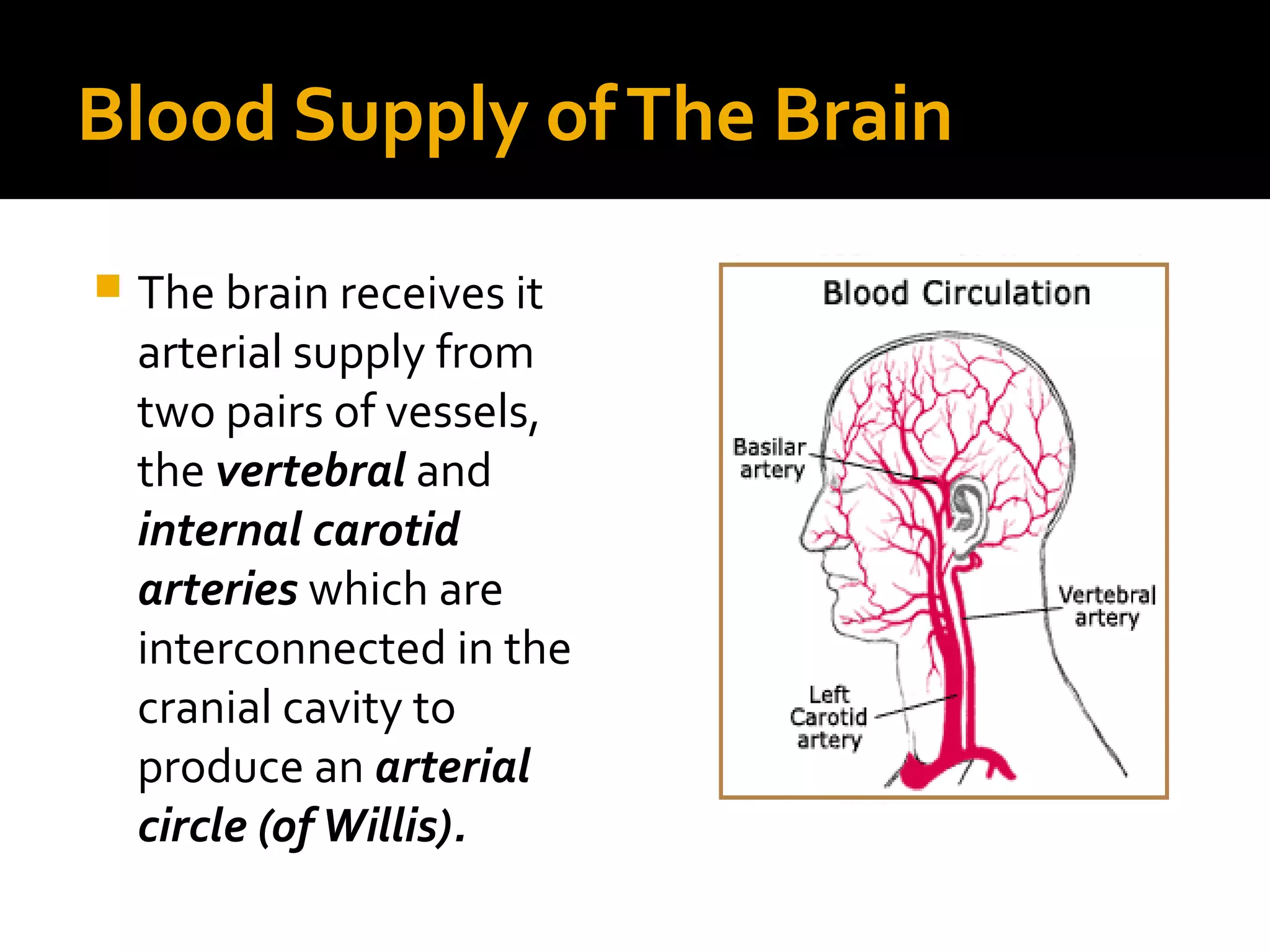
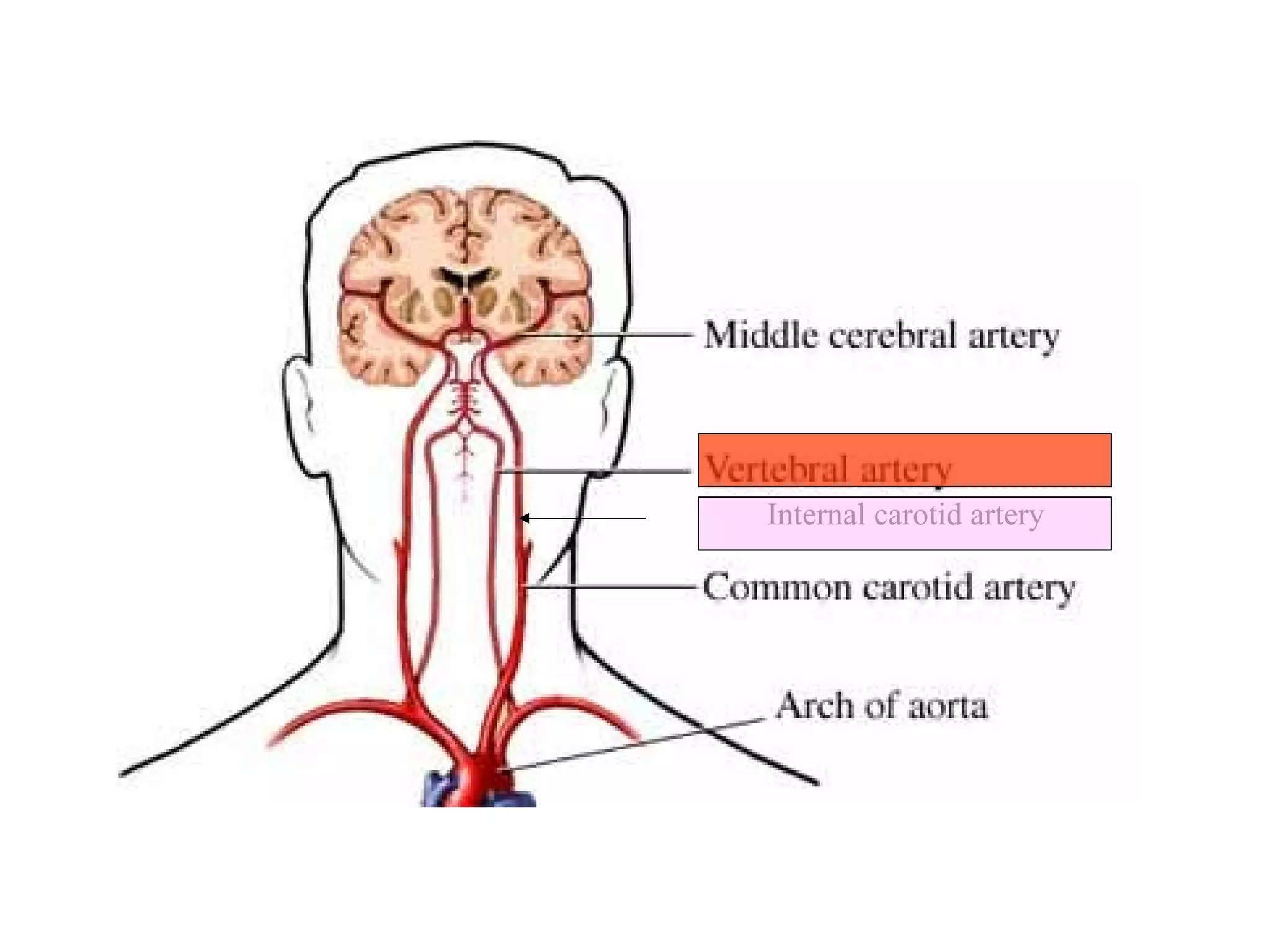
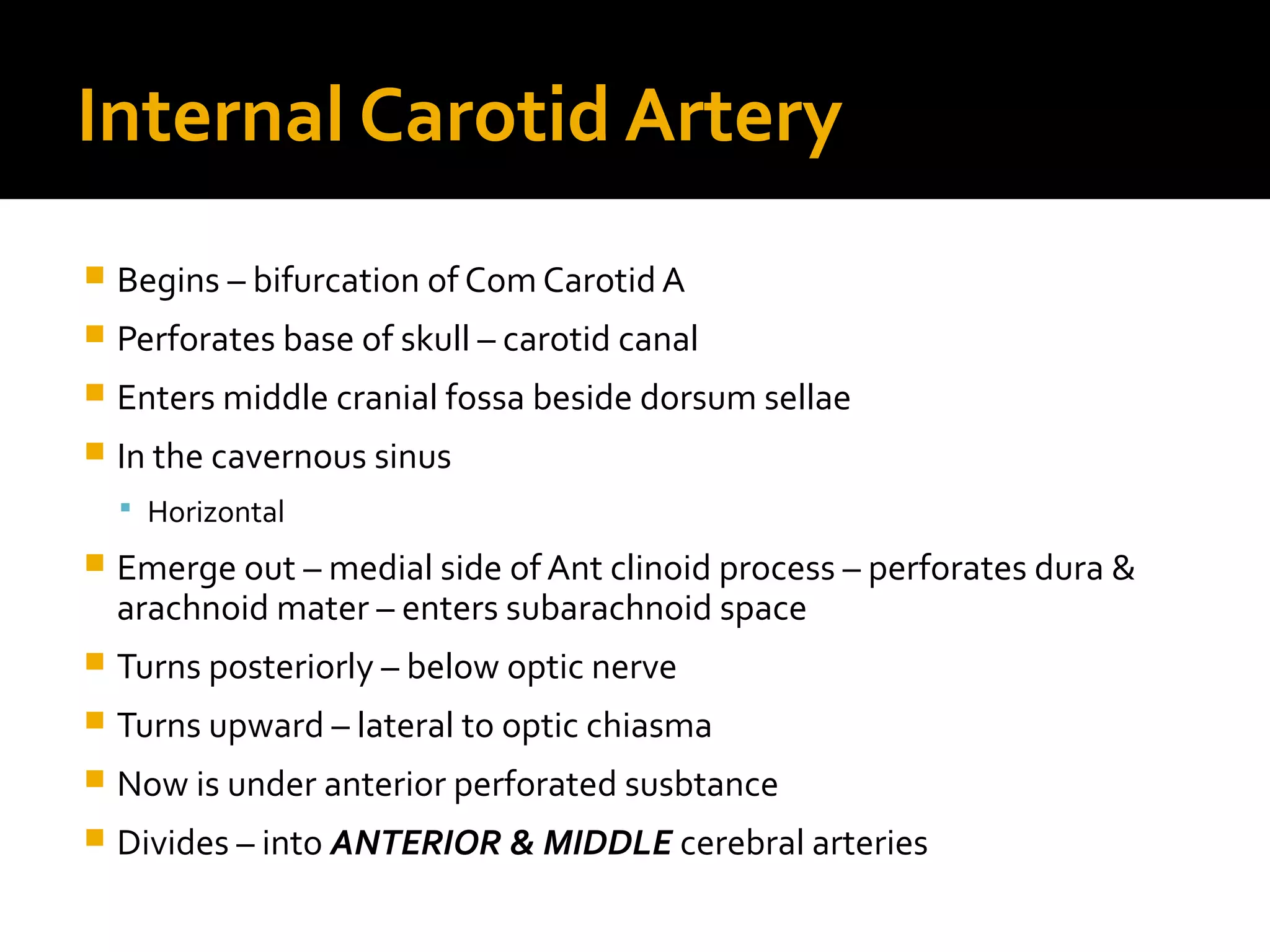
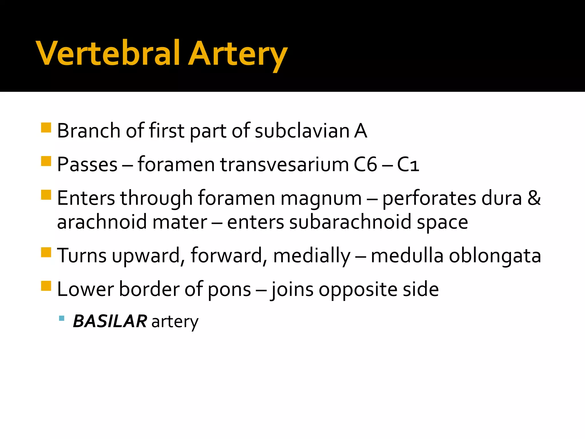
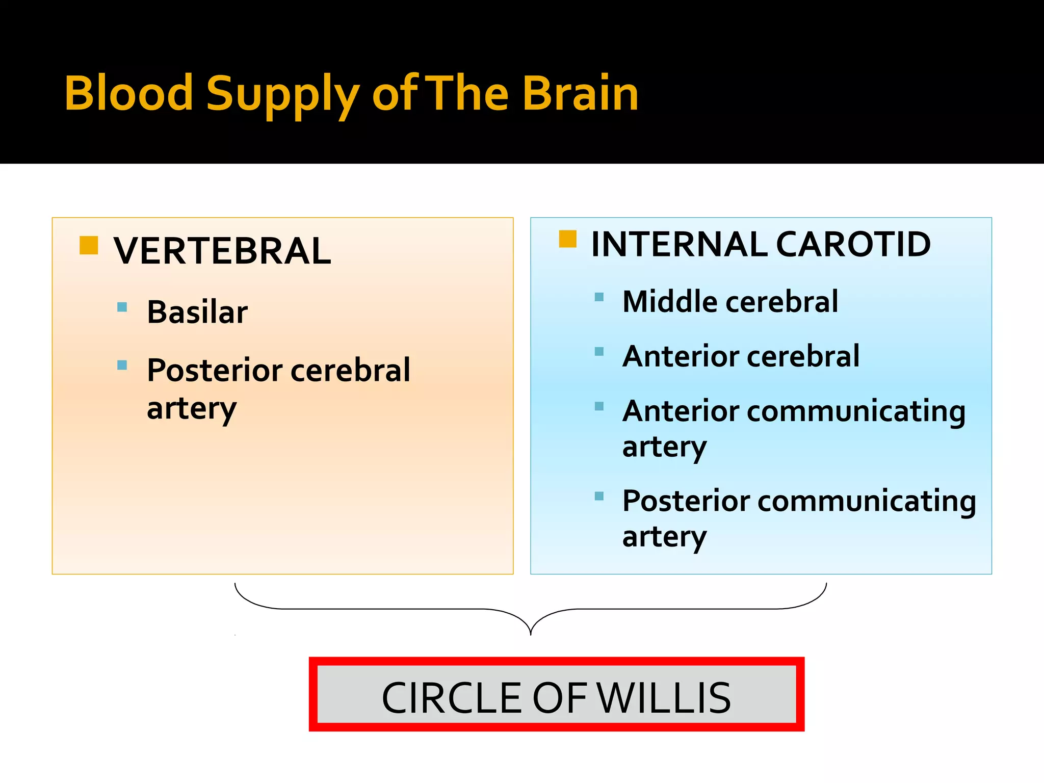
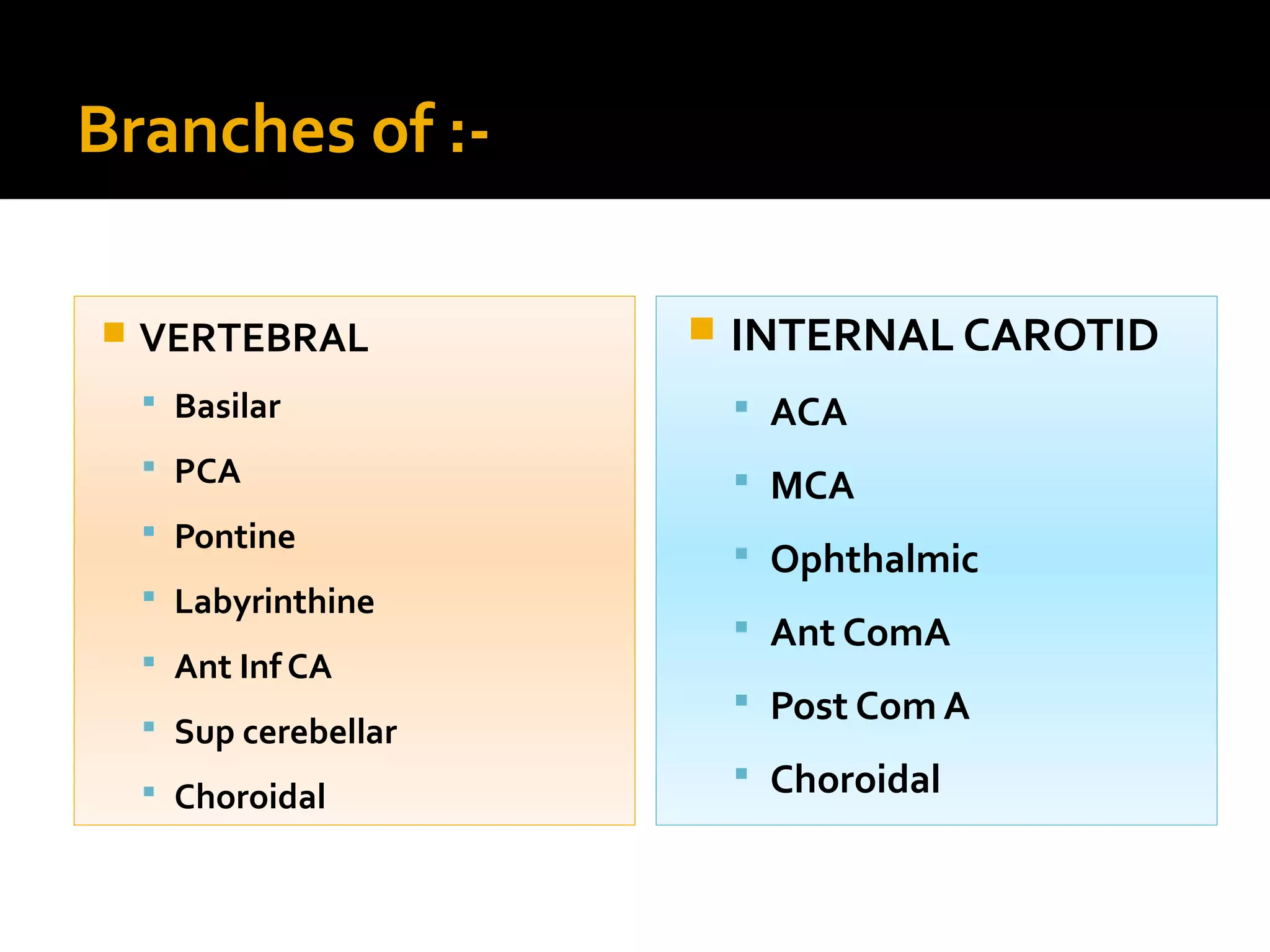
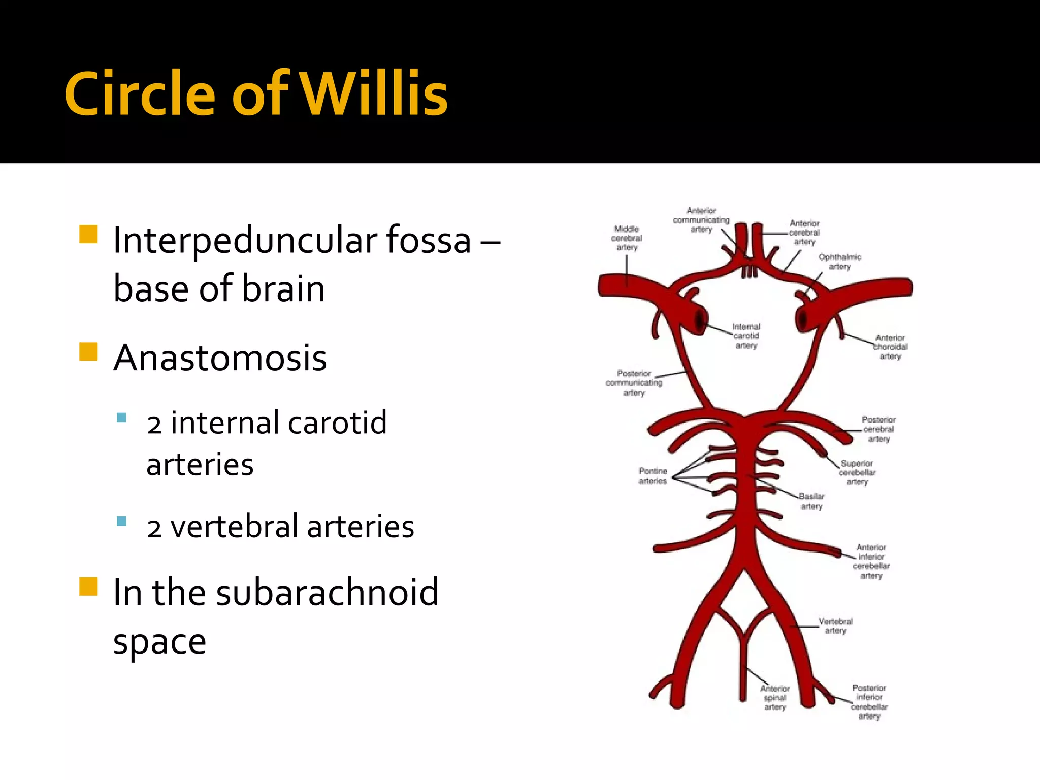
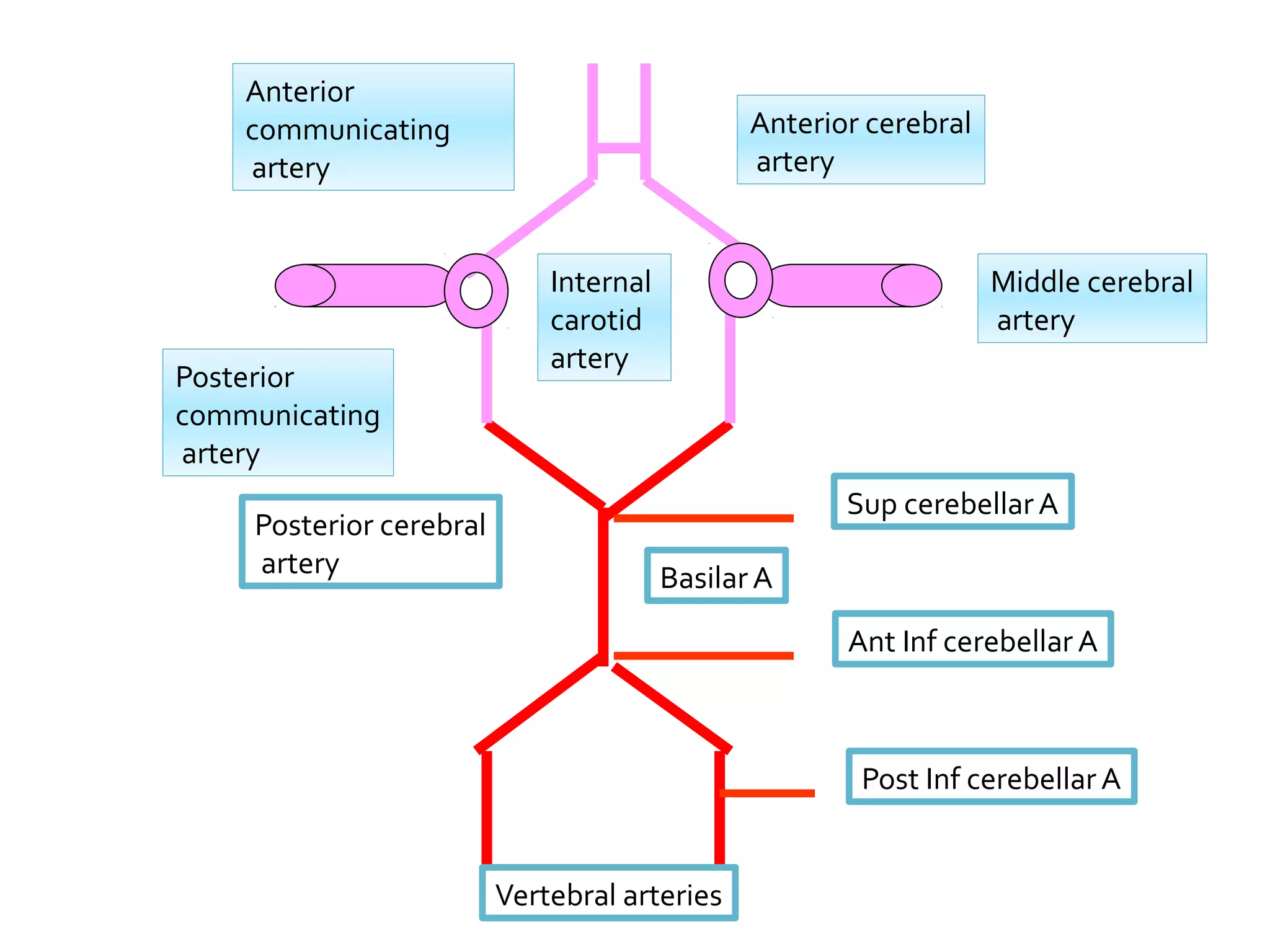
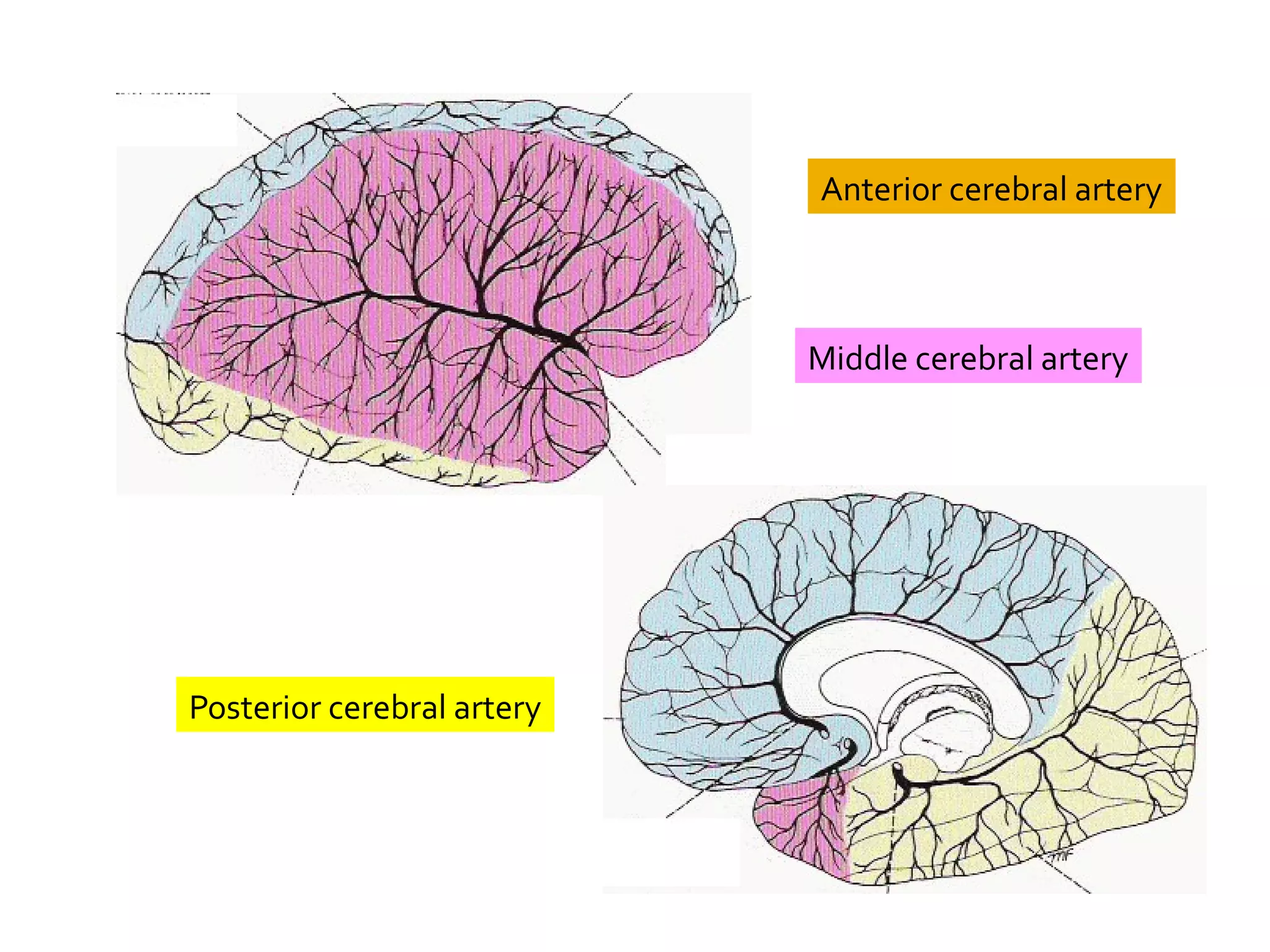
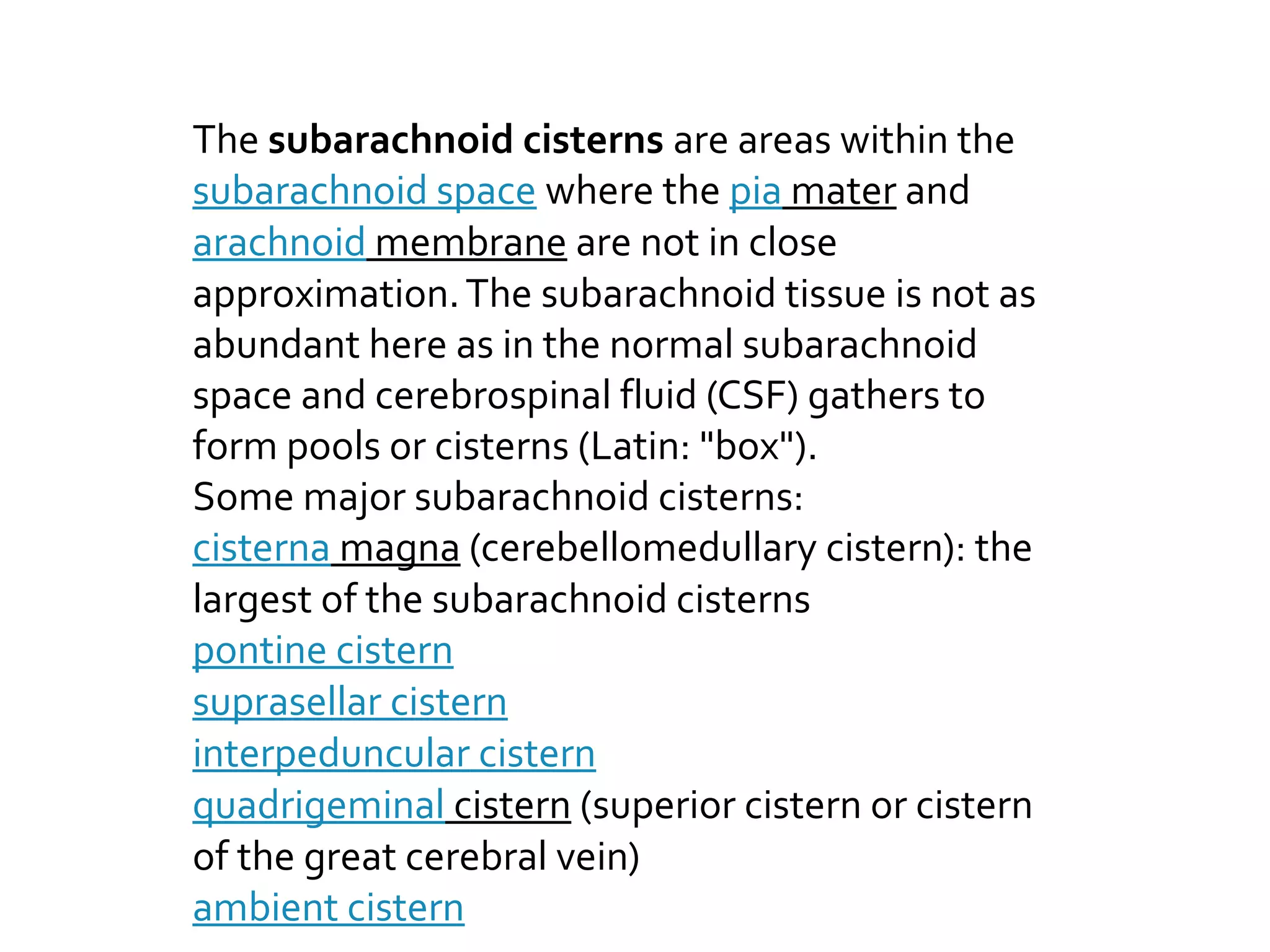


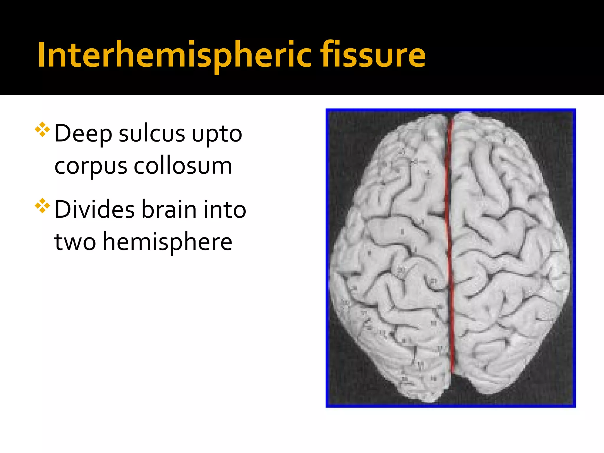

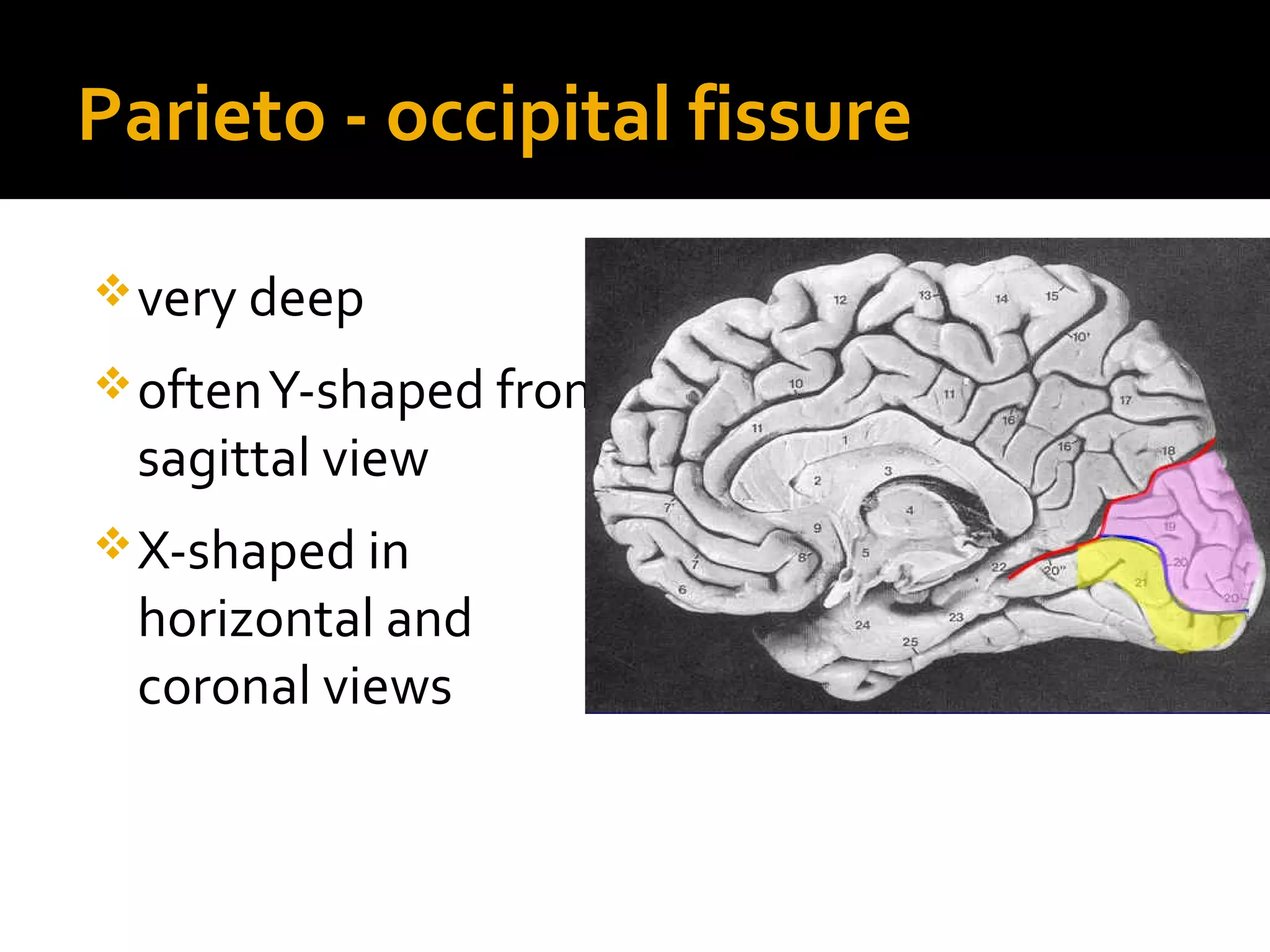

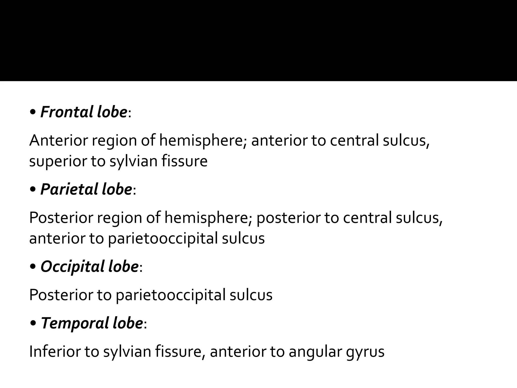
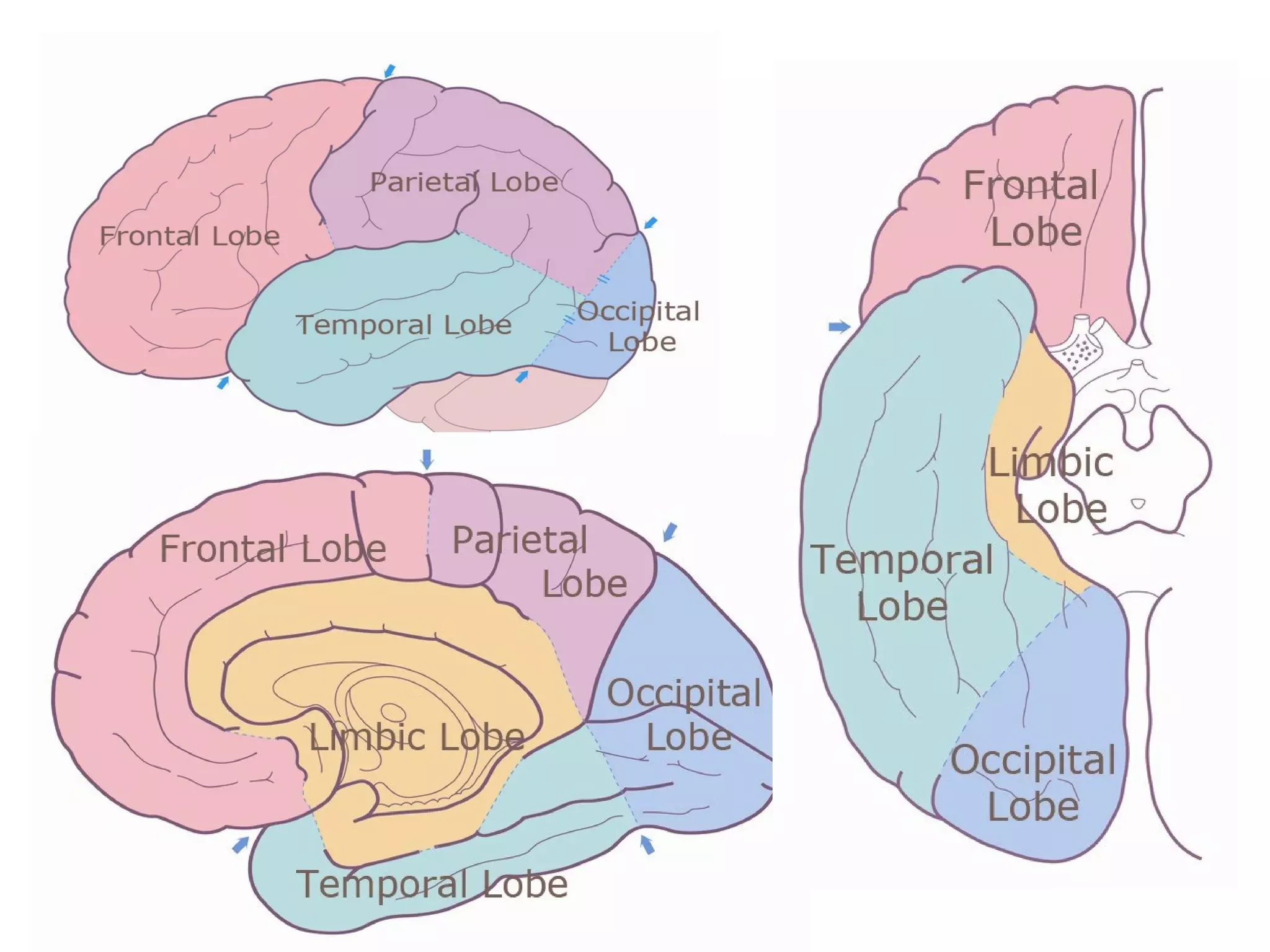
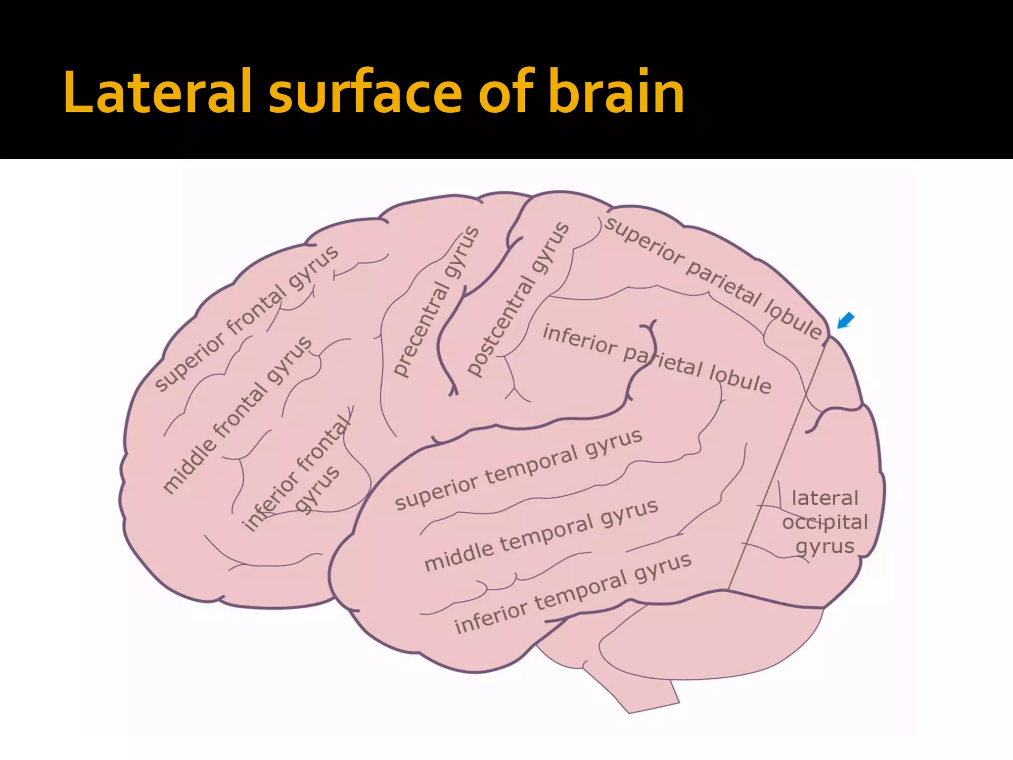
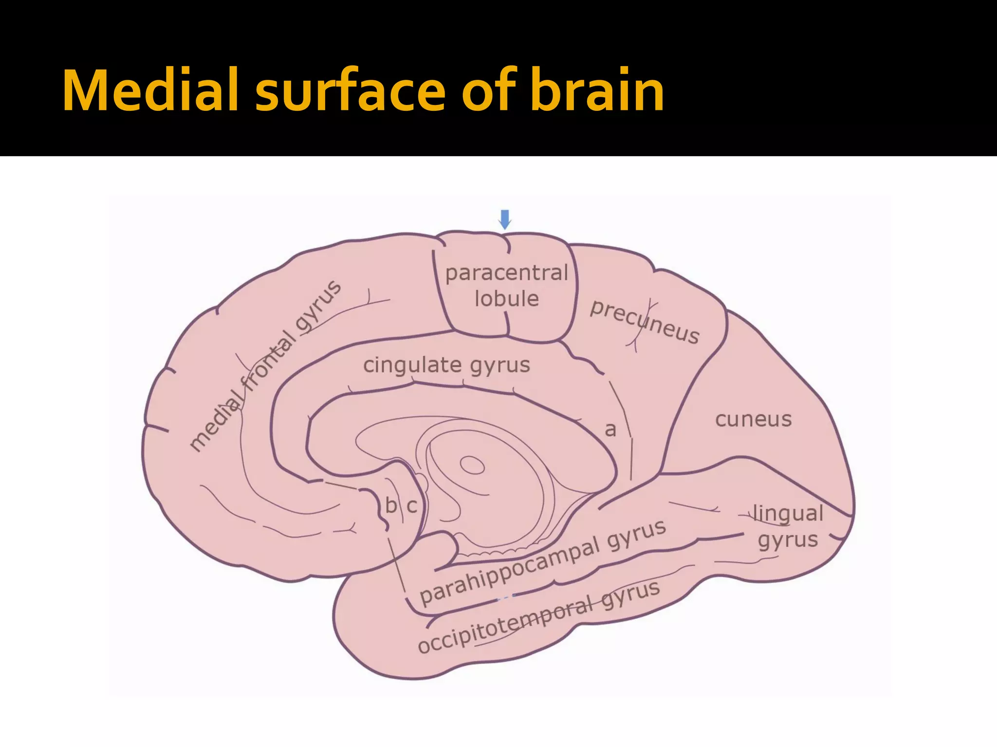
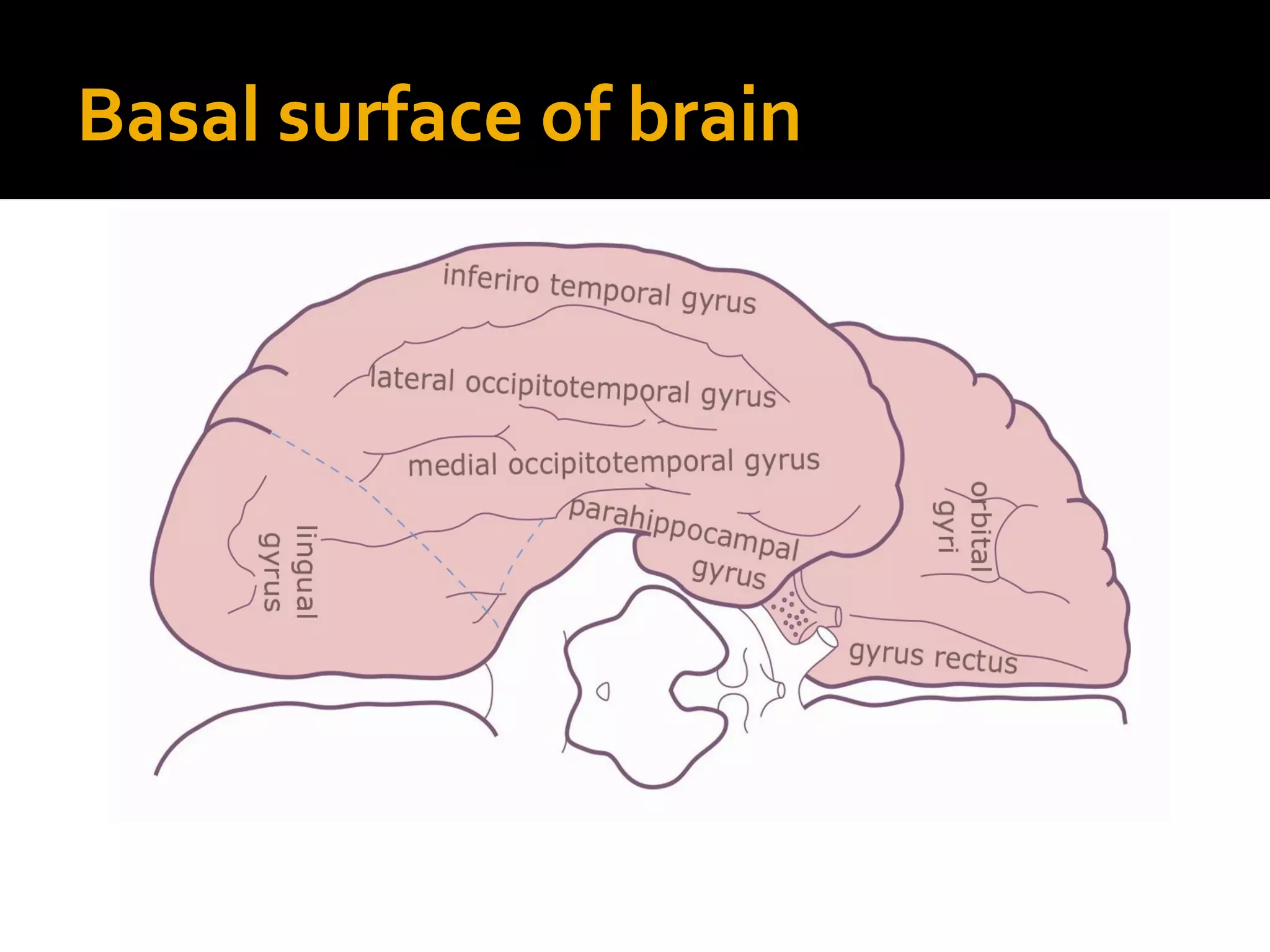

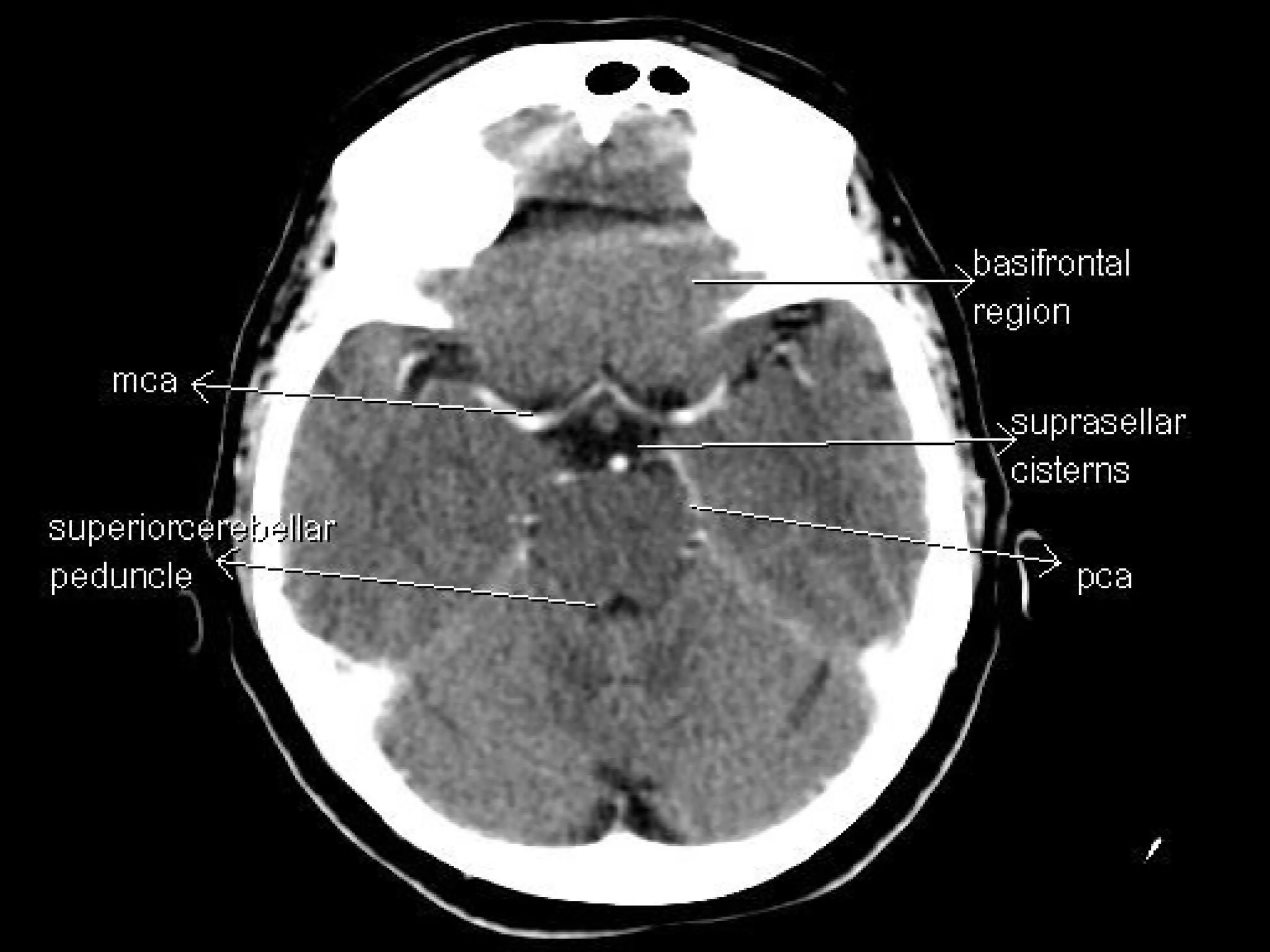
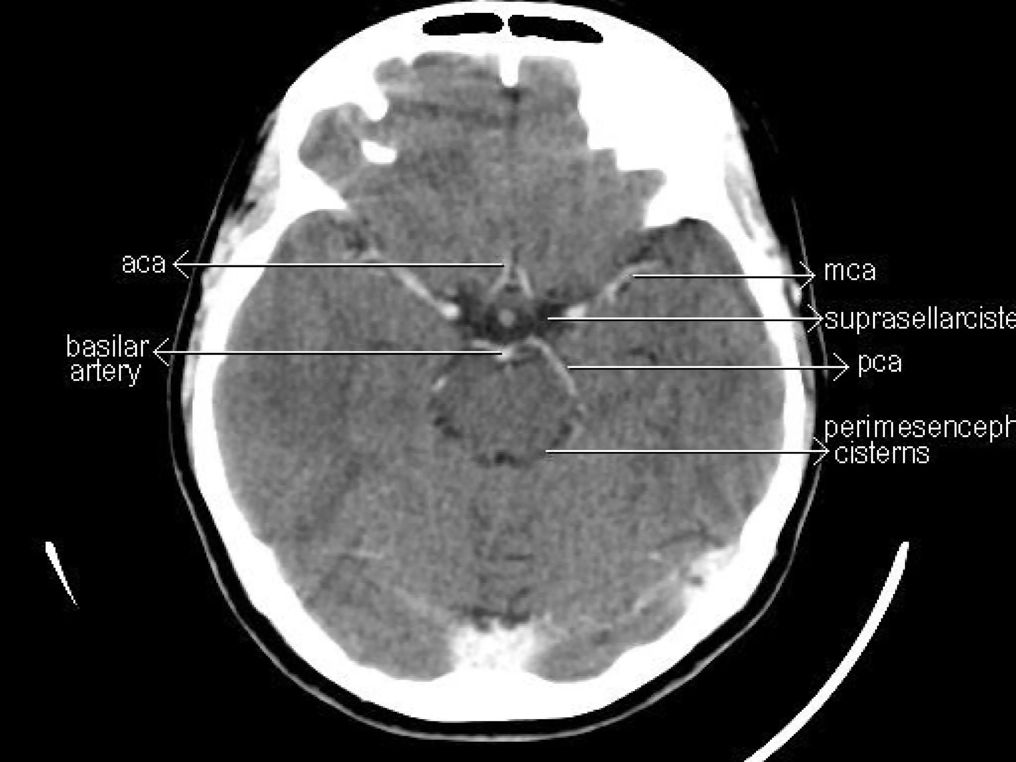
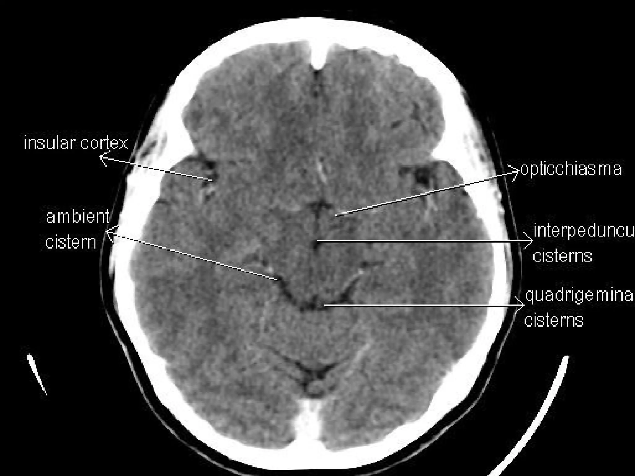
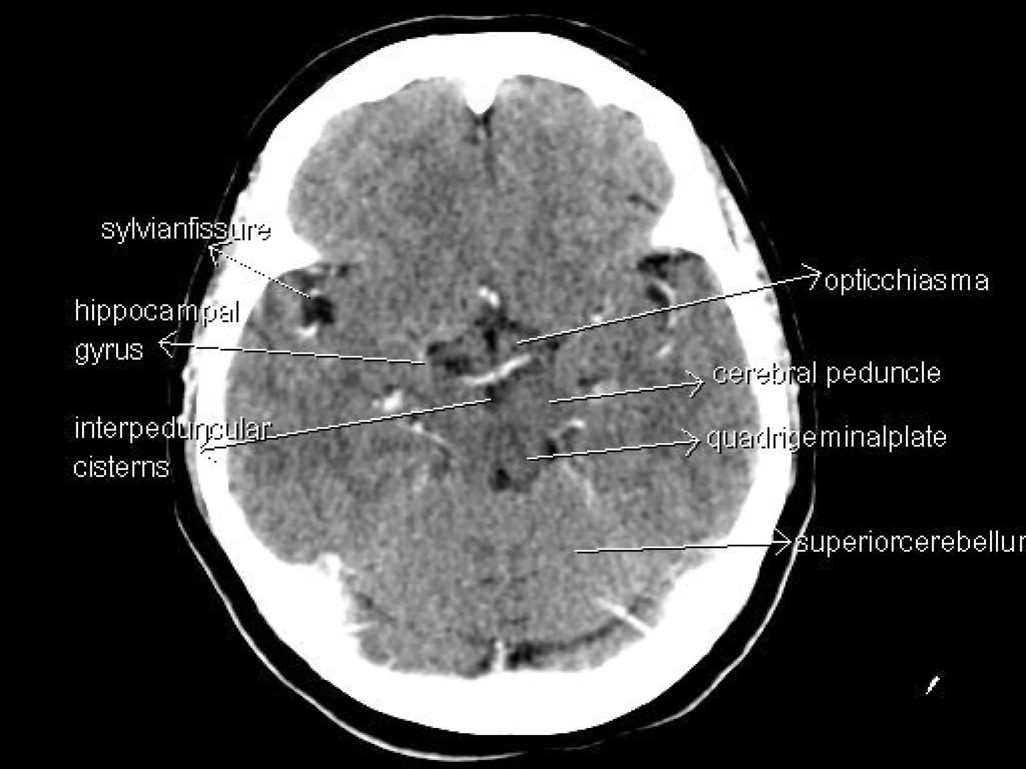
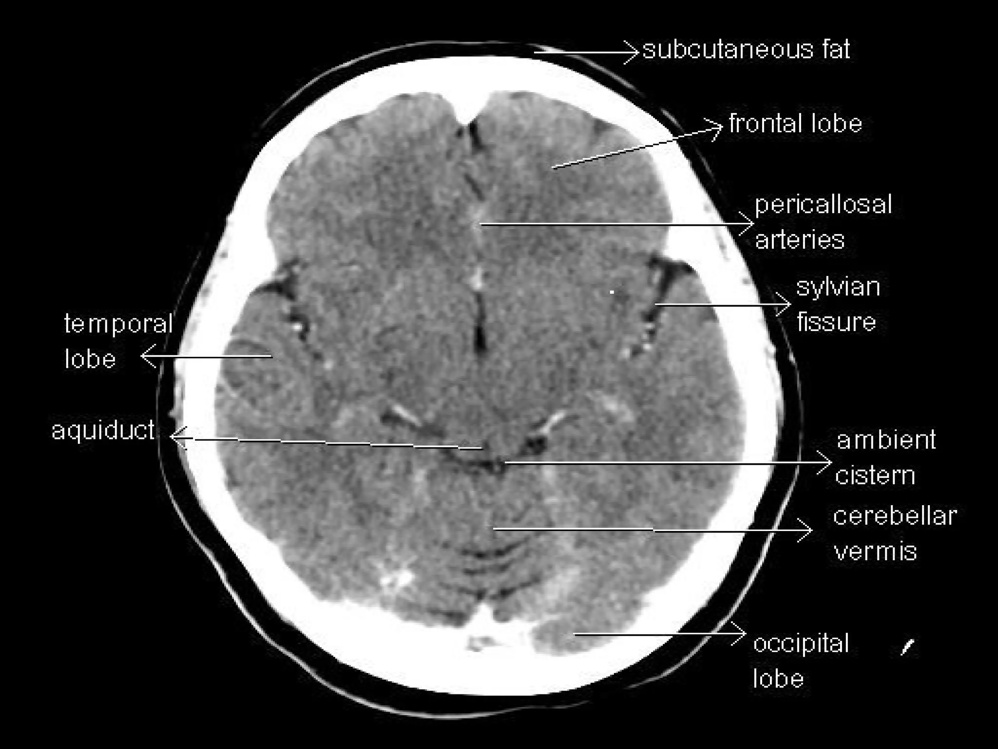
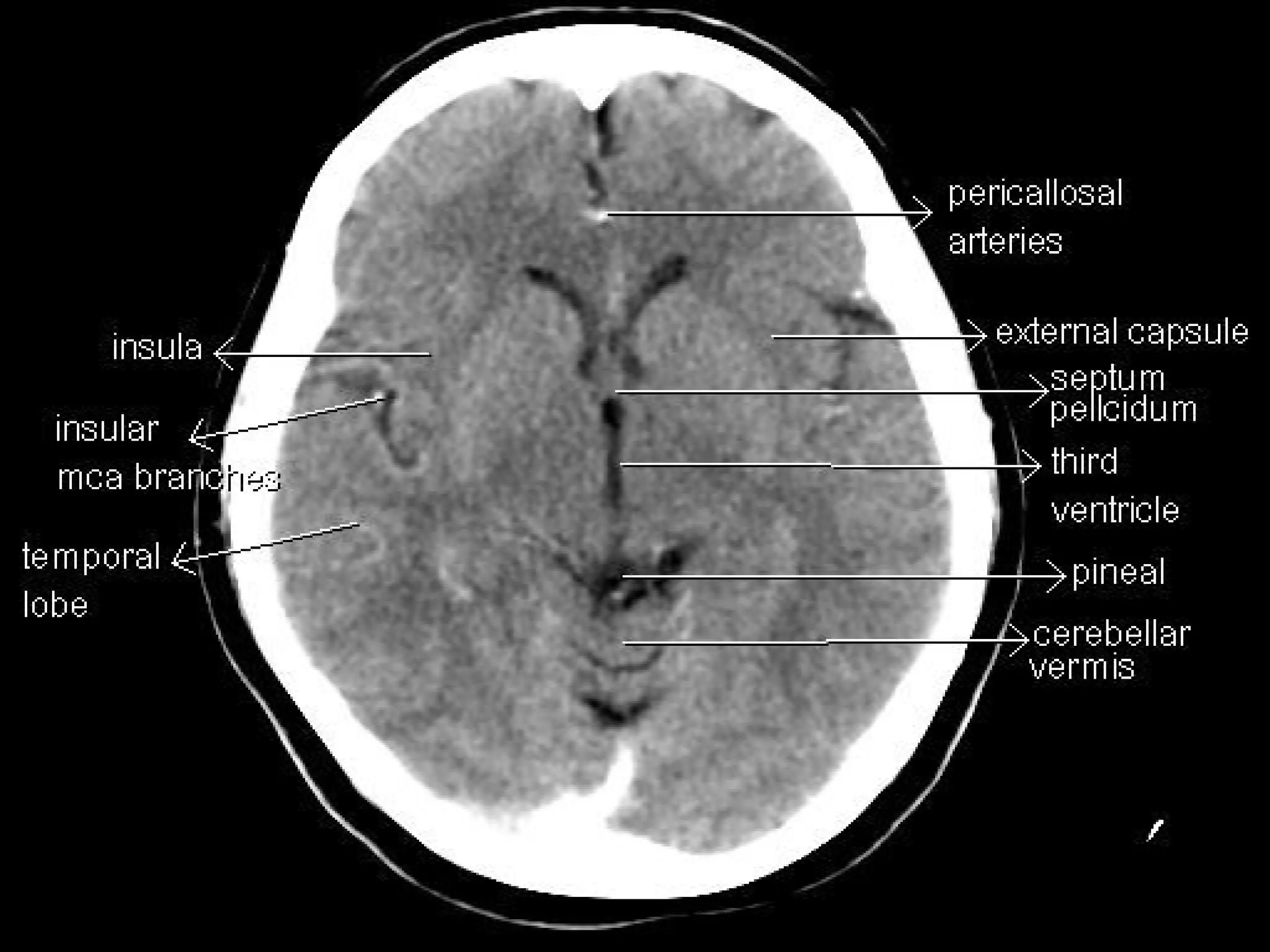
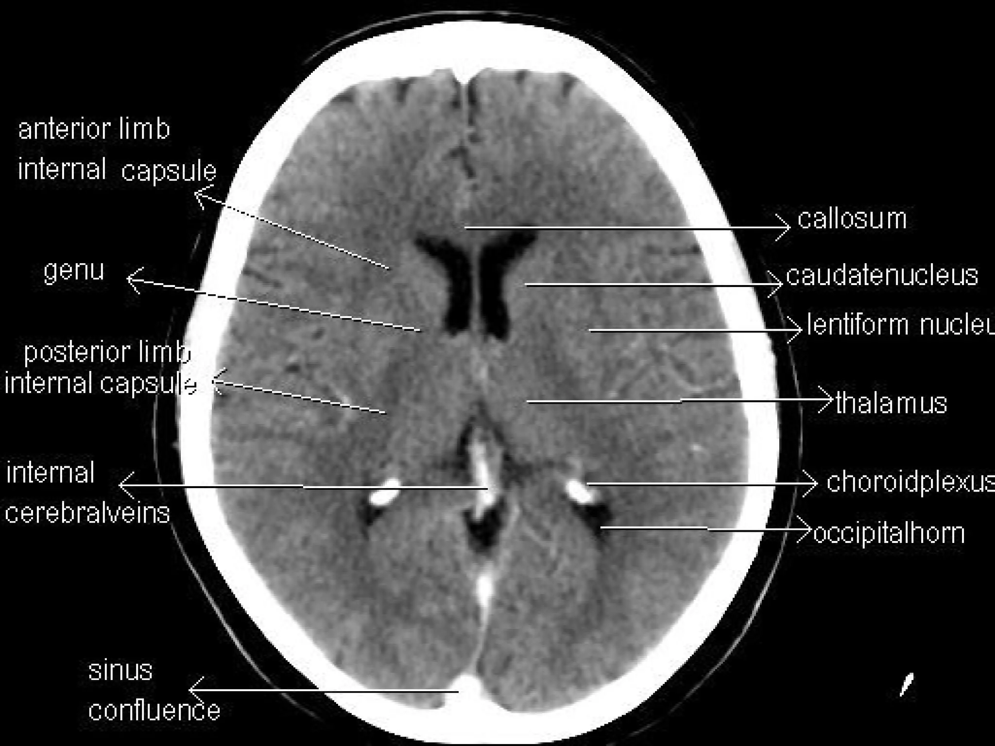
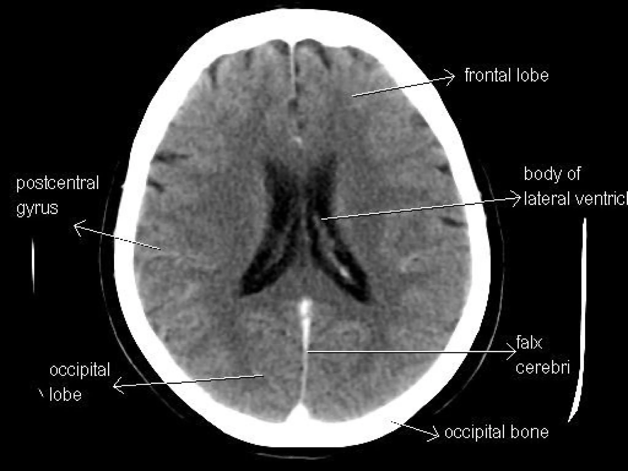
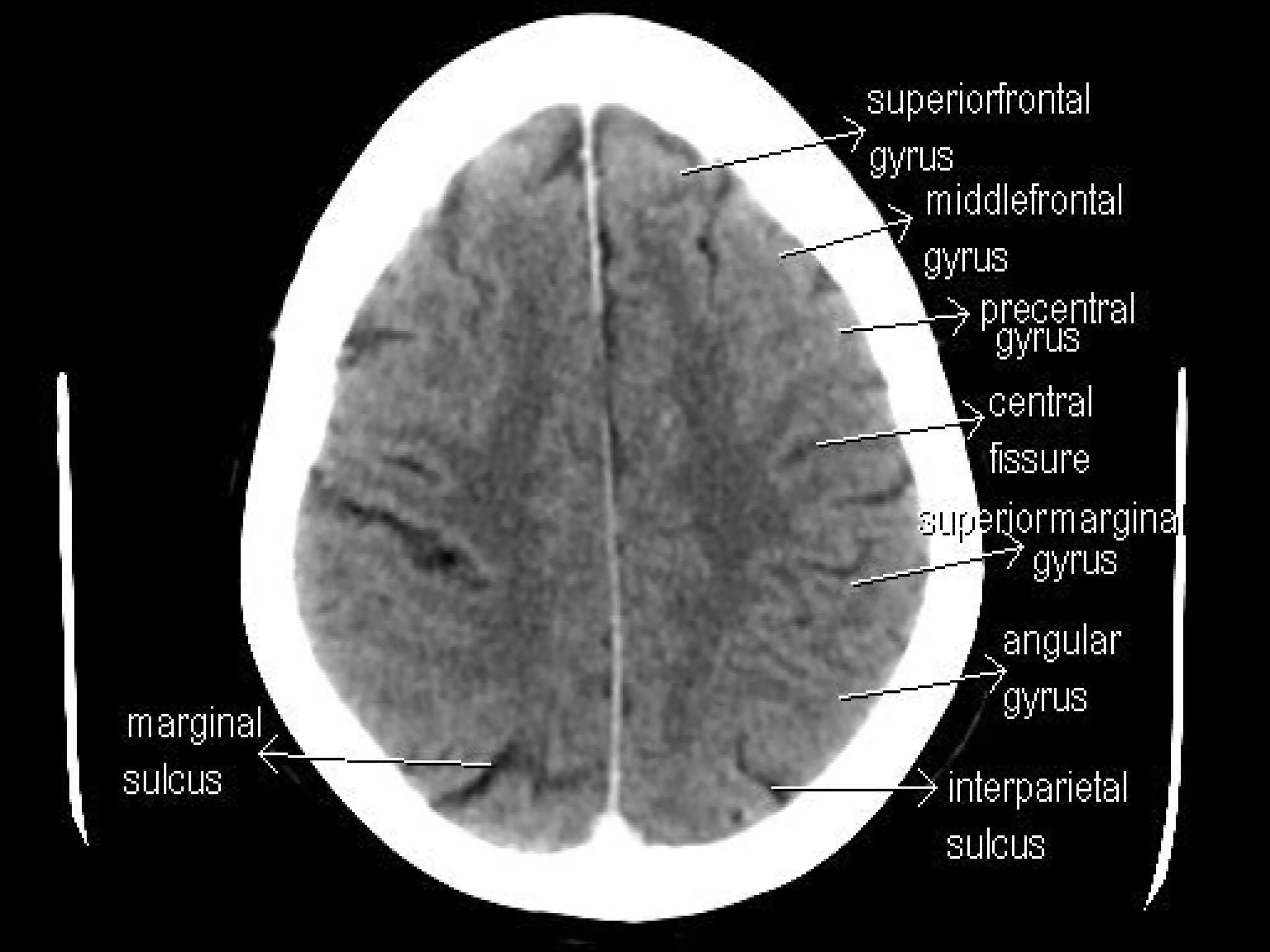
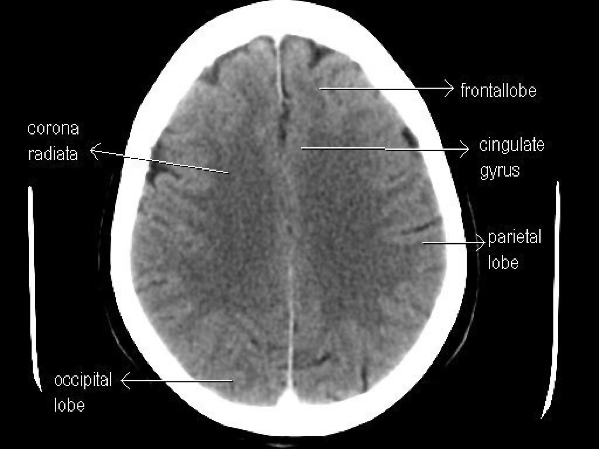
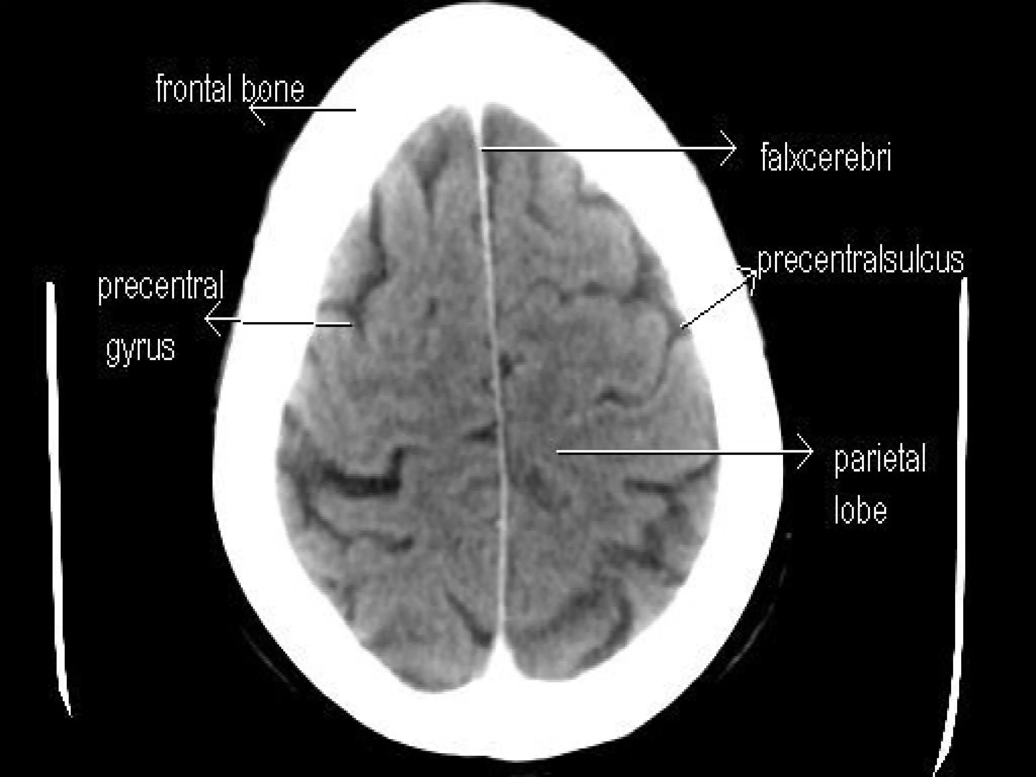
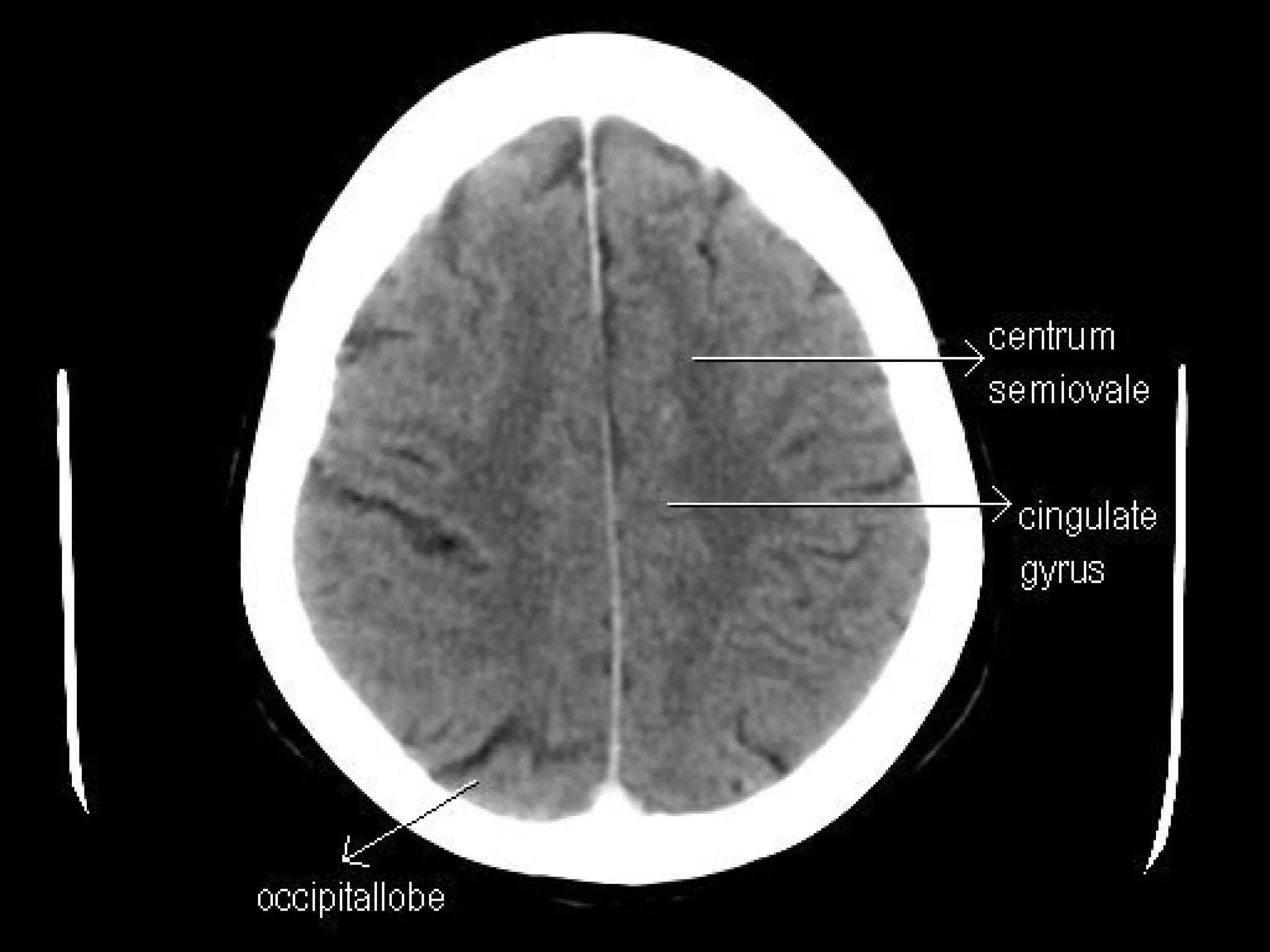
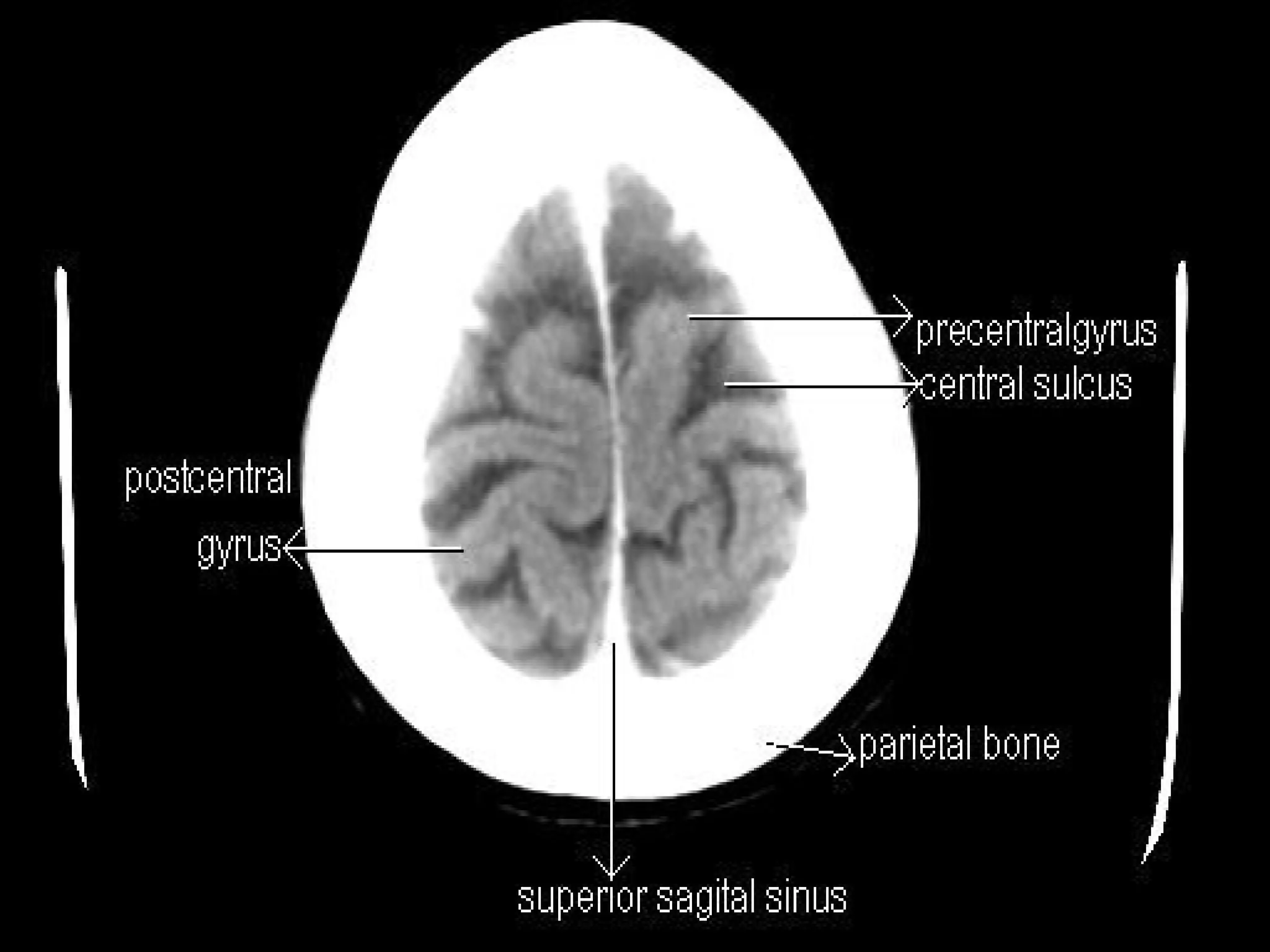


The document details the anatomy and function of the brain's ventricles and cerebrospinal fluid (CSF). It describes the structure of the ventricular system, the production and circulation of CSF, and highlights the brain's blood supply through the Circle of Willis. The text also outlines the major subarachnoid cisterns and the organization of the brain's lobes, gyri, and sulci.


