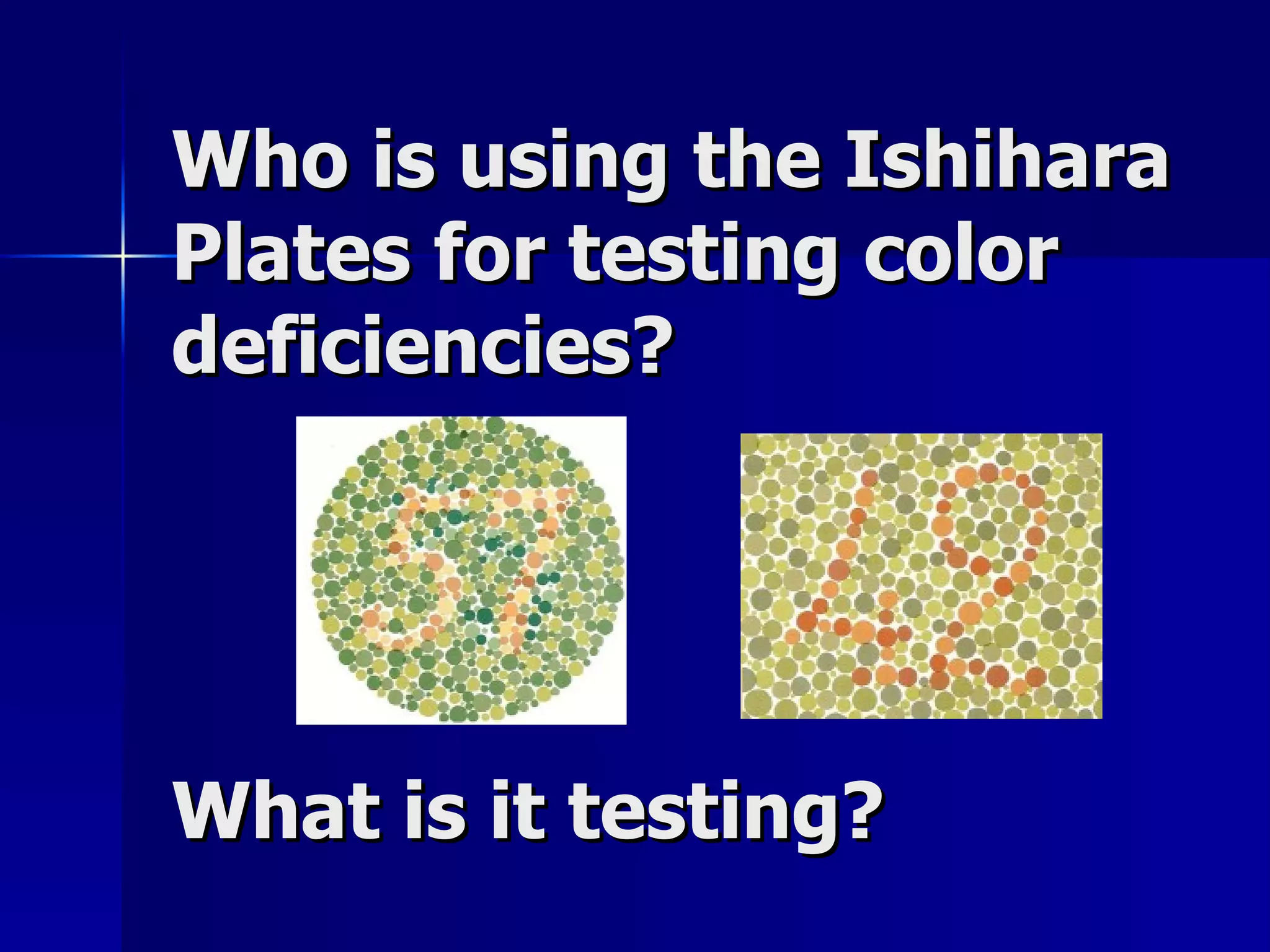
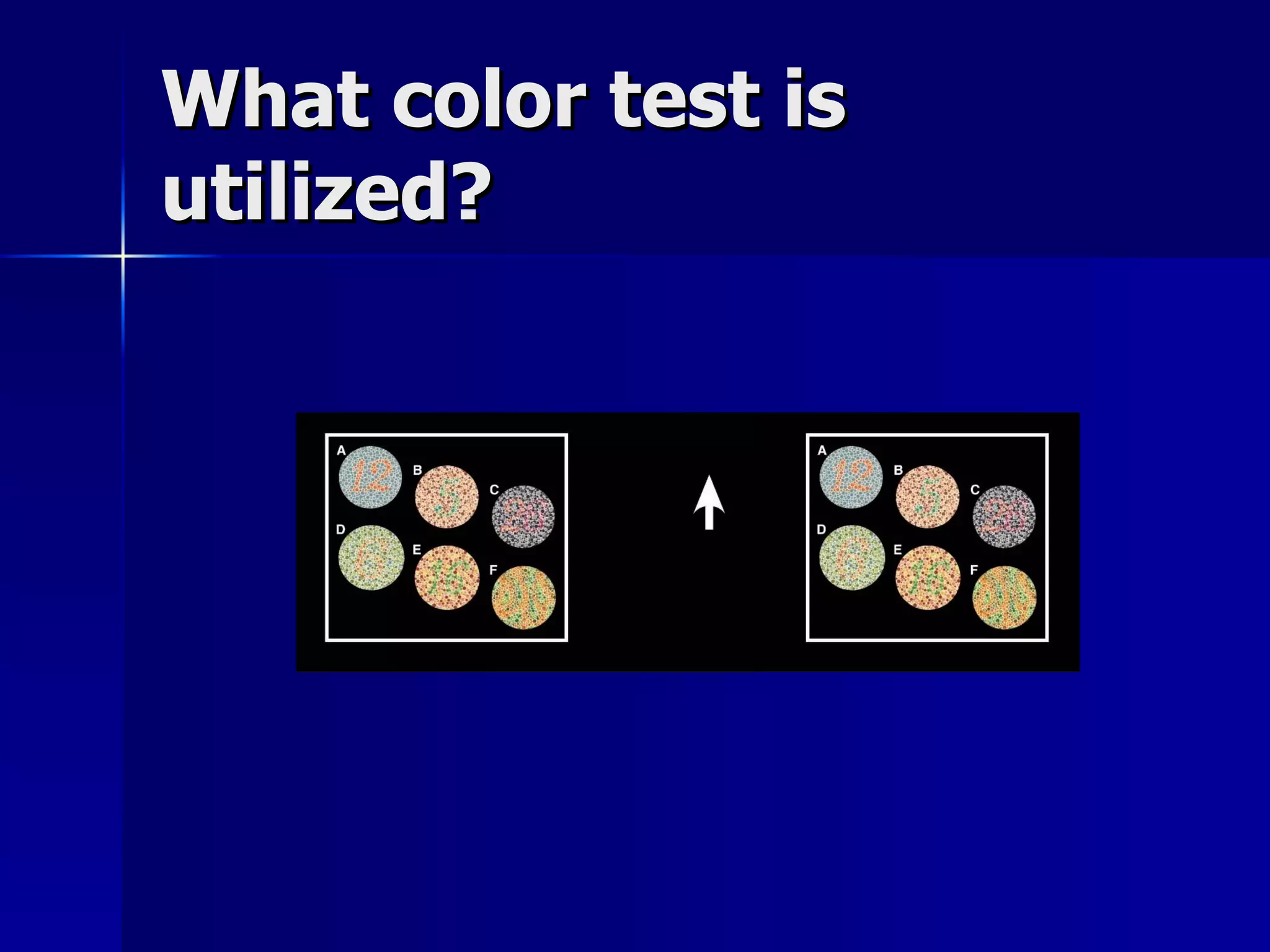
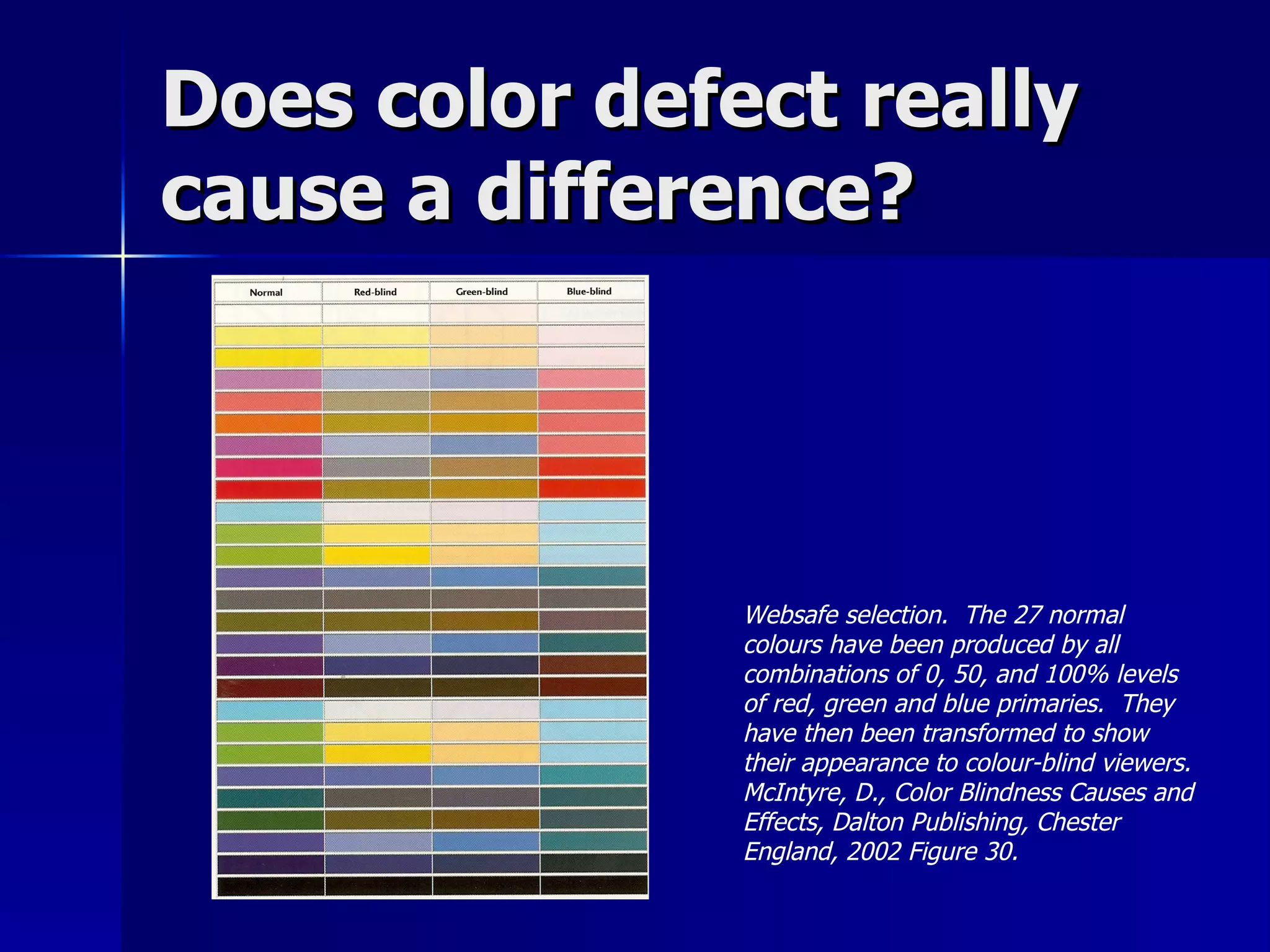
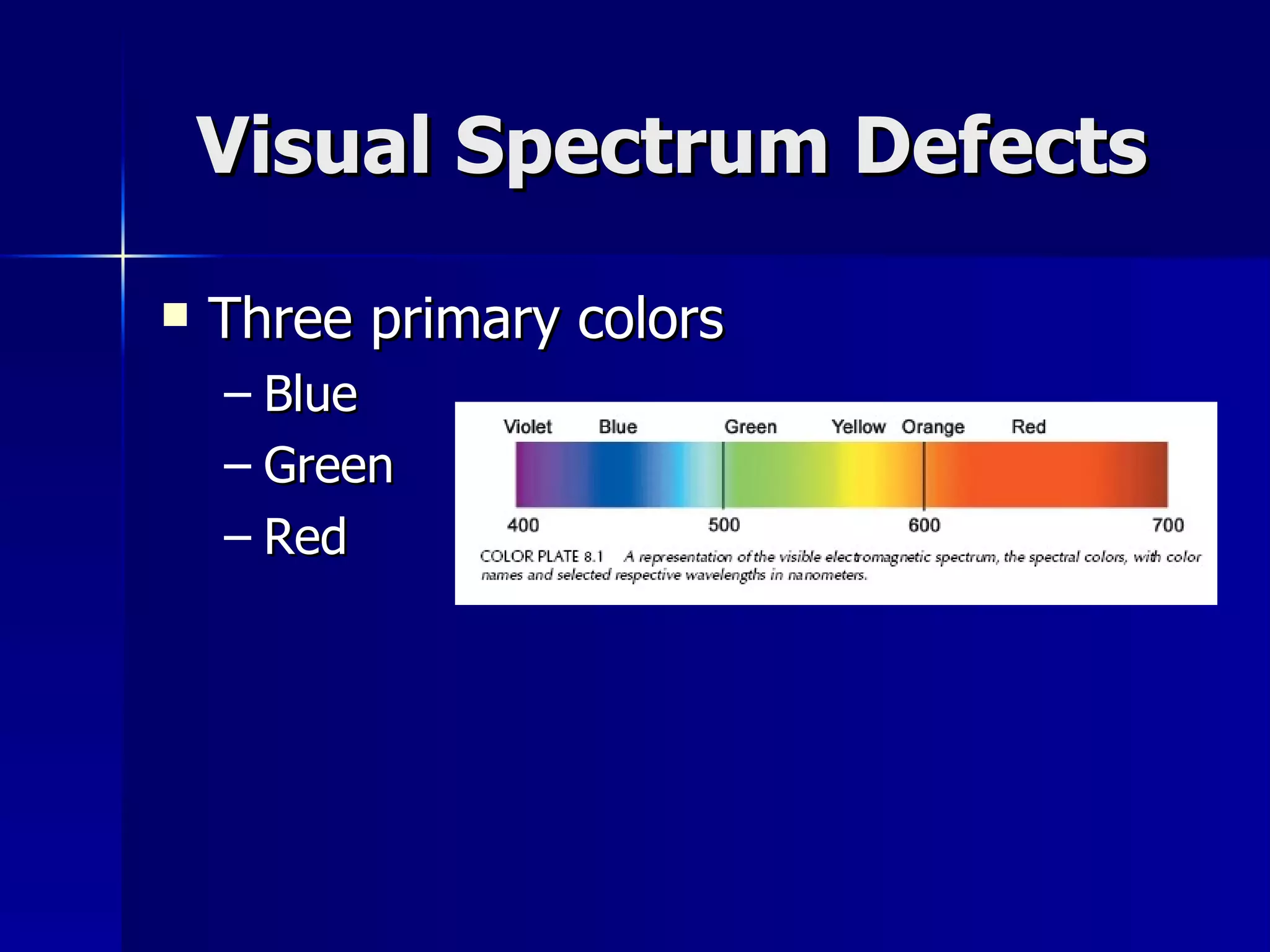
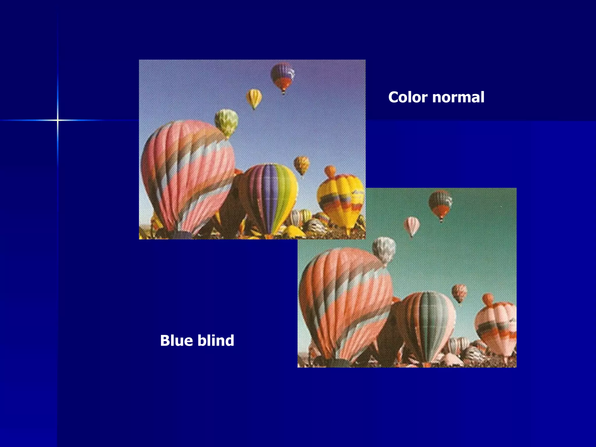
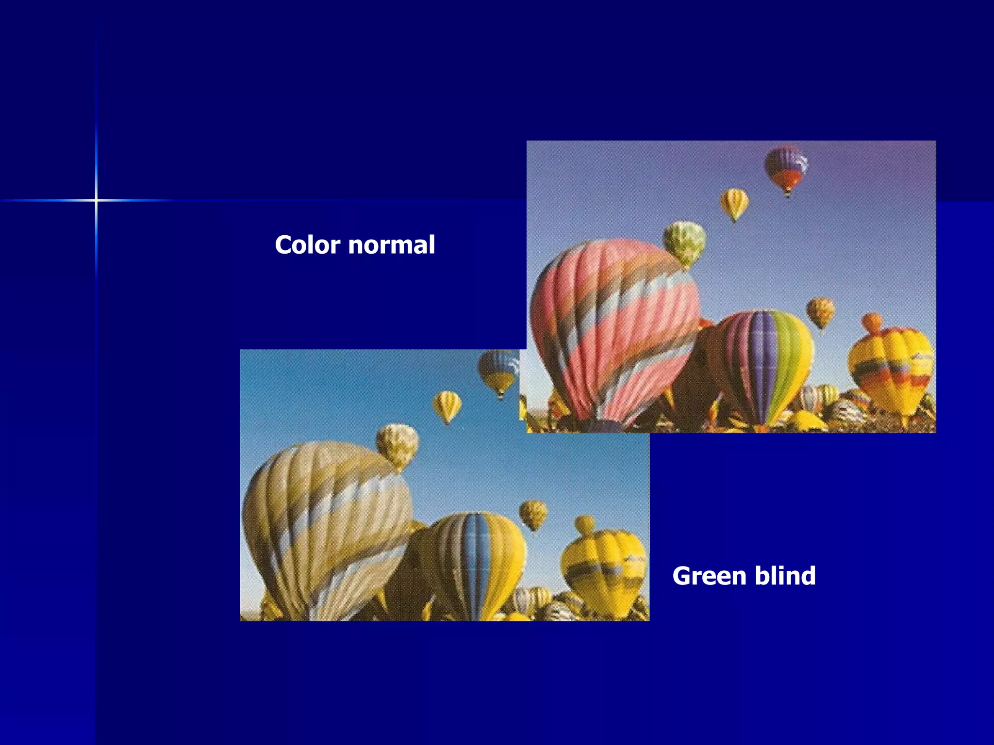
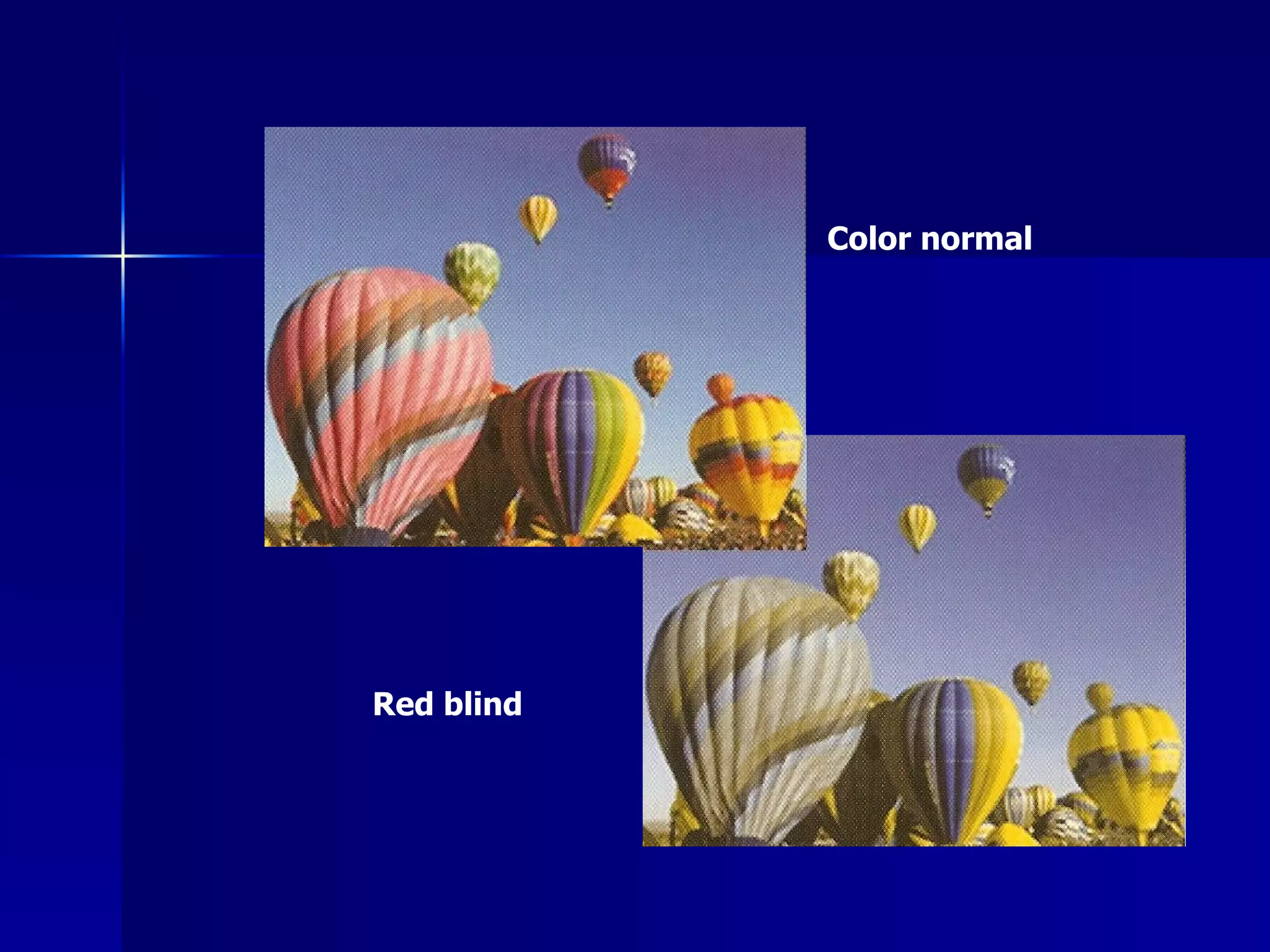
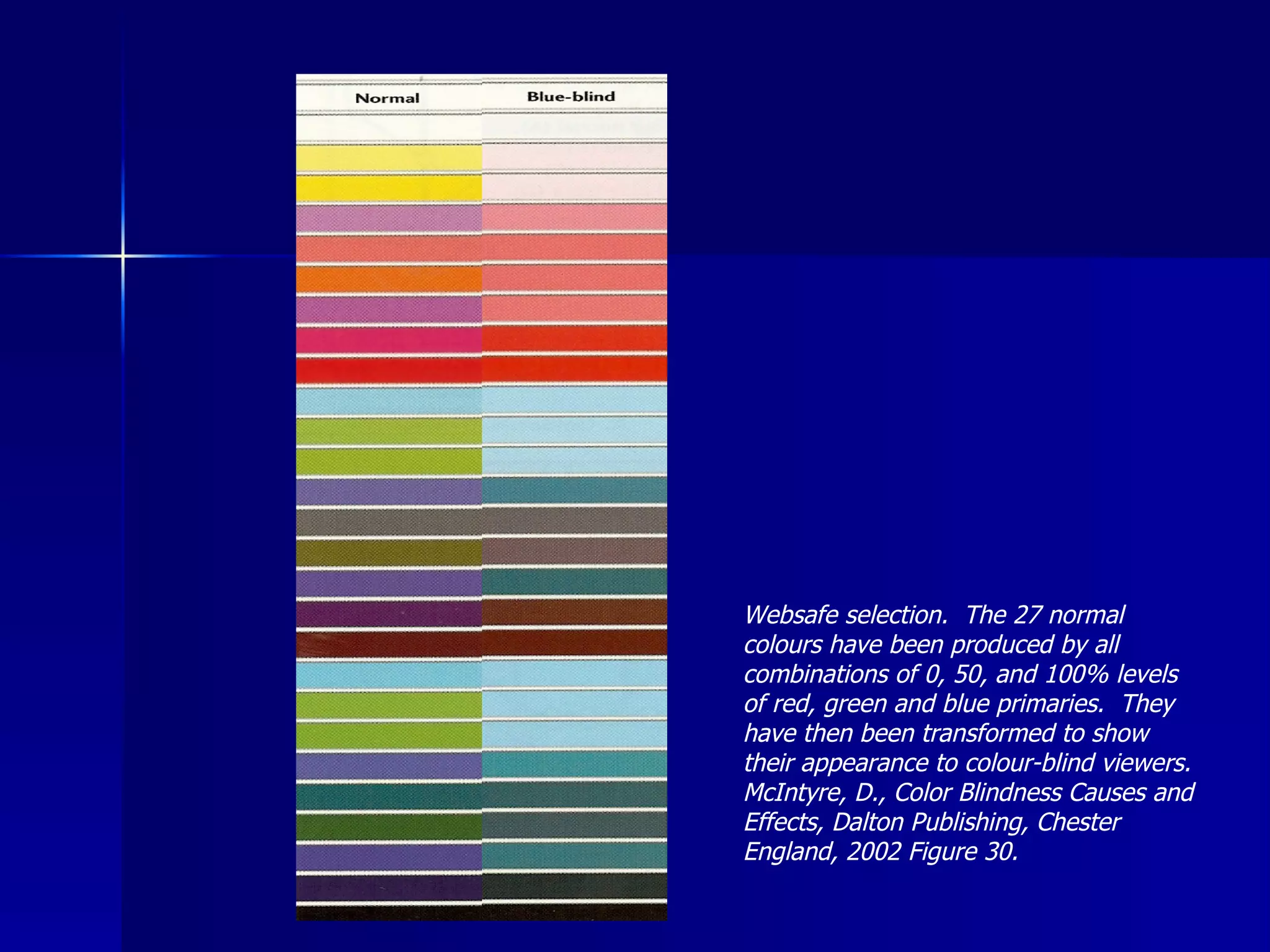
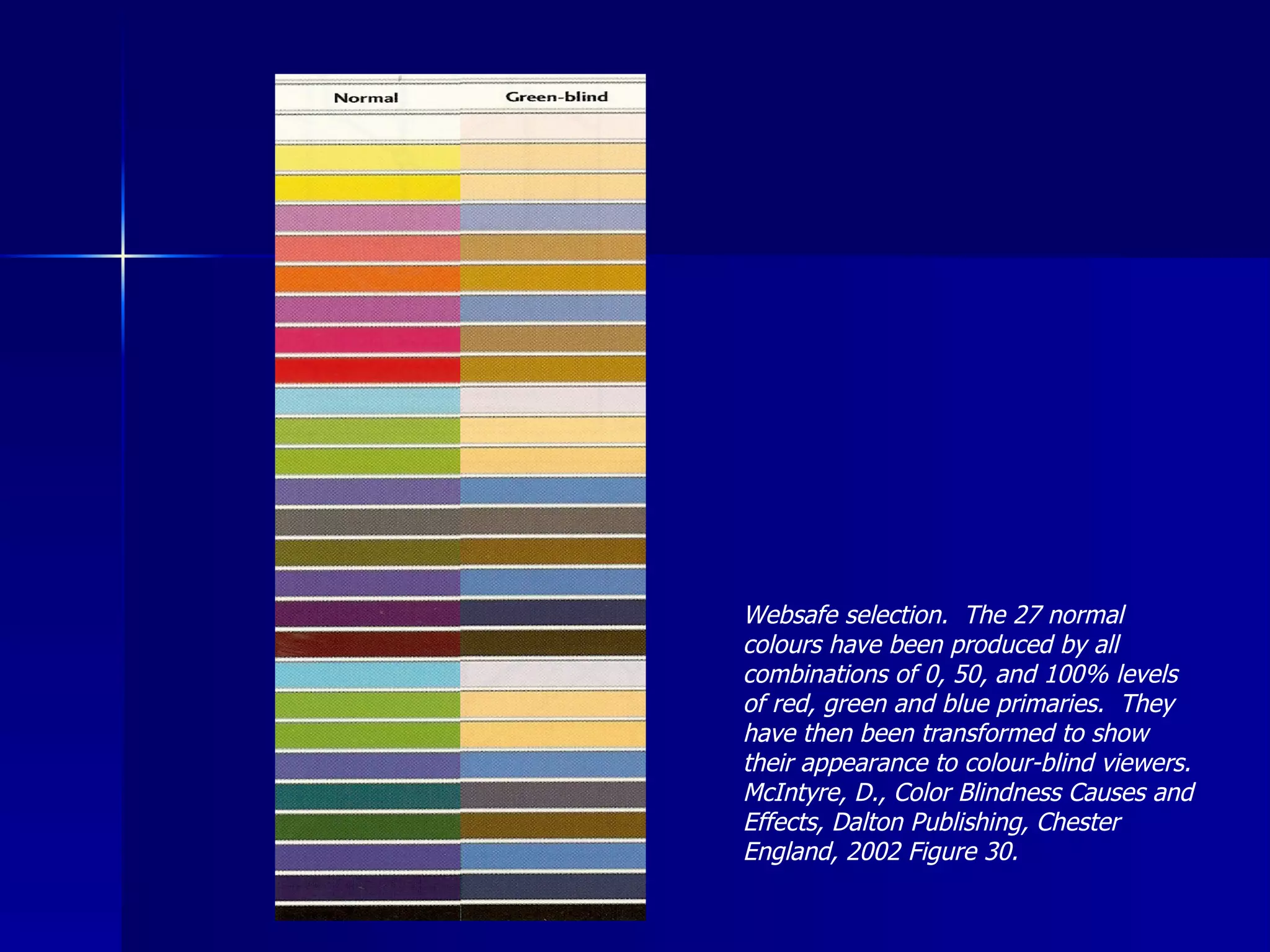
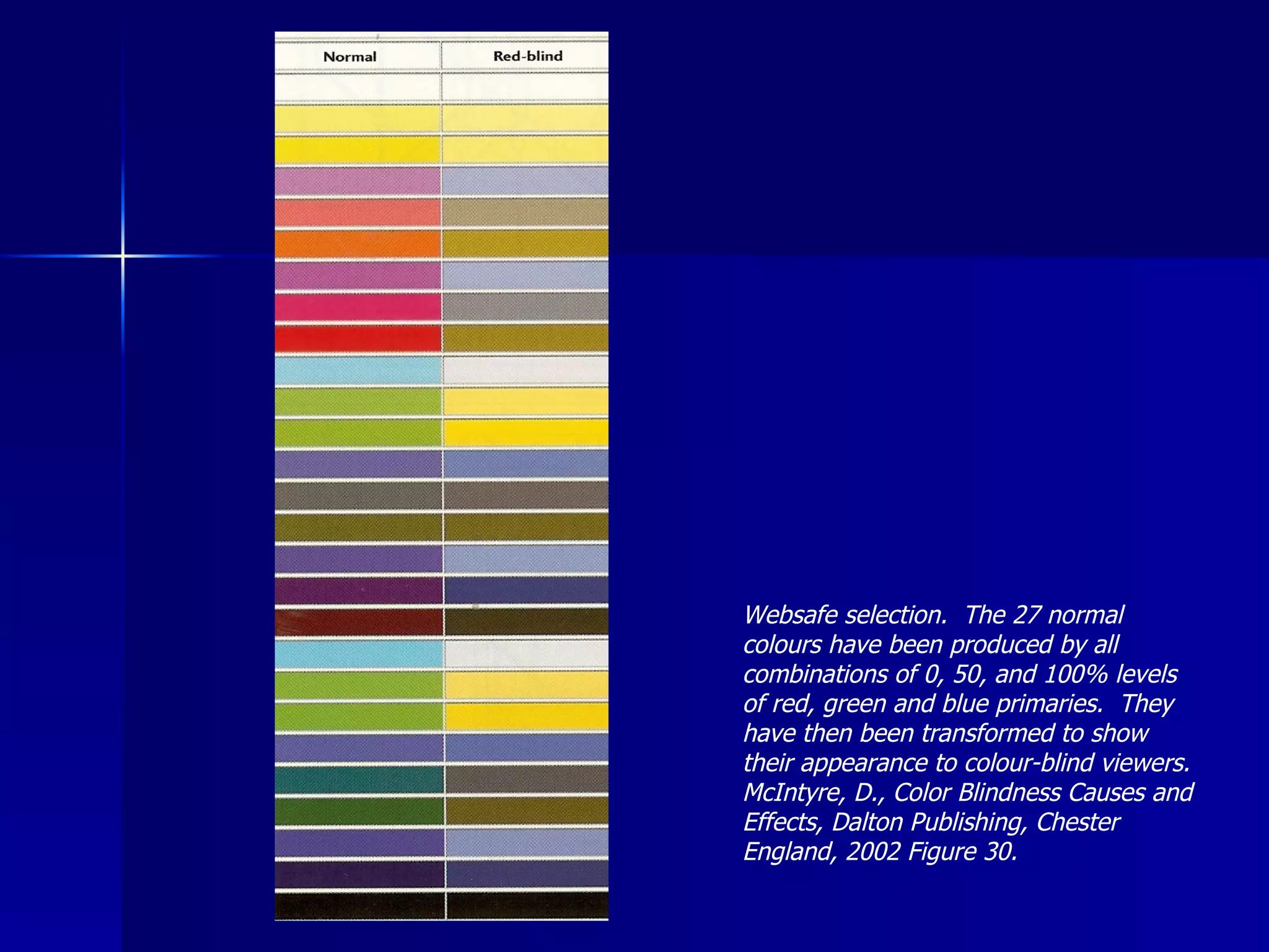
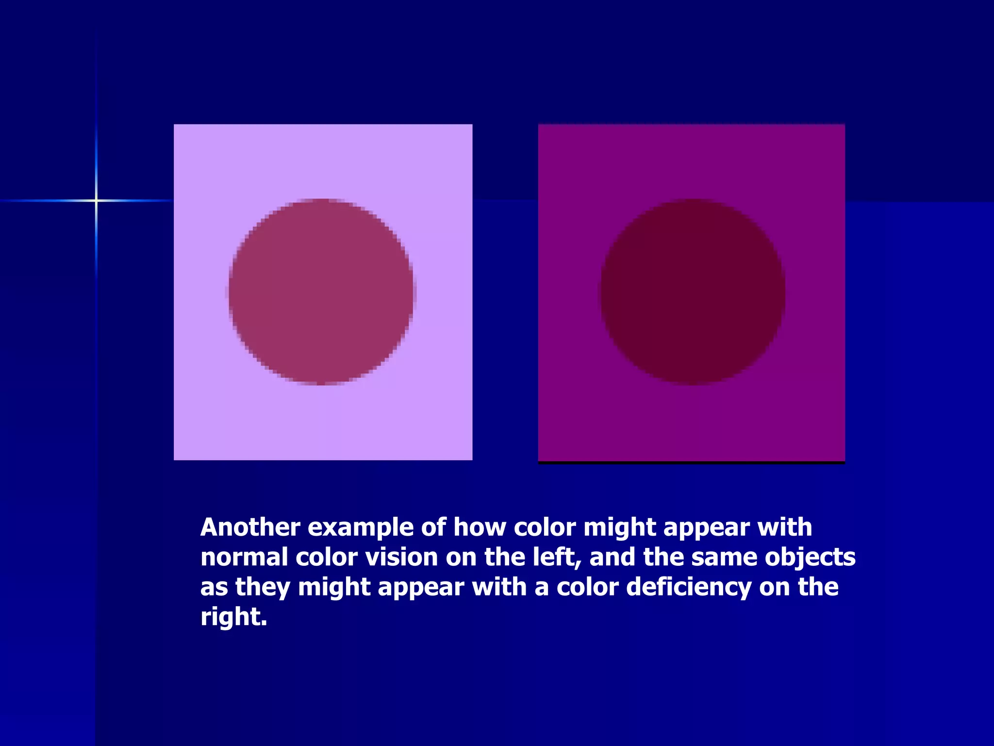
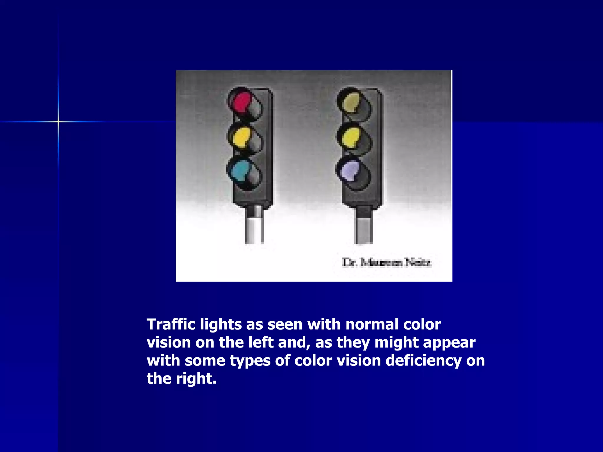
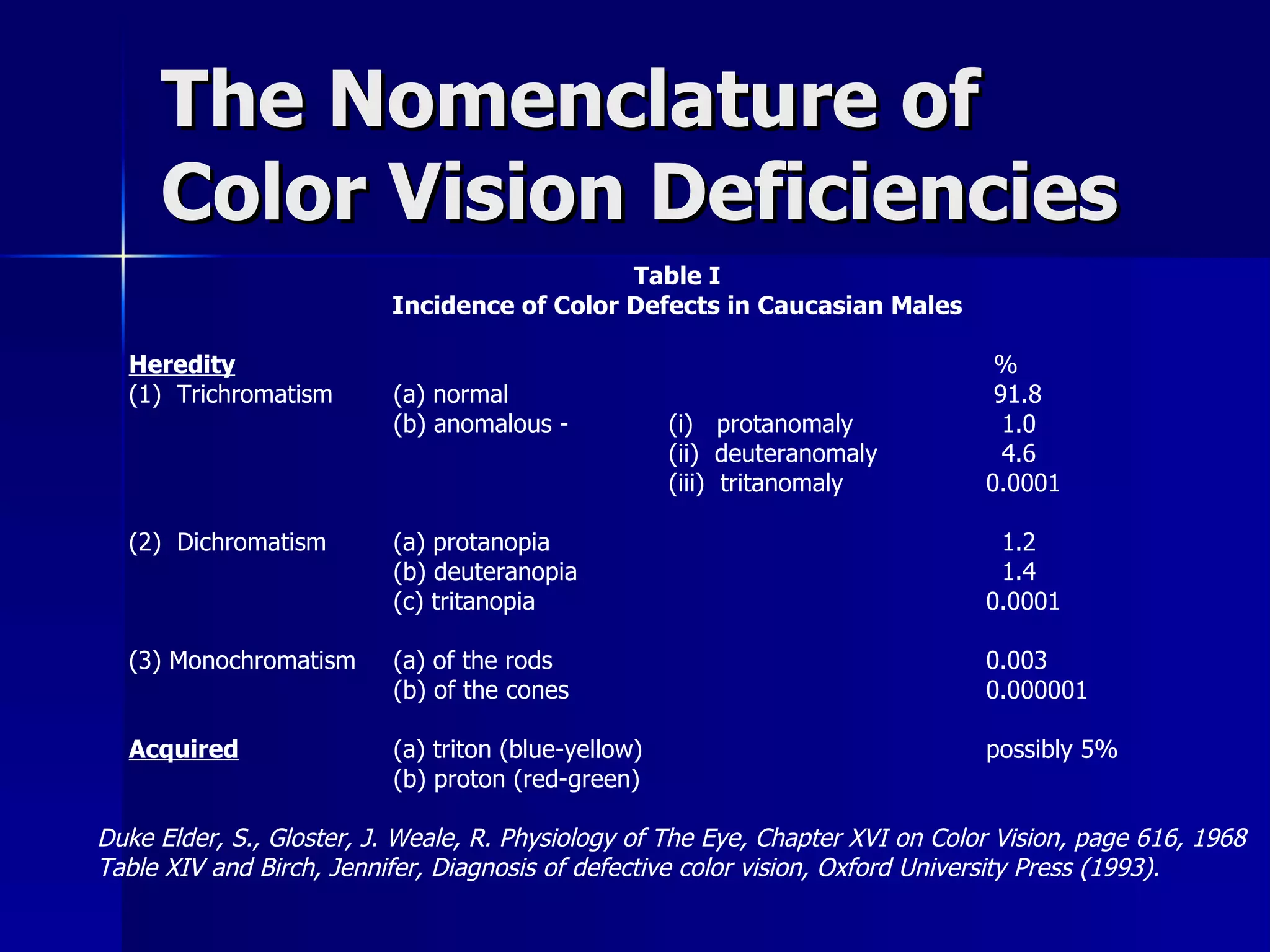
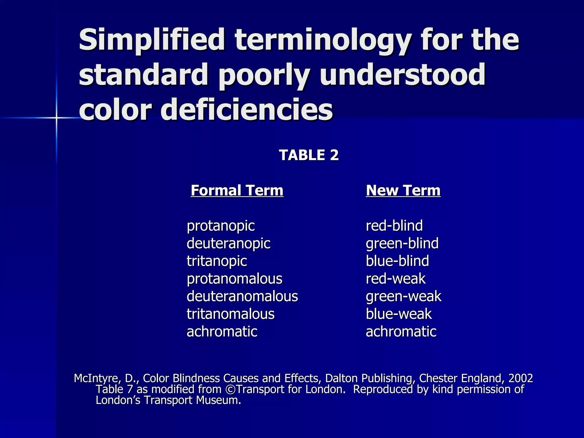
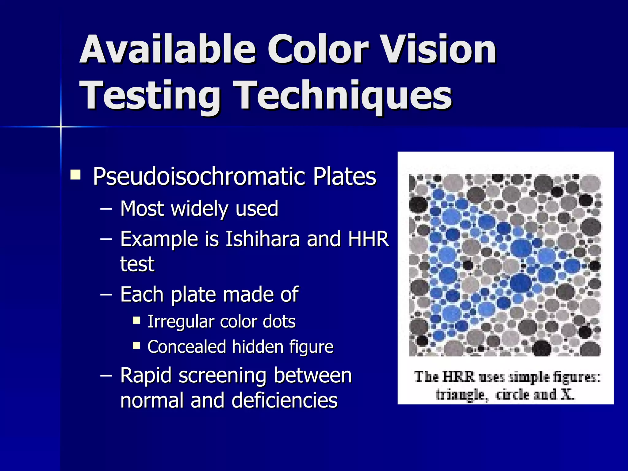
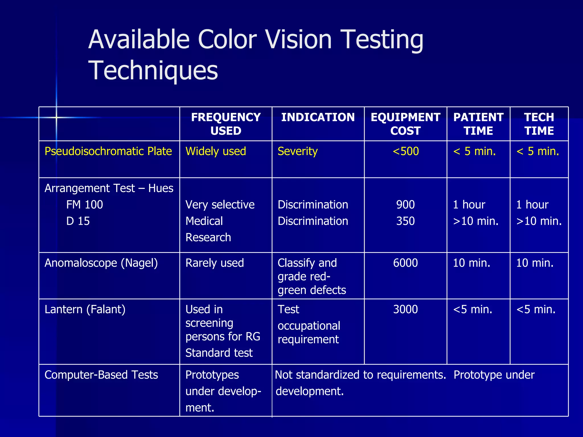
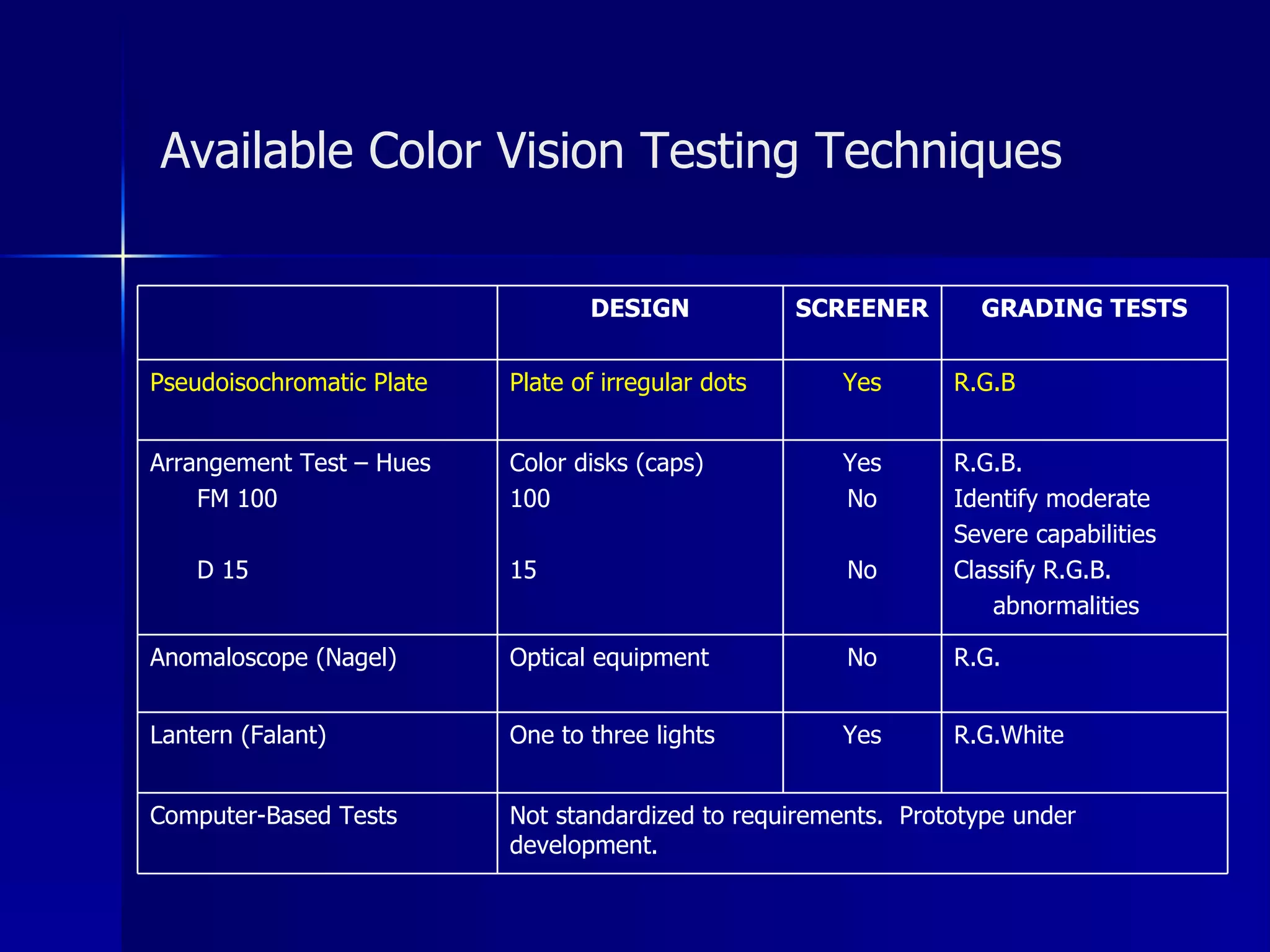
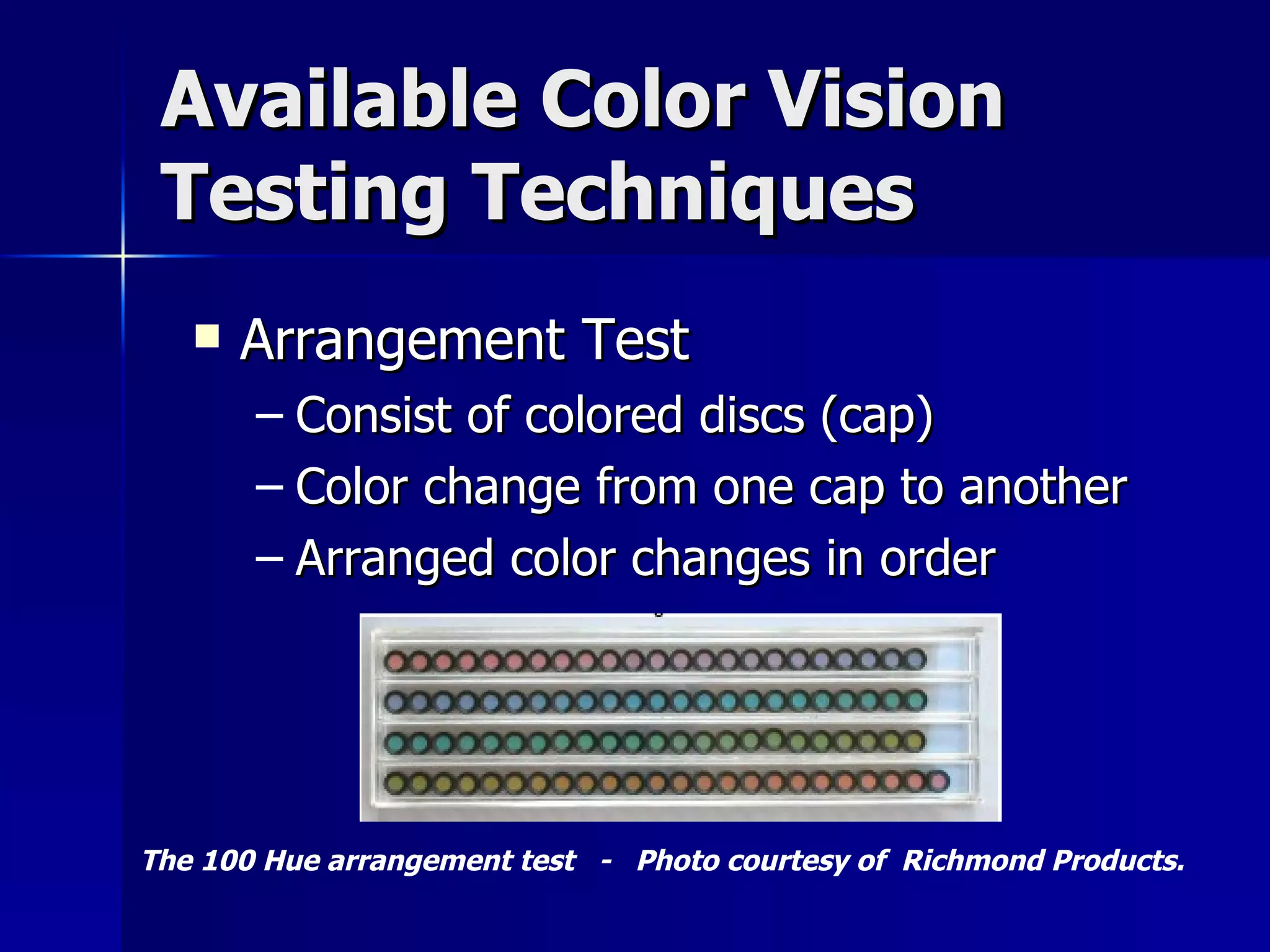
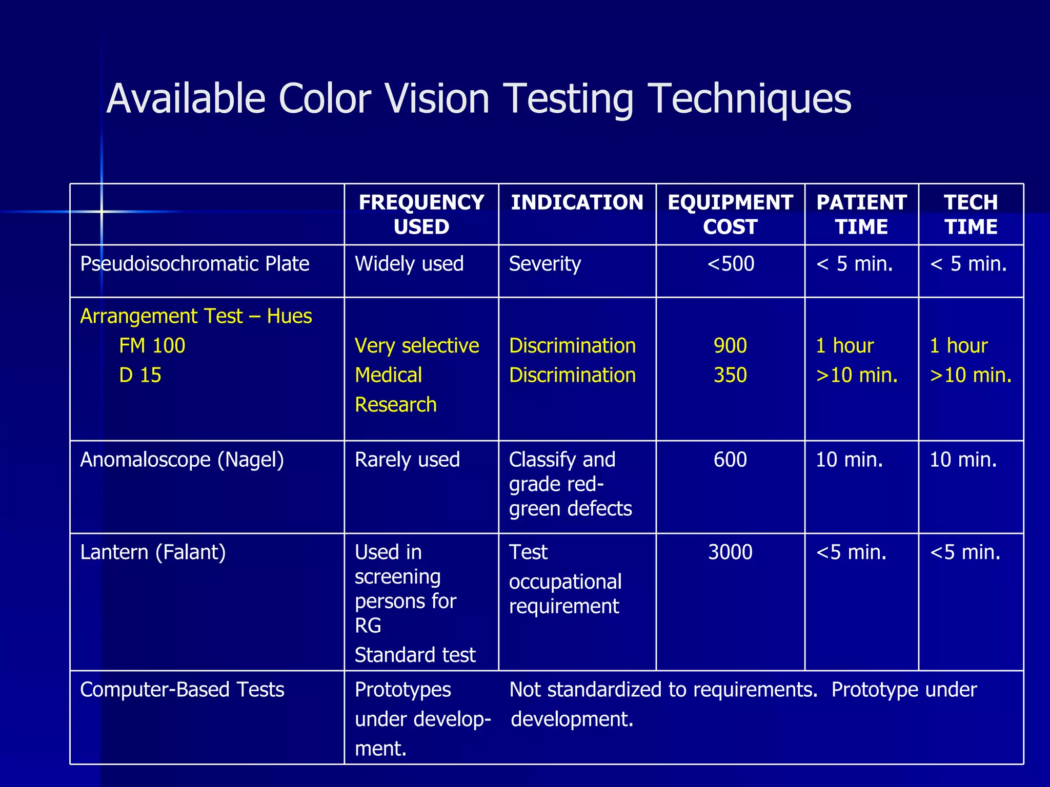
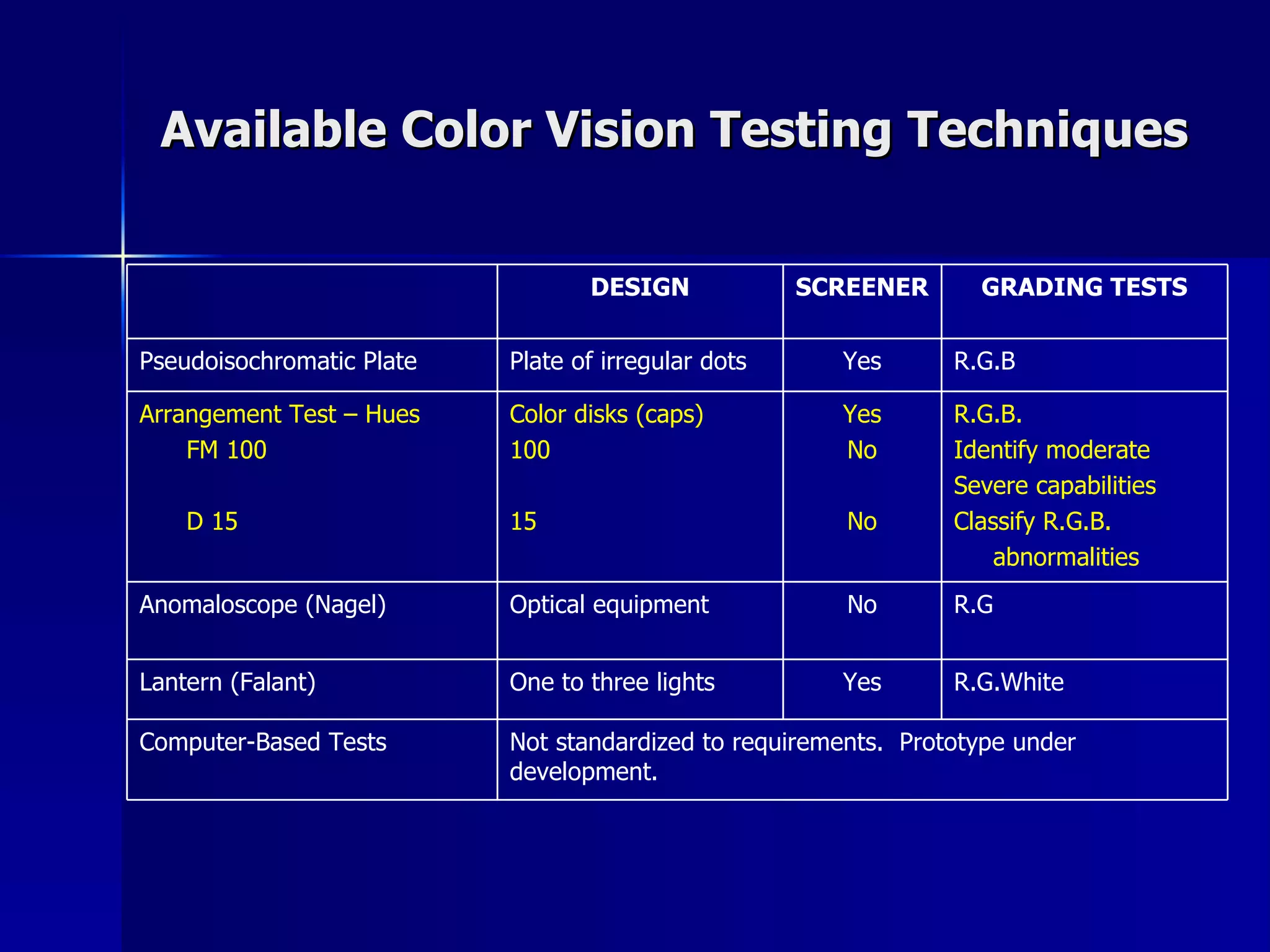
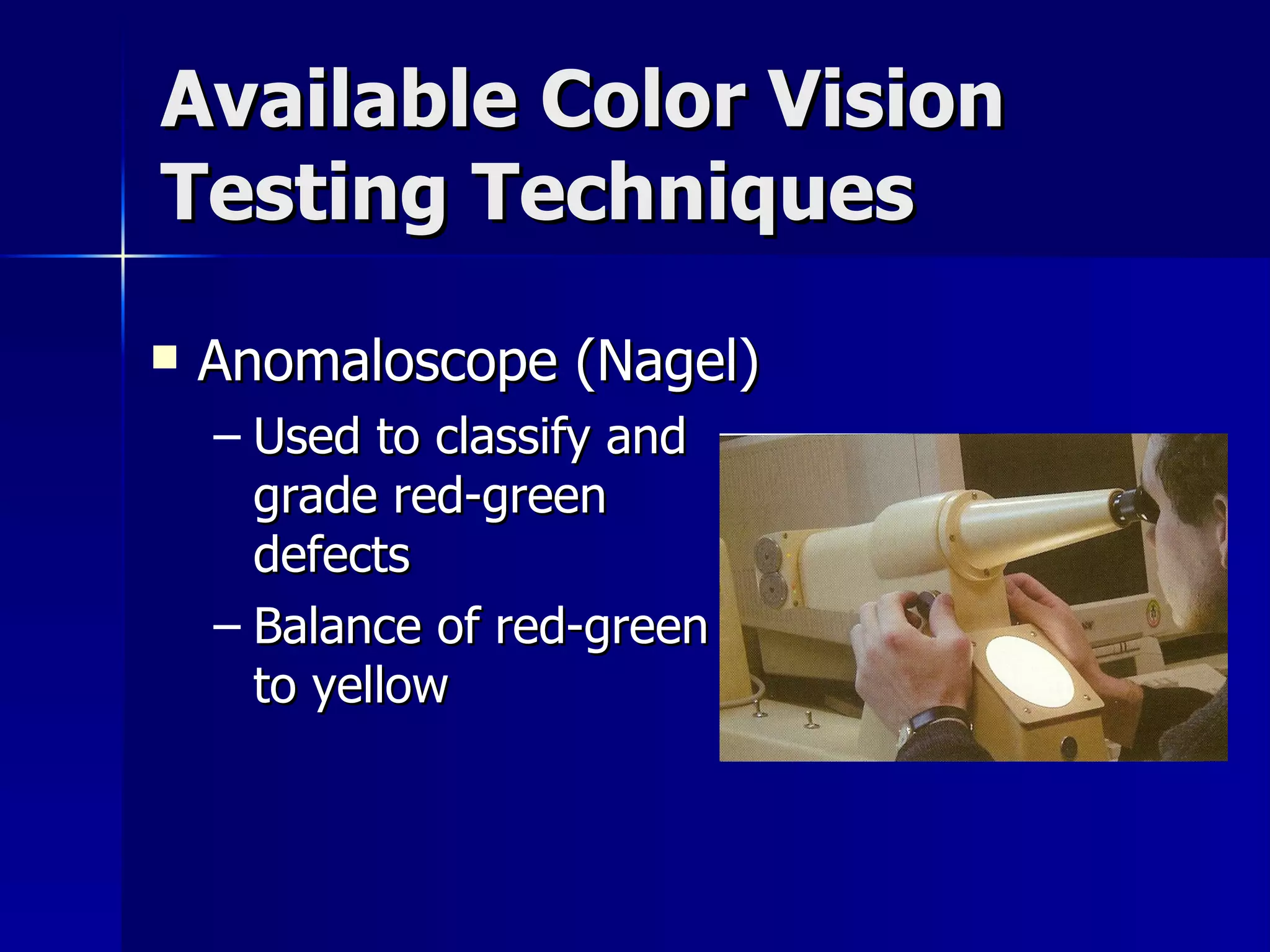
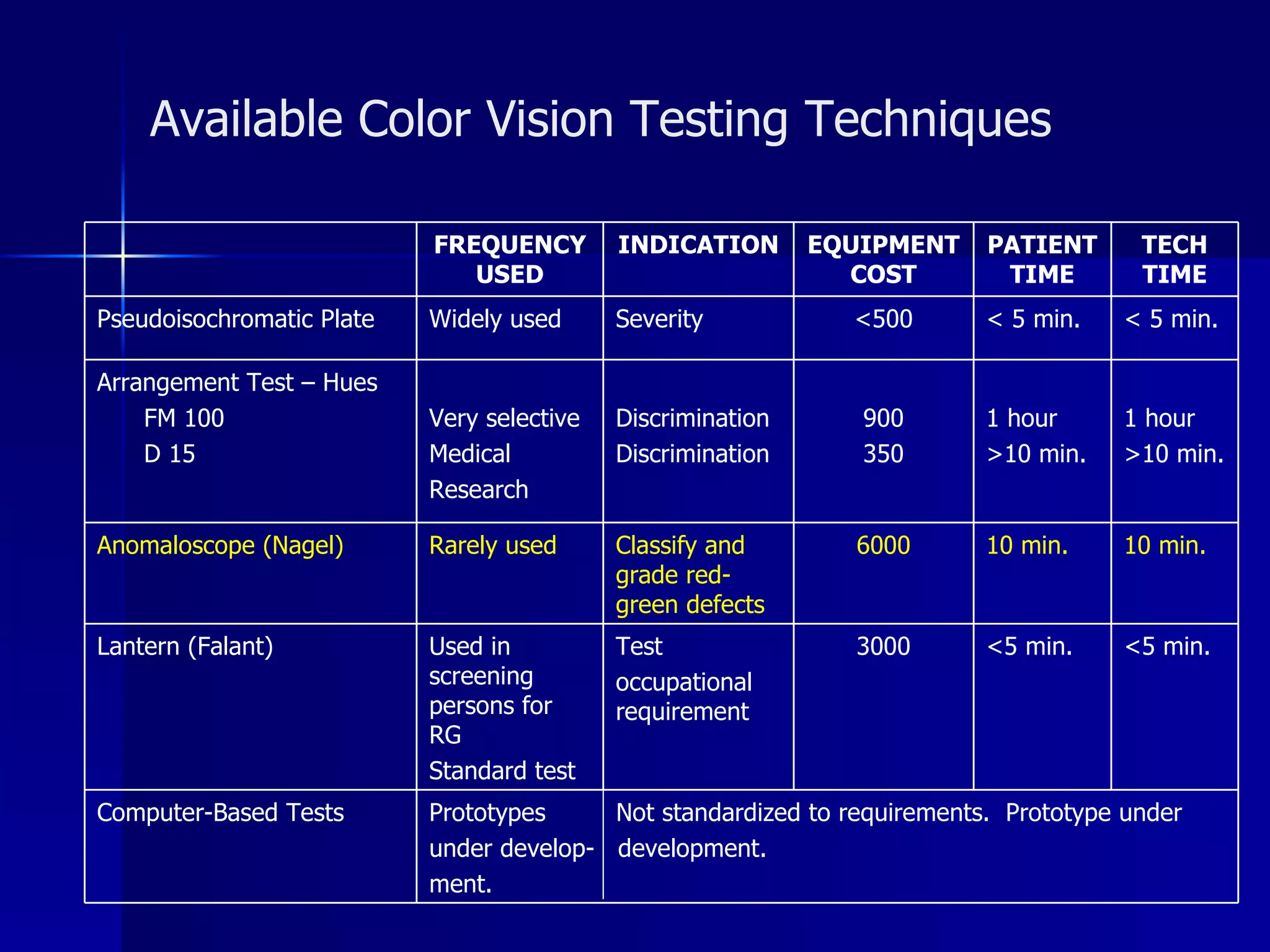
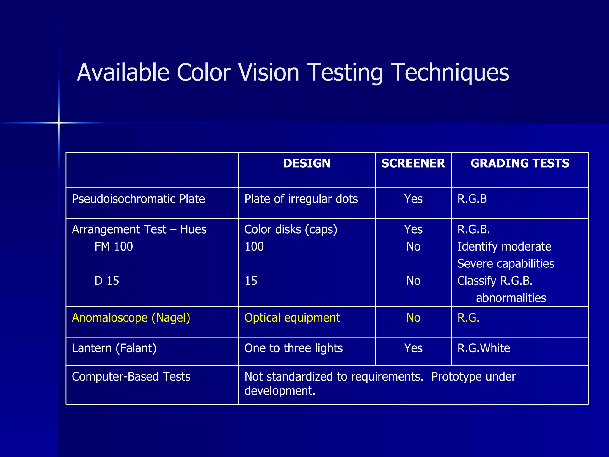
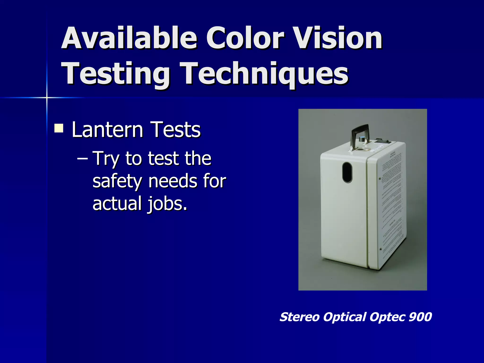
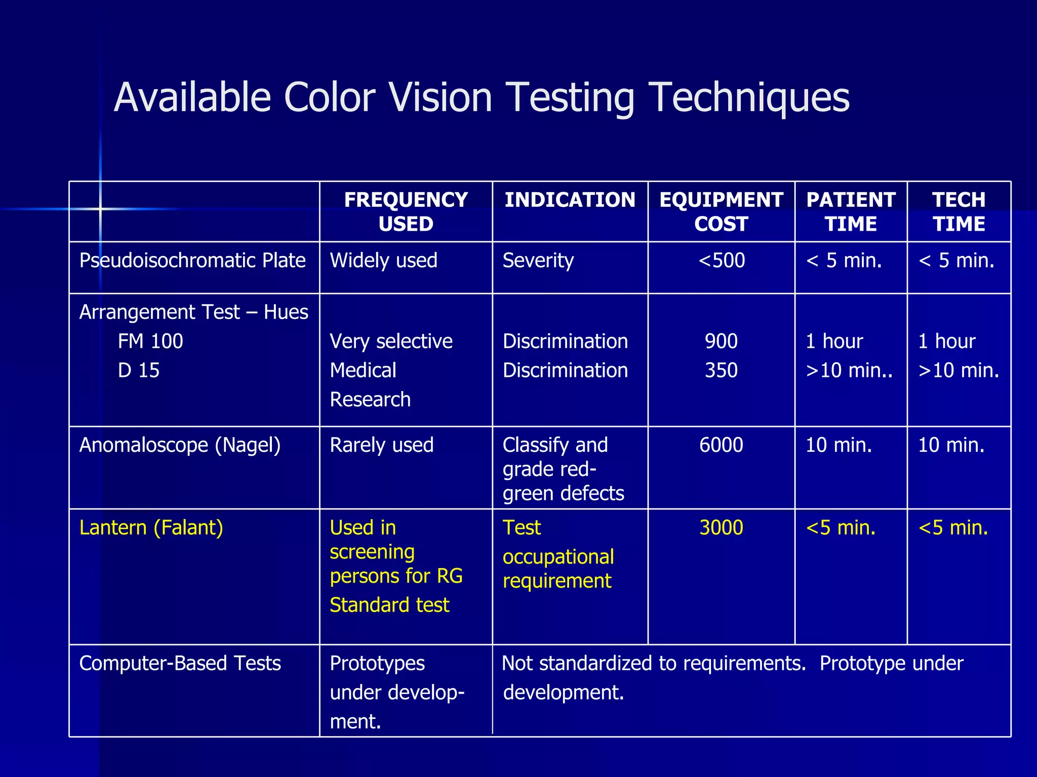
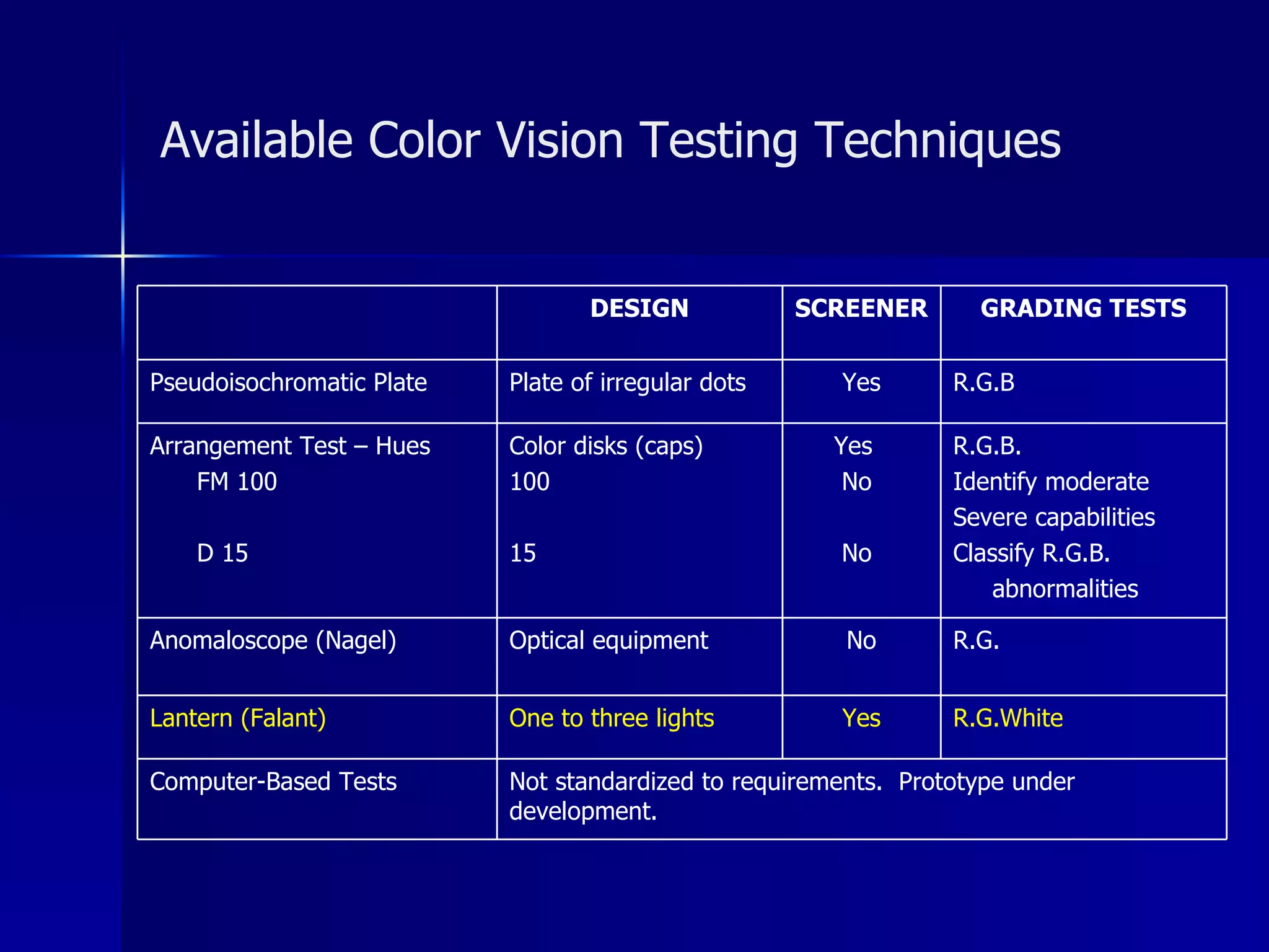
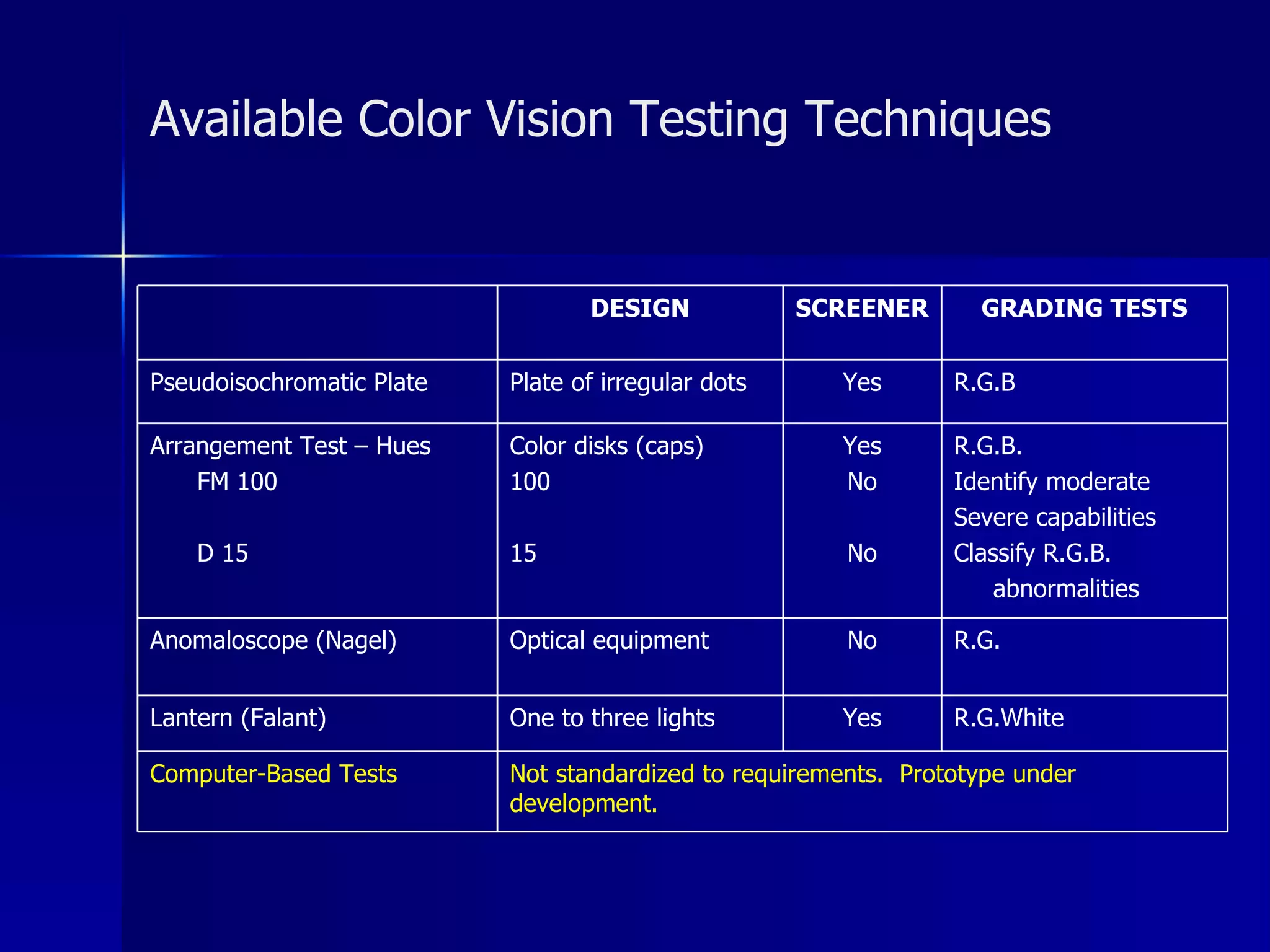
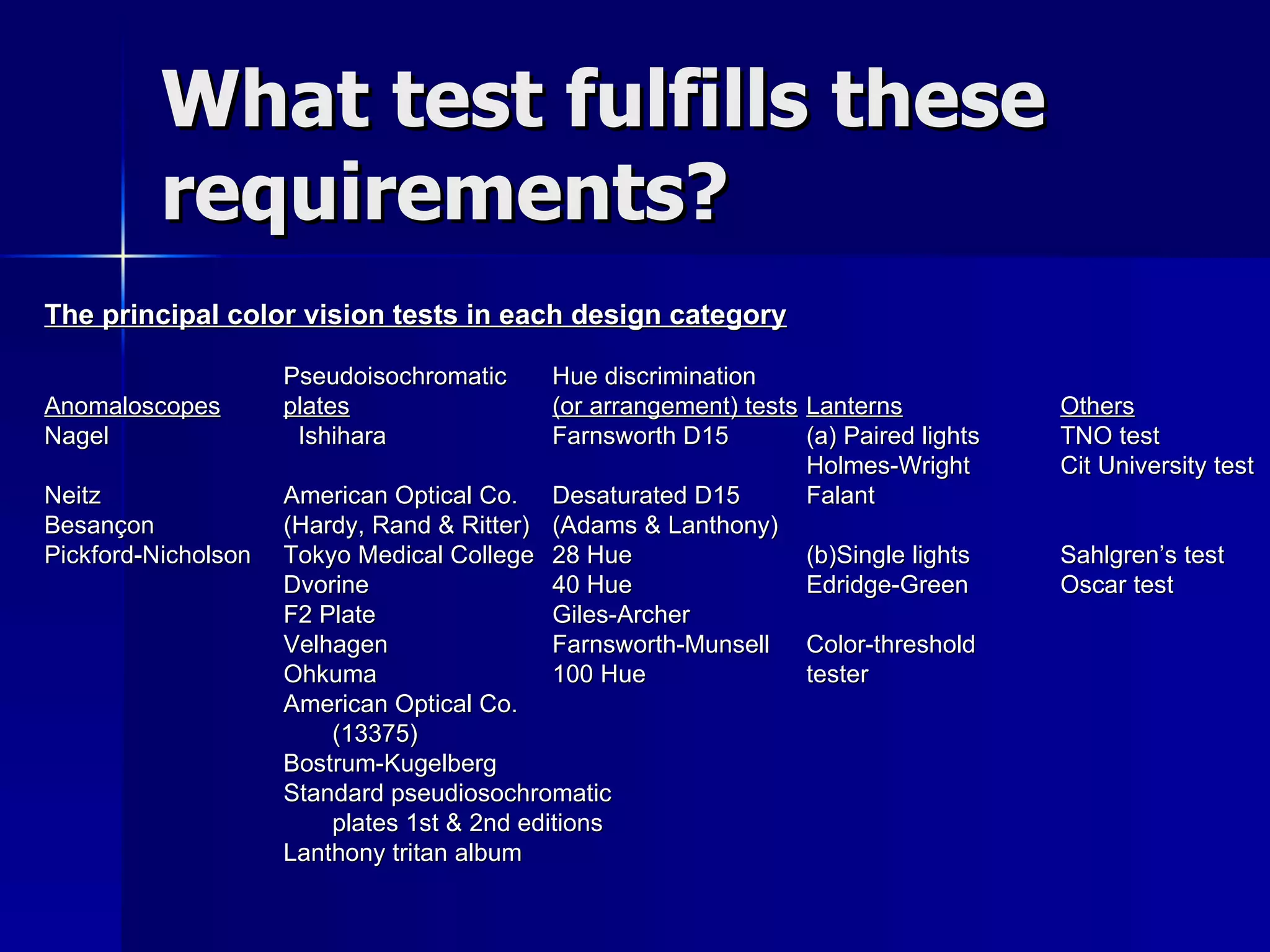
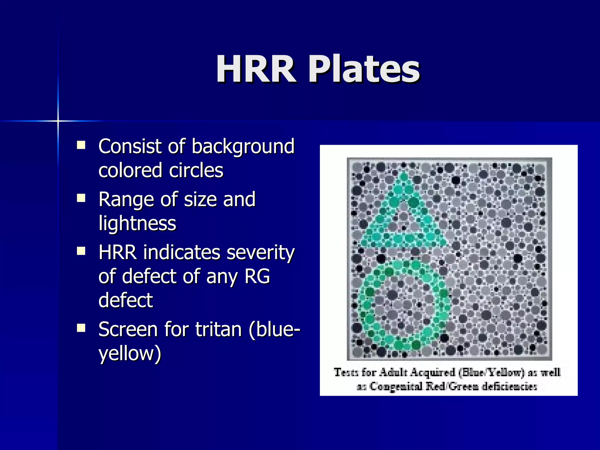
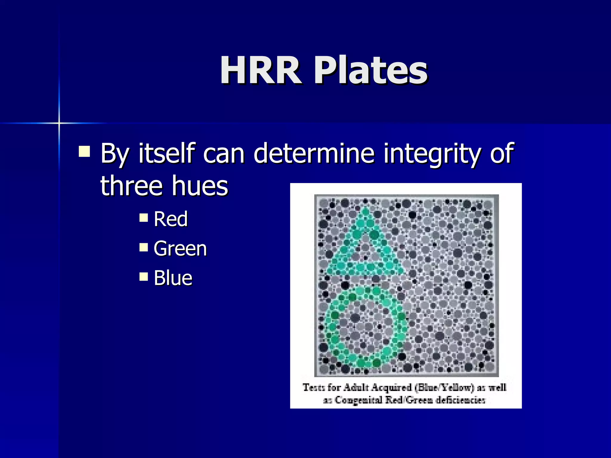
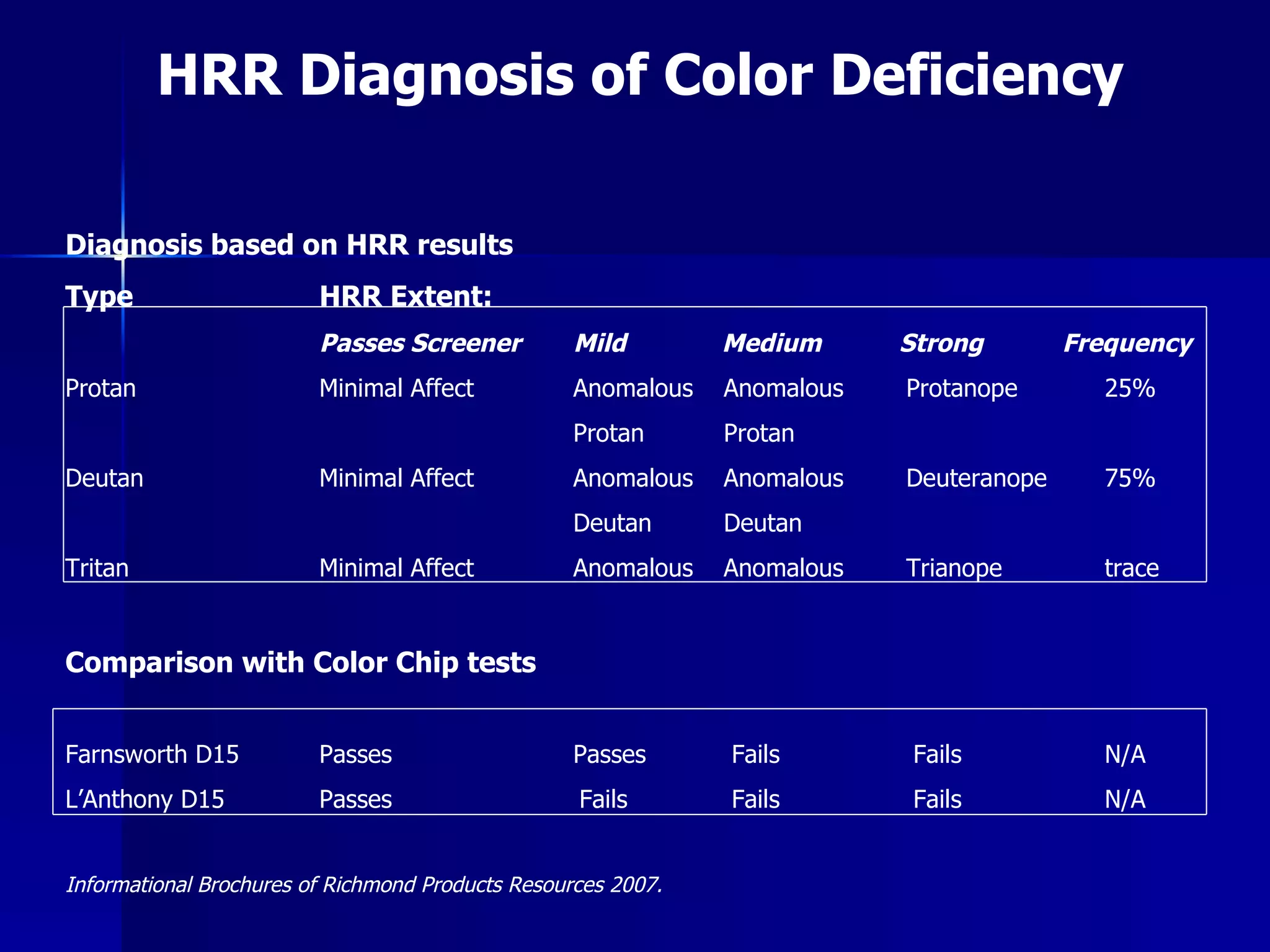
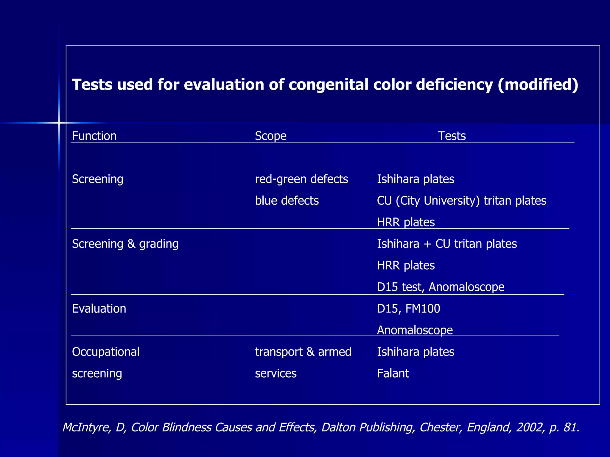
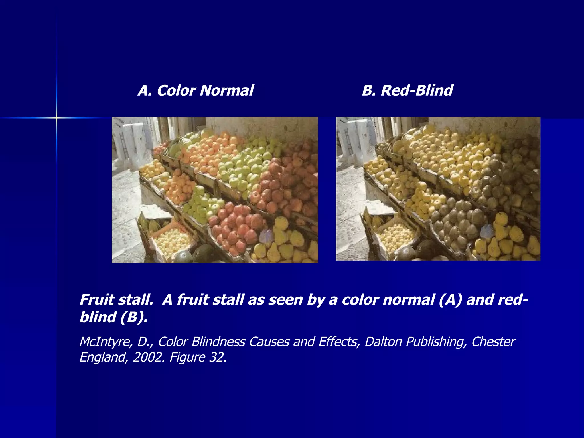
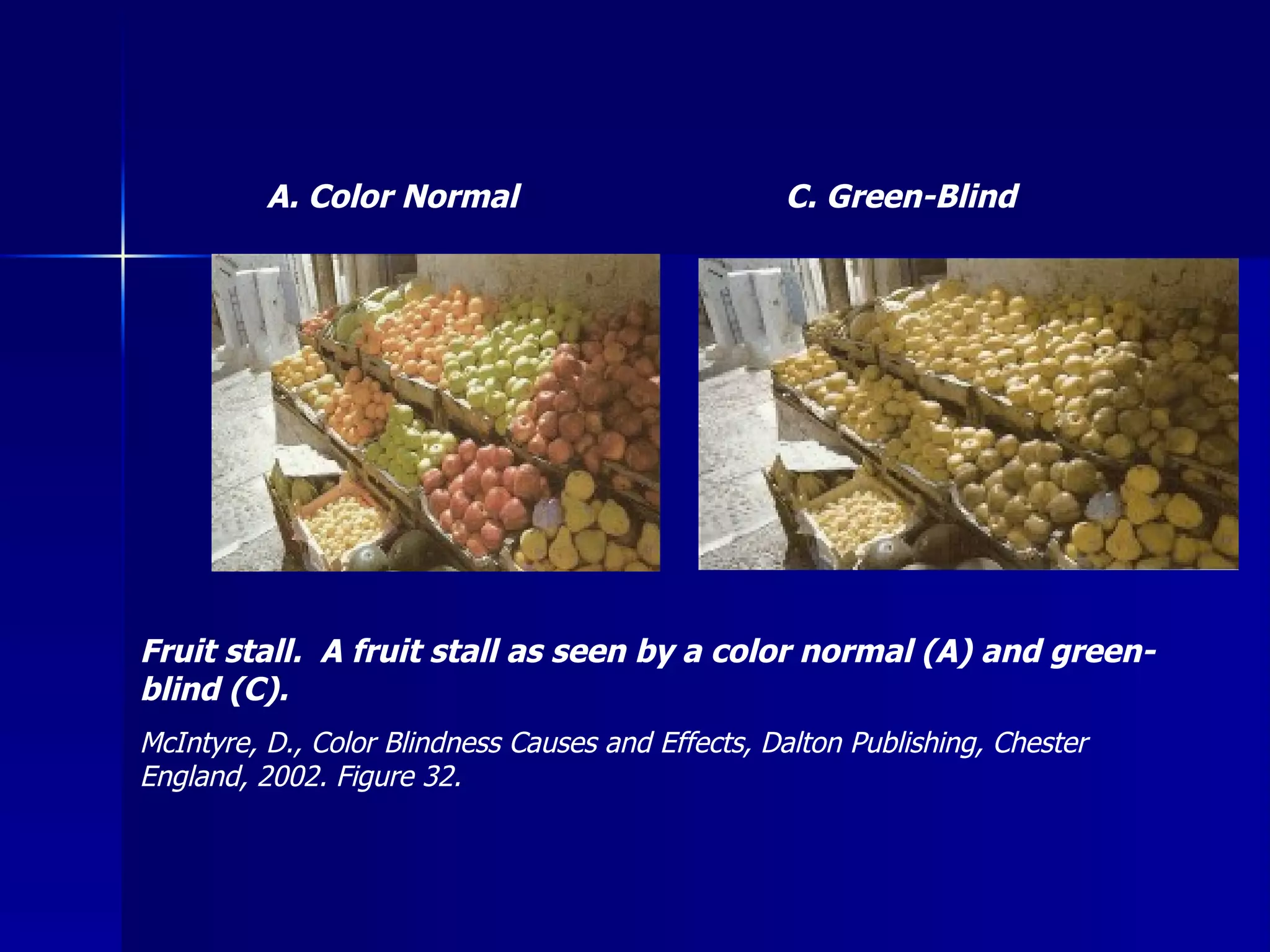
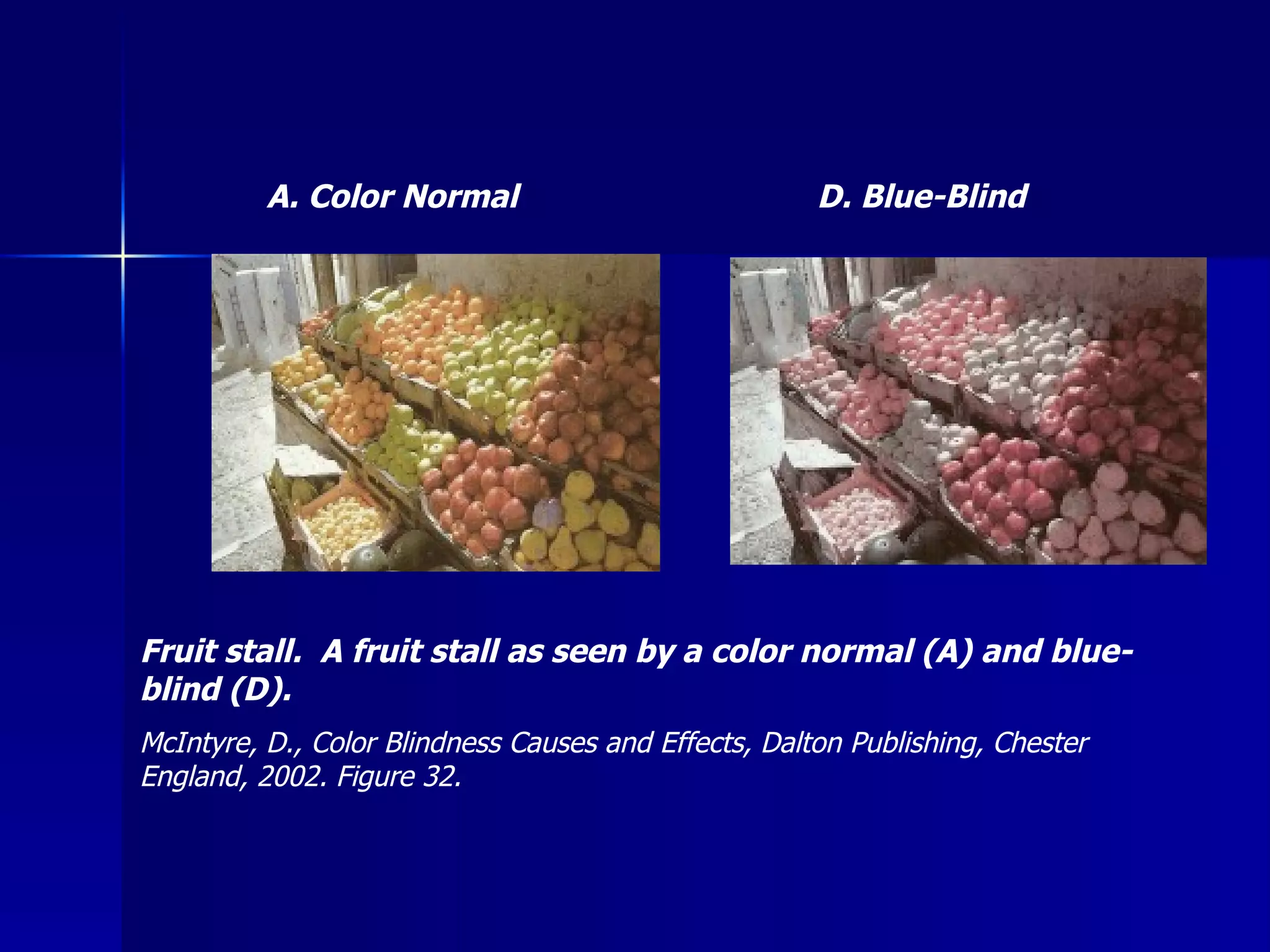
Color Vision Testing Bernard R. Blais, M.D. Clinical Professor Albany Medical College Albany, New York Blais Consulting, Ltd. 4 Innisbrook Drive Clifton Park, NY  12065 Phone:  (518) 371-8147 Fax:  (518) 373-9347 Email: bblais@nycap.rr.com  Disclosure:  Book Royalty Publisher New Color Vision Book, RPI Publishers, Atlanta, GA
Who is doing color vision testing?
What are you testing for? Congenital deficiencies
What are you testing for? Acquired deficiencies
What are you testing for? Both
Who is using the Ishihara Plates for testing color deficiencies? What is it testing?
Who utilizes the color vision testing in the Titmus Equipment Stereo Optical Equipment Armed Forces Screening
What color test is utilized?
Ishihara plates
What do they test for? Red deficiencies Green deficiencies
Does color defect really cause a difference? Websafe selection.  The 27 normal colours have been produced by all combinations of 0, 50, and 100% levels of red, green and blue primaries.  They have then been transformed to show their appearance to colour-blind viewers.  McIntyre, D., Color Blindness Causes and Effects, Dalton Publishing, Chester England, 2002 Figure 30.
Visual Spectrum Defects Three primary colors Blue Green Red
Retinal cones Contain classes of photo pigment Overlapping spectral sensitivity Have maximum sensitivity in the spectral regions Classic photo pigment
Color Defects Trichromatic Theory All spectral hues matched by mixture of three primary colors Short Wave – Blue  Medium Wave – Green Long Wave – Red
Trichromatic Theory Mariotte (1681) Newton (1704) All colors could be matched by three others. Cut-away view of the Munsell solid. McIntyre, D., Color Blindness Causes and Effects, Dalton Publishing, Chester England, 2002 Figure 6 as modified from Britannica® DVD2000 © 1994-2000 Encyclopedia Britannica, Inc.
So what does one actually see with each of these deficiencies?
Color normal Blue blind
Color normal Green blind
Color normal Red blind
Websafe selection.  The 27 normal colours have been produced by all combinations of 0, 50, and 100% levels of red, green and blue primaries.  They have then been transformed to show their appearance to colour-blind viewers.  McIntyre, D., Color Blindness Causes and Effects, Dalton Publishing, Chester England, 2002 Figure 30.
How does this affect the  individual’s ability?
Another example of how color might appear with normal color vision on the left, and the same objects as they might appear with a color deficiency on the right.
Traffic lights as seen with normal color vision on the left and, as they might appear with some types of color vision deficiency on the right.
The Nomenclature of Color Vision Deficiencies Table I Incidence of Color Defects in Caucasian Males Heredity      % (1)  Trichromatism (a) normal    91.8 (b) anomalous - (i)  protanomaly   1.0 (ii)  deuteranomaly   4.6 (iii)  tritanomaly 0.0001 (2)  Dichromatism (a) protanopia     1.2 (b) deuteranopia   1.4 (c) tritanopia 0.0001 (3) Monochromatism (a) of the rods 0.003 (b) of the cones 0.000001 Acquired (a) triton (blue-yellow) possibly 5% (b) proton (red-green) Duke Elder, S., Gloster, J. Weale, R. Physiology of The Eye, Chapter XVI on Color Vision, page 616, 1968 Table XIV and Birch, Jennifer, Diagnosis of defective color vision, Oxford University Press (1993).
Simplified terminology for the standard poorly understood color deficiencies TABLE 2 Formal Term New Term protanopic red-blind deuteranopic green-blind tritanopic blue-blind protanomalous red-weak deuteranomalous green-weak tritanomalous blue-weak achromatic achromatic McIntyre, D., Color Blindness Causes and Effects, Dalton Publishing, Chester England, 2002 Table 7 as modified from ©Transport for London.  Reproduced by kind permission of London’s Transport Museum.
Available Color Vision Testing Techniques Pseudoisochromatic Plates Most widely used Example is Ishihara and HHR test Each plate made of Irregular color dots Concealed hidden figure Rapid screening between normal and deficiencies
Available Color Vision Testing Techniques 10 min. 10 min. 6000 Classify and grade red-green defects Rarely used Anomaloscope (Nagel) Prototypes  Not standardized to requirements.  Prototype under  under develop-  development. ment. Computer-Based Tests <5 min. 1 hour >10 min. < 5 min. PATIENT TIME Test  occupational requirement Discrimination Discrimination Severity INDICATION < 5 min. <500 Widely used Pseudoisochromatic Plate <5 min. 3000 Used in screening persons for RG  Standard test Lantern (Falant) 1 hour >10 min. 900 350 Very selective Medical  Research Arrangement Test – Hues FM 100 D 15 TECH TIME EQUIPMENT COST FREQUENCY USED
Available Color Vision Testing Techniques R.G. No Optical equipment Anomaloscope (Nagel) R.G.B Yes Plate of irregular dots Pseudoisochromatic Plate Not standardized to requirements.  Prototype under development. Computer-Based Tests R.G.White Yes One to three lights Lantern (Falant) R.G.B. Identify moderate Severe capabilities Classify R.G.B.  abnormalities Yes No No Color disks (caps) 100 15 Arrangement Test – Hues FM 100 D 15 GRADING TESTS SCREENER DESIGN
Available Color Vision Testing Techniques Arrangement Test Consist of colored discs (cap) Color change from one cap to another Arranged color changes in order The 100 Hue arrangement test  -  Photo courtesy of  Richmond Products.
Available Color Vision Testing Techniques 10 min. 10 min. 600 Classify and grade red-green defects Rarely used Anomaloscope (Nagel) Prototypes  Not standardized to requirements.  Prototype under  under develop-  development. ment. Computer-Based Tests <5 min. 1 hour >10 min. < 5 min. PATIENT TIME Test  occupational requirement Discrimination Discrimination Severity INDICATION < 5 min. <500 Widely used Pseudoisochromatic Plate <5 min. 3000 Used in screening persons for RG  Standard test Lantern (Falant) 1 hour >10 min. 900 350 Very selective Medical Research Arrangement Test – Hues FM 100 D 15 TECH TIME EQUIPMENT COST FREQUENCY USED
Illumination Hue Discrimination Arrangements, PIC Tests Appearance pigment colors change by illumination. Ishihara plates results altered by Tungsten light Pass unqualified CIE – standard source required. Insert Figure 20 from manuscript
Available Color Vision Testing Techniques R.G No Optical equipment Anomaloscope (Nagel) R.G.B Yes Plate of irregular dots Pseudoisochromatic Plate Not standardized to requirements.  Prototype under development. Computer-Based Tests R.G.White Yes One to three lights Lantern (Falant) R.G.B. Identify moderate Severe capabilities Classify R.G.B.  abnormalities Yes No No Color disks (caps) 100 15 Arrangement Test – Hues FM 100 D 15 GRADING TESTS SCREENER DESIGN
Available Color Vision Testing Techniques Anomaloscope (Nagel) Used to classify and grade red-green  defects Balance of red-green to yellow
Available Color Vision Testing Techniques 10 min. 10 min. 6000 Classify and grade red-green defects Rarely used Anomaloscope (Nagel) Prototypes  Not standardized to requirements.  Prototype under  under develop-  development. ment. Computer-Based Tests <5 min. 1 hour >10 min. < 5 min. PATIENT TIME Test  occupational requirement Discrimination Discrimination Severity INDICATION < 5 min. <500 Widely used Pseudoisochromatic Plate <5 min. 3000 Used in screening persons for RG  Standard test Lantern (Falant) 1 hour >10 min. 900 350 Very selective Medical Research Arrangement Test – Hues FM 100 D 15 TECH TIME EQUIPMENT COST FREQUENCY USED
Available Color Vision Testing Techniques R.G. No Optical equipment Anomaloscope (Nagel) R.G.B Yes Plate of irregular dots Pseudoisochromatic Plate Not standardized to requirements.  Prototype under development. Computer-Based Tests R.G.White Yes One to three lights Lantern (Falant) R.G.B. Identify moderate Severe capabilities Classify R.G.B.  abnormalities Yes No No Color disks (caps) 100 15 Arrangement Test – Hues FM 100 D 15 GRADING TESTS SCREENER DESIGN
Available Color Vision Testing Techniques Lantern Tests Try to test the safety needs for actual jobs. Stereo Optical Optec 900
Available Color Vision Testing Techniques 10 min. 10 min. 6000 Classify and grade red-green defects Rarely used Anomaloscope (Nagel) Prototypes  Not standardized to requirements.  Prototype under  under develop-  development. ment. Computer-Based Tests <5 min. 1 hour >10 min.. < 5 min. PATIENT TIME Test  occupational requirement Discrimination Discrimination Severity INDICATION < 5 min. <500 Widely used Pseudoisochromatic Plate <5 min. 3000 Used in screening persons for RG  Standard test Lantern (Falant) 1 hour >10 min. 900 350 Very selective Medical Research Arrangement Test – Hues FM 100 D 15 TECH TIME EQUIPMENT COST FREQUENCY USED
Available Color Vision Testing Techniques R.G. No Optical equipment Anomaloscope (Nagel) R.G.B Yes Plate of irregular dots Pseudoisochromatic Plate Not standardized to requirements.  Prototype under development. Computer-Based Tests R.G.White Yes One to three lights Lantern (Falant) R.G.B. Identify moderate Severe capabilities Classify R.G.B.  abnormalities Yes No No Color disks (caps) 100 15 Arrangement Test – Hues FM 100 D 15 GRADING TESTS SCREENER DESIGN
Available Color Vision Testing Techniques Computer-Based Tests Just being developed in several laboratories Some based on Conventional diagnostic tests Others explain the new medium
Available Color Vision Testing Techniques 10 min. 10 min. 6000 Classify and grade red-green defects Rarely used Anomaloscope (Nagel) Prototypes  Not standardized to requirements.  Prototype under  under develop-  development. ment. Computer-Based Tests <5 min. 1 hour >10 min. < 5 min. PATIENT TIME Test  occupational requirement Discrimination Discrimination Severity INDICATION < 5 min. <500 Widely used Pseudoisochromatic Plate <5 min. 3000 Used in screening persons for RG  Standard test Lantern (Falant) 1 hour >10 min. 900 350 Very selective Medical Research Arrangement Test – Hues FM 100 D 15 TECH TIME EQUIPMENT COST FREQUENCY USED
Available Color Vision Testing Techniques R.G. No Optical equipment Anomaloscope (Nagel) R.G.B Yes Plate of irregular dots Pseudoisochromatic Plate Not standardized to requirements.  Prototype under development. Computer-Based Tests R.G.White Yes One to three lights Lantern (Falant) R.G.B. Identify moderate Severe capabilities Classify R.G.B.  abnormalities Yes No No Color disks (caps) 100 15 Arrangement Test – Hues FM 100 D 15 GRADING TESTS SCREENER DESIGN
Acquired Color Vision Defects Resulting from Ocular Pathology Intracranial injury Excessive use of therapeutic drugs Chemical toxicological effects
Chemical  Chemical toxicological effects seen Xylene, toluene toxicity Blue-yellow color deficiency present prior to findings of peripheral neuropathy At what toxic level does the color vision defect found more research needed?
Basic Requirements of Color Vision Screening Must be able to evaluate Red.Green.Blue hues Must be technically simple to operate Must take short period to complete  (1-2 minutes maximum) Technician/nurse time normally 5 minutes. Must be inexpensive to purchase test and special illumination
What test fulfills these requirements? The function of different types of color vision tests   Classification and Screening tests grading tests Diagnostic tests   Vocational tests 1.  Anomaloscopes 1.  Anomaloscopes 1.  Anomaloscopes and   psychophysical tests 2.  Pseudoisochromatic 2.  Some pseudo- plates     isochromatic plates 3.  Hue discrimination   1. Hue discrimination   (or arrangement) tests   (or arrangement)    tests   2.  Lanterns
What test fulfills these requirements? The principal color vision tests in each design category Pseudoisochromatic Hue discrimination Anomaloscopes plates (or arrangement) tests Lanterns Others Nagel Ishihara Farnsworth D15 (a) Paired lights TNO test Holmes-Wright Cit University test Neitz American Optical Co. Desaturated D15 Falant Besançon (Hardy, Rand & Ritter) (Adams & Lanthony) Pickford-Nicholson Tokyo Medical College 28 Hue (b)Single lights Sahlgren’s test Dvorine 40 Hue Edridge-Green Oscar test F2 Plate Giles-Archer Velhagen Farnsworth-Munsell Color-threshold Ohkuma 100 Hue   tester American Optical Co.     (13375) Bostrum-Kugelberg Standard pseudiosochromatic   plates 1st & 2nd editions Lanthony tritan album
Printed Pseudoisochromatic Plates Most widely used screening for color vision Most design to detect congenital Red-Green Only four tests – detects tritan (blue defects)
Goal in Occupational Medicine A screening test that screens for Red Green Tritan (blue – yellow)
Screening Test in Vision Screeners  Armed Forces Titmus Stereo Optical Ishihara Test Screens for only red and green
AO HRR Plates American Optical’s original Hardy, Rand, Ritter Plates
HRR Plates Consist of background colored circles Range of size and lightness HRR indicates severity of defect of any RG defect Screen for tritan (blue-yellow)
HRR Plates By itself can determine integrity of three hues Red Green Blue
Two HRR Plates Acceptable Original  American Optical Plates (1954) Richmond Products, Inc. Fourth Edition only (2002)
HRR Diagnosis of Color Deficiency Diagnosis based on HRR results Type HRR Extent: Passes Screener  Mild   Medium   Strong  Frequency Protan Minimal Affect Anomalous   Anomalous  Protanope   25% Protan   Protan Deutan Minimal Affect Anomalous   Anomalous  Deuteranope   75% Deutan   Deutan Tritan Minimal Affect Anomalous   Anomalous  Trianope   trace Comparison with Color Chip tests Farnsworth D15 Passes Passes   Fails   Fails   N/A L’Anthony D15 Passes   Fails   Fails   Fails   N/A Informational Brochures of Richmond Products Resources 2007.
Alternate Test Two tests needed to identify Red-Green defects and tritan (blue-yellow)
Tests used for evaluation of congenital color deficiency (modified) Function Scope Tests Screening red-green defects Ishihara plates blue defects CU (City University) tritan plates HRR plates Screening & grading Ishihara + CU tritan plates HRR plates D15 test, Anomaloscope Evaluation D15, FM100 Anomaloscope Occupational transport & armed Ishihara plates screening services Falant McIntyre, D, Color Blindness Causes and Effects, Dalton Publishing, Chester, England, 2002, p. 81.
Confused? Yes – When I started to evaluate this subject, books were all too complicated for occupational medicine. Tasking of dyschromatopsia by ACOEM’s Council on Scientific Affairs to Sensory Perception Committee to explore the evidence for toxicological acquired color deficiency.
Do you want your employees having the “activities of daily living” working with these defects?
What do you want the employees to see where color vision is a mandatory requirement?
B. Red-Blind A. Color Normal Fruit stall.  A fruit stall as seen by a color normal (A) and red-blind (B). McIntyre, D., Color Blindness Causes and Effects, Dalton Publishing, Chester England, 2002. Figure 32.
C. Green-Blind A. Color Normal Fruit stall.  A fruit stall as seen by a color normal (A) and green-blind (C). McIntyre, D., Color Blindness Causes and Effects, Dalton Publishing, Chester England, 2002. Figure 32.
D. Blue-Blind A. Color Normal Fruit stall.  A fruit stall as seen by a color normal (A) and blue-blind (D). McIntyre, D., Color Blindness Causes and Effects, Dalton Publishing, Chester England, 2002. Figure 32.
Dr. Maureen Neitz

Color Vision Testing

  • 1.
    Color Vision TestingBernard R. Blais, M.D. Clinical Professor Albany Medical College Albany, New York Blais Consulting, Ltd. 4 Innisbrook Drive Clifton Park, NY 12065 Phone: (518) 371-8147 Fax: (518) 373-9347 Email: bblais@nycap.rr.com Disclosure: Book Royalty Publisher New Color Vision Book, RPI Publishers, Atlanta, GA
  • 2.
    Who is doingcolor vision testing?
  • 3.
    What are youtesting for? Congenital deficiencies
  • 4.
    What are youtesting for? Acquired deficiencies
  • 5.
    What are youtesting for? Both
  • 6.
    Who is usingthe Ishihara Plates for testing color deficiencies? What is it testing?
  • 7.
    Who utilizes thecolor vision testing in the Titmus Equipment Stereo Optical Equipment Armed Forces Screening
  • 8.
    What color testis utilized?
  • 9.
  • 10.
    What do theytest for? Red deficiencies Green deficiencies
  • 11.
    Does color defectreally cause a difference? Websafe selection. The 27 normal colours have been produced by all combinations of 0, 50, and 100% levels of red, green and blue primaries. They have then been transformed to show their appearance to colour-blind viewers. McIntyre, D., Color Blindness Causes and Effects, Dalton Publishing, Chester England, 2002 Figure 30.
  • 12.
    Visual Spectrum DefectsThree primary colors Blue Green Red
  • 13.
    Retinal cones Containclasses of photo pigment Overlapping spectral sensitivity Have maximum sensitivity in the spectral regions Classic photo pigment
  • 14.
    Color Defects TrichromaticTheory All spectral hues matched by mixture of three primary colors Short Wave – Blue Medium Wave – Green Long Wave – Red
  • 15.
    Trichromatic Theory Mariotte(1681) Newton (1704) All colors could be matched by three others. Cut-away view of the Munsell solid. McIntyre, D., Color Blindness Causes and Effects, Dalton Publishing, Chester England, 2002 Figure 6 as modified from Britannica® DVD2000 © 1994-2000 Encyclopedia Britannica, Inc.
  • 16.
    So what doesone actually see with each of these deficiencies?
  • 17.
  • 18.
  • 19.
  • 20.
    Websafe selection. The 27 normal colours have been produced by all combinations of 0, 50, and 100% levels of red, green and blue primaries. They have then been transformed to show their appearance to colour-blind viewers. McIntyre, D., Color Blindness Causes and Effects, Dalton Publishing, Chester England, 2002 Figure 30.
  • 21.
    Websafe selection. The 27 normal colours have been produced by all combinations of 0, 50, and 100% levels of red, green and blue primaries. They have then been transformed to show their appearance to colour-blind viewers. McIntyre, D., Color Blindness Causes and Effects, Dalton Publishing, Chester England, 2002 Figure 30.
  • 22.
    Websafe selection. The 27 normal colours have been produced by all combinations of 0, 50, and 100% levels of red, green and blue primaries. They have then been transformed to show their appearance to colour-blind viewers. McIntyre, D., Color Blindness Causes and Effects, Dalton Publishing, Chester England, 2002 Figure 30.
  • 23.
    How does thisaffect the individual’s ability?
  • 24.
    Another example ofhow color might appear with normal color vision on the left, and the same objects as they might appear with a color deficiency on the right.
  • 25.
    Traffic lights asseen with normal color vision on the left and, as they might appear with some types of color vision deficiency on the right.
  • 26.
    The Nomenclature ofColor Vision Deficiencies Table I Incidence of Color Defects in Caucasian Males Heredity % (1) Trichromatism (a) normal 91.8 (b) anomalous - (i) protanomaly 1.0 (ii) deuteranomaly 4.6 (iii) tritanomaly 0.0001 (2) Dichromatism (a) protanopia 1.2 (b) deuteranopia 1.4 (c) tritanopia 0.0001 (3) Monochromatism (a) of the rods 0.003 (b) of the cones 0.000001 Acquired (a) triton (blue-yellow) possibly 5% (b) proton (red-green) Duke Elder, S., Gloster, J. Weale, R. Physiology of The Eye, Chapter XVI on Color Vision, page 616, 1968 Table XIV and Birch, Jennifer, Diagnosis of defective color vision, Oxford University Press (1993).
  • 27.
    Simplified terminology forthe standard poorly understood color deficiencies TABLE 2 Formal Term New Term protanopic red-blind deuteranopic green-blind tritanopic blue-blind protanomalous red-weak deuteranomalous green-weak tritanomalous blue-weak achromatic achromatic McIntyre, D., Color Blindness Causes and Effects, Dalton Publishing, Chester England, 2002 Table 7 as modified from ©Transport for London. Reproduced by kind permission of London’s Transport Museum.
  • 28.
    Available Color VisionTesting Techniques Pseudoisochromatic Plates Most widely used Example is Ishihara and HHR test Each plate made of Irregular color dots Concealed hidden figure Rapid screening between normal and deficiencies
  • 29.
    Available Color VisionTesting Techniques 10 min. 10 min. 6000 Classify and grade red-green defects Rarely used Anomaloscope (Nagel) Prototypes Not standardized to requirements. Prototype under under develop- development. ment. Computer-Based Tests <5 min. 1 hour >10 min. < 5 min. PATIENT TIME Test occupational requirement Discrimination Discrimination Severity INDICATION < 5 min. <500 Widely used Pseudoisochromatic Plate <5 min. 3000 Used in screening persons for RG Standard test Lantern (Falant) 1 hour >10 min. 900 350 Very selective Medical Research Arrangement Test – Hues FM 100 D 15 TECH TIME EQUIPMENT COST FREQUENCY USED
  • 30.
    Available Color VisionTesting Techniques R.G. No Optical equipment Anomaloscope (Nagel) R.G.B Yes Plate of irregular dots Pseudoisochromatic Plate Not standardized to requirements. Prototype under development. Computer-Based Tests R.G.White Yes One to three lights Lantern (Falant) R.G.B. Identify moderate Severe capabilities Classify R.G.B. abnormalities Yes No No Color disks (caps) 100 15 Arrangement Test – Hues FM 100 D 15 GRADING TESTS SCREENER DESIGN
  • 31.
    Available Color VisionTesting Techniques Arrangement Test Consist of colored discs (cap) Color change from one cap to another Arranged color changes in order The 100 Hue arrangement test - Photo courtesy of Richmond Products.
  • 32.
    Available Color VisionTesting Techniques 10 min. 10 min. 600 Classify and grade red-green defects Rarely used Anomaloscope (Nagel) Prototypes Not standardized to requirements. Prototype under under develop- development. ment. Computer-Based Tests <5 min. 1 hour >10 min. < 5 min. PATIENT TIME Test occupational requirement Discrimination Discrimination Severity INDICATION < 5 min. <500 Widely used Pseudoisochromatic Plate <5 min. 3000 Used in screening persons for RG Standard test Lantern (Falant) 1 hour >10 min. 900 350 Very selective Medical Research Arrangement Test – Hues FM 100 D 15 TECH TIME EQUIPMENT COST FREQUENCY USED
  • 33.
    Illumination Hue DiscriminationArrangements, PIC Tests Appearance pigment colors change by illumination. Ishihara plates results altered by Tungsten light Pass unqualified CIE – standard source required. Insert Figure 20 from manuscript
  • 34.
    Available Color VisionTesting Techniques R.G No Optical equipment Anomaloscope (Nagel) R.G.B Yes Plate of irregular dots Pseudoisochromatic Plate Not standardized to requirements. Prototype under development. Computer-Based Tests R.G.White Yes One to three lights Lantern (Falant) R.G.B. Identify moderate Severe capabilities Classify R.G.B. abnormalities Yes No No Color disks (caps) 100 15 Arrangement Test – Hues FM 100 D 15 GRADING TESTS SCREENER DESIGN
  • 35.
    Available Color VisionTesting Techniques Anomaloscope (Nagel) Used to classify and grade red-green defects Balance of red-green to yellow
  • 36.
    Available Color VisionTesting Techniques 10 min. 10 min. 6000 Classify and grade red-green defects Rarely used Anomaloscope (Nagel) Prototypes Not standardized to requirements. Prototype under under develop- development. ment. Computer-Based Tests <5 min. 1 hour >10 min. < 5 min. PATIENT TIME Test occupational requirement Discrimination Discrimination Severity INDICATION < 5 min. <500 Widely used Pseudoisochromatic Plate <5 min. 3000 Used in screening persons for RG Standard test Lantern (Falant) 1 hour >10 min. 900 350 Very selective Medical Research Arrangement Test – Hues FM 100 D 15 TECH TIME EQUIPMENT COST FREQUENCY USED
  • 37.
    Available Color VisionTesting Techniques R.G. No Optical equipment Anomaloscope (Nagel) R.G.B Yes Plate of irregular dots Pseudoisochromatic Plate Not standardized to requirements. Prototype under development. Computer-Based Tests R.G.White Yes One to three lights Lantern (Falant) R.G.B. Identify moderate Severe capabilities Classify R.G.B. abnormalities Yes No No Color disks (caps) 100 15 Arrangement Test – Hues FM 100 D 15 GRADING TESTS SCREENER DESIGN
  • 38.
    Available Color VisionTesting Techniques Lantern Tests Try to test the safety needs for actual jobs. Stereo Optical Optec 900
  • 39.
    Available Color VisionTesting Techniques 10 min. 10 min. 6000 Classify and grade red-green defects Rarely used Anomaloscope (Nagel) Prototypes Not standardized to requirements. Prototype under under develop- development. ment. Computer-Based Tests <5 min. 1 hour >10 min.. < 5 min. PATIENT TIME Test occupational requirement Discrimination Discrimination Severity INDICATION < 5 min. <500 Widely used Pseudoisochromatic Plate <5 min. 3000 Used in screening persons for RG Standard test Lantern (Falant) 1 hour >10 min. 900 350 Very selective Medical Research Arrangement Test – Hues FM 100 D 15 TECH TIME EQUIPMENT COST FREQUENCY USED
  • 40.
    Available Color VisionTesting Techniques R.G. No Optical equipment Anomaloscope (Nagel) R.G.B Yes Plate of irregular dots Pseudoisochromatic Plate Not standardized to requirements. Prototype under development. Computer-Based Tests R.G.White Yes One to three lights Lantern (Falant) R.G.B. Identify moderate Severe capabilities Classify R.G.B. abnormalities Yes No No Color disks (caps) 100 15 Arrangement Test – Hues FM 100 D 15 GRADING TESTS SCREENER DESIGN
  • 41.
    Available Color VisionTesting Techniques Computer-Based Tests Just being developed in several laboratories Some based on Conventional diagnostic tests Others explain the new medium
  • 42.
    Available Color VisionTesting Techniques 10 min. 10 min. 6000 Classify and grade red-green defects Rarely used Anomaloscope (Nagel) Prototypes Not standardized to requirements. Prototype under under develop- development. ment. Computer-Based Tests <5 min. 1 hour >10 min. < 5 min. PATIENT TIME Test occupational requirement Discrimination Discrimination Severity INDICATION < 5 min. <500 Widely used Pseudoisochromatic Plate <5 min. 3000 Used in screening persons for RG Standard test Lantern (Falant) 1 hour >10 min. 900 350 Very selective Medical Research Arrangement Test – Hues FM 100 D 15 TECH TIME EQUIPMENT COST FREQUENCY USED
  • 43.
    Available Color VisionTesting Techniques R.G. No Optical equipment Anomaloscope (Nagel) R.G.B Yes Plate of irregular dots Pseudoisochromatic Plate Not standardized to requirements. Prototype under development. Computer-Based Tests R.G.White Yes One to three lights Lantern (Falant) R.G.B. Identify moderate Severe capabilities Classify R.G.B. abnormalities Yes No No Color disks (caps) 100 15 Arrangement Test – Hues FM 100 D 15 GRADING TESTS SCREENER DESIGN
  • 44.
    Acquired Color VisionDefects Resulting from Ocular Pathology Intracranial injury Excessive use of therapeutic drugs Chemical toxicological effects
  • 45.
    Chemical Chemicaltoxicological effects seen Xylene, toluene toxicity Blue-yellow color deficiency present prior to findings of peripheral neuropathy At what toxic level does the color vision defect found more research needed?
  • 46.
    Basic Requirements ofColor Vision Screening Must be able to evaluate Red.Green.Blue hues Must be technically simple to operate Must take short period to complete (1-2 minutes maximum) Technician/nurse time normally 5 minutes. Must be inexpensive to purchase test and special illumination
  • 47.
    What test fulfillsthese requirements? The function of different types of color vision tests Classification and Screening tests grading tests Diagnostic tests Vocational tests 1. Anomaloscopes 1. Anomaloscopes 1. Anomaloscopes and psychophysical tests 2. Pseudoisochromatic 2. Some pseudo- plates isochromatic plates 3. Hue discrimination 1. Hue discrimination (or arrangement) tests (or arrangement) tests 2. Lanterns
  • 48.
    What test fulfillsthese requirements? The principal color vision tests in each design category Pseudoisochromatic Hue discrimination Anomaloscopes plates (or arrangement) tests Lanterns Others Nagel Ishihara Farnsworth D15 (a) Paired lights TNO test Holmes-Wright Cit University test Neitz American Optical Co. Desaturated D15 Falant Besançon (Hardy, Rand & Ritter) (Adams & Lanthony) Pickford-Nicholson Tokyo Medical College 28 Hue (b)Single lights Sahlgren’s test Dvorine 40 Hue Edridge-Green Oscar test F2 Plate Giles-Archer Velhagen Farnsworth-Munsell Color-threshold Ohkuma 100 Hue tester American Optical Co. (13375) Bostrum-Kugelberg Standard pseudiosochromatic plates 1st & 2nd editions Lanthony tritan album
  • 49.
    Printed Pseudoisochromatic PlatesMost widely used screening for color vision Most design to detect congenital Red-Green Only four tests – detects tritan (blue defects)
  • 50.
    Goal in OccupationalMedicine A screening test that screens for Red Green Tritan (blue – yellow)
  • 51.
    Screening Test inVision Screeners Armed Forces Titmus Stereo Optical Ishihara Test Screens for only red and green
  • 52.
    AO HRR PlatesAmerican Optical’s original Hardy, Rand, Ritter Plates
  • 53.
    HRR Plates Consistof background colored circles Range of size and lightness HRR indicates severity of defect of any RG defect Screen for tritan (blue-yellow)
  • 54.
    HRR Plates Byitself can determine integrity of three hues Red Green Blue
  • 55.
    Two HRR PlatesAcceptable Original American Optical Plates (1954) Richmond Products, Inc. Fourth Edition only (2002)
  • 56.
    HRR Diagnosis ofColor Deficiency Diagnosis based on HRR results Type HRR Extent: Passes Screener Mild Medium Strong Frequency Protan Minimal Affect Anomalous Anomalous Protanope 25% Protan Protan Deutan Minimal Affect Anomalous Anomalous Deuteranope 75% Deutan Deutan Tritan Minimal Affect Anomalous Anomalous Trianope trace Comparison with Color Chip tests Farnsworth D15 Passes Passes Fails Fails N/A L’Anthony D15 Passes Fails Fails Fails N/A Informational Brochures of Richmond Products Resources 2007.
  • 57.
    Alternate Test Twotests needed to identify Red-Green defects and tritan (blue-yellow)
  • 58.
    Tests used forevaluation of congenital color deficiency (modified) Function Scope Tests Screening red-green defects Ishihara plates blue defects CU (City University) tritan plates HRR plates Screening & grading Ishihara + CU tritan plates HRR plates D15 test, Anomaloscope Evaluation D15, FM100 Anomaloscope Occupational transport & armed Ishihara plates screening services Falant McIntyre, D, Color Blindness Causes and Effects, Dalton Publishing, Chester, England, 2002, p. 81.
  • 59.
    Confused? Yes –When I started to evaluate this subject, books were all too complicated for occupational medicine. Tasking of dyschromatopsia by ACOEM’s Council on Scientific Affairs to Sensory Perception Committee to explore the evidence for toxicological acquired color deficiency.
  • 60.
    Do you wantyour employees having the “activities of daily living” working with these defects?
  • 61.
    What do youwant the employees to see where color vision is a mandatory requirement?
  • 62.
    B. Red-Blind A.Color Normal Fruit stall. A fruit stall as seen by a color normal (A) and red-blind (B). McIntyre, D., Color Blindness Causes and Effects, Dalton Publishing, Chester England, 2002. Figure 32.
  • 63.
    C. Green-Blind A.Color Normal Fruit stall. A fruit stall as seen by a color normal (A) and green-blind (C). McIntyre, D., Color Blindness Causes and Effects, Dalton Publishing, Chester England, 2002. Figure 32.
  • 64.
    D. Blue-Blind A.Color Normal Fruit stall. A fruit stall as seen by a color normal (A) and blue-blind (D). McIntyre, D., Color Blindness Causes and Effects, Dalton Publishing, Chester England, 2002. Figure 32.
  • 65.