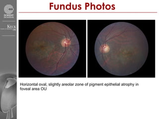
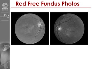
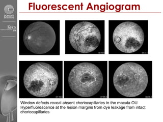
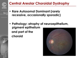
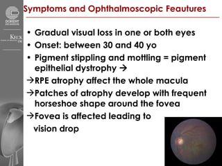
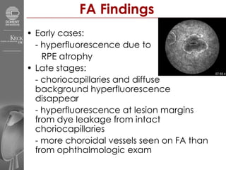
Doheny Eye Institute Grand Rounds Presenter Irina Bykhovskaya, MD Discussant SriniVas Sadda, MD 4/20/2007
Patient Presentation 31yo Vietnamese male with c/o gradual decrease in vision OU since adolescence  He has been using magnifying glass in the past 3 years for reading FHx: mother and brother also with “visual problems” PMH: h/o malaria 2 years ago treated with oral medications SHx: recent immigrant from Vietnam
Patient Examination VA: 8ft/200 OD and 6ft/200 OS Pupils: RR, no RAPD Ta 18 OU LLL: wnl OU S/C: w/q OU K: clear OU I: brown, flat OU AC: d/q OU L: 2+ PSC OU
Fundus Photos Horizontal oval, slightly areolar zone of pigment epithelial atrophy in  foveal area OU
Red Free Fundus Photos
Fluorescent Angiogram Window defects reveal absent choriocapillaries in the macula OU Hyperfluorescence at the lesion margins from dye leakage from intact choriocapillaries
Central Areolar Choroidal Dystrophy   Rare Autosomal Dominant (rarely recessive, occasionally sporadic) Pathology: atrophy of neuroepithelium, pigment epithelium  and part of the  choroid
Symptoms and Ophthalmoscopic Feautures Gradual visual loss in one or both eyes Onset: between 30 and 40 yo Pigment stippling and mottling = pigment epithelial dystrophy     RPE atrophy affect the whole macula  Patches of atrophy develop with frequent horseshoe shape around the fovea Fovea is affected leading to  vision drop
Copyright restrictions may apply. Keilhauer, C. N. et al. Arch Ophthalmol 2006;124:1020-1027. Clinical Findings
FA Findings Early cases:  - hyperfluorescence due to  RPE atrophy Late stages:  - choriocapillaries and diffuse background hyperfluorescence disappear - hyperfluorescence at lesion margins from dye leakage from intact choriocapillaries - more choroidal vessels seen on FA than from ophthalmologic exam
Visual Functions Visual Acuity   –  severely affected (CF) Visual Fields   –  normal in the periphery with central or paracentral scotoma Color Vision   –  moderately affected Dark Adaptation  - normal Electroretinography  – normal full-field, abnormal multifocal Electro-oculography  – usually normal
Histology Well-demarkated avascular zone in posterior choroid Atrophy and fibrosis No outer layers of retina or RPE corresponding to underlying choroidal atrophy Usually no breaks in Bruch’s membrane
Pathogenesis Hypothesis Likely primary dystrophy of the choroidal vessels Vs Primary tapetochoroidal dystrophy or primary dystrophy of RPE with secondary choroidal involvement
Differential Diagnosis Pseudoinflammatory dystrophy – Sorsby Choroideremia Myopia gravior Macular coloboma Angioid streaks Gyrate atrophy Peripapillary and circinate choroidal dystrophy End stages of Stargardt’s disease  Cone dystrophy Progressive bifocal chorioretinal atrophy Multifocal placoid pigment epitheliopathy
Mode of Inheritance  Autosomal Dominant - mutation in the Peripherin/RDS gene on chromosome 6 - plays role in   assembly, orientation, and structural stability of outer   segment discs and accounts for an increased turnover of instable   membranous segments  Autosomal Recessive  Sporadic
References K. Zhang, D.C. Garabaldi, R.E. Carr and J.S. Sunness , Hereditary choroidal disease. In: S.J. Ryan, Editor,  Retina  (3rd ed.), Mosby, St. Louis (2001), pp. 462–463.  Hoyng CB, Deutman AF. The development of central areolar choroidal dystrophy.  Graefes Arch Clin Exp Ophthalmol.  1996;234:87-93.  Castori M, Valente EM, Maurizio C, Tormene AP, Brancati F, Caputo V, Dallapiccola B. A Novel Locus for Autosomal Dominant Cone and Cone–Rod Dystrophies Maps to the 6p Gene Cluster of Retinal Dystrophies. Invest Ophthalmol Vis Sci. 2005 Oct;46(10):3539-44.  Keilhauer CN, Meigen T, Weber BH. Clinical findings in a multigeneration family with autosomal dominant central areolar choroidal dystrophy associated with an Arg195Leu mutation in the peripherin/RDS gene. Arch Ophthalmol. 2006 Jul;124(7):1020-7.  Nagasaka K, Horiauchi M, Shimada Y, Yuzawa M. Multifocal electroretinograms in cases of central areolar choroidal dystrophy. Invest Ophthalmol Vis Sci. 2003 Apr;44(4):1673-9.

Areolar Dystrophy

  • 1.
    Doheny Eye InstituteGrand Rounds Presenter Irina Bykhovskaya, MD Discussant SriniVas Sadda, MD 4/20/2007
  • 2.
    Patient Presentation 31yoVietnamese male with c/o gradual decrease in vision OU since adolescence He has been using magnifying glass in the past 3 years for reading FHx: mother and brother also with “visual problems” PMH: h/o malaria 2 years ago treated with oral medications SHx: recent immigrant from Vietnam
  • 3.
    Patient Examination VA:8ft/200 OD and 6ft/200 OS Pupils: RR, no RAPD Ta 18 OU LLL: wnl OU S/C: w/q OU K: clear OU I: brown, flat OU AC: d/q OU L: 2+ PSC OU
  • 4.
    Fundus Photos Horizontaloval, slightly areolar zone of pigment epithelial atrophy in foveal area OU
  • 5.
  • 6.
    Fluorescent Angiogram Windowdefects reveal absent choriocapillaries in the macula OU Hyperfluorescence at the lesion margins from dye leakage from intact choriocapillaries
  • 7.
    Central Areolar ChoroidalDystrophy Rare Autosomal Dominant (rarely recessive, occasionally sporadic) Pathology: atrophy of neuroepithelium, pigment epithelium and part of the choroid
  • 8.
    Symptoms and OphthalmoscopicFeautures Gradual visual loss in one or both eyes Onset: between 30 and 40 yo Pigment stippling and mottling = pigment epithelial dystrophy  RPE atrophy affect the whole macula Patches of atrophy develop with frequent horseshoe shape around the fovea Fovea is affected leading to vision drop
  • 9.
    Copyright restrictions mayapply. Keilhauer, C. N. et al. Arch Ophthalmol 2006;124:1020-1027. Clinical Findings
  • 10.
    FA Findings Earlycases: - hyperfluorescence due to RPE atrophy Late stages: - choriocapillaries and diffuse background hyperfluorescence disappear - hyperfluorescence at lesion margins from dye leakage from intact choriocapillaries - more choroidal vessels seen on FA than from ophthalmologic exam
  • 11.
    Visual Functions VisualAcuity – severely affected (CF) Visual Fields – normal in the periphery with central or paracentral scotoma Color Vision – moderately affected Dark Adaptation - normal Electroretinography – normal full-field, abnormal multifocal Electro-oculography – usually normal
  • 12.
    Histology Well-demarkated avascularzone in posterior choroid Atrophy and fibrosis No outer layers of retina or RPE corresponding to underlying choroidal atrophy Usually no breaks in Bruch’s membrane
  • 13.
    Pathogenesis Hypothesis Likelyprimary dystrophy of the choroidal vessels Vs Primary tapetochoroidal dystrophy or primary dystrophy of RPE with secondary choroidal involvement
  • 14.
    Differential Diagnosis Pseudoinflammatorydystrophy – Sorsby Choroideremia Myopia gravior Macular coloboma Angioid streaks Gyrate atrophy Peripapillary and circinate choroidal dystrophy End stages of Stargardt’s disease Cone dystrophy Progressive bifocal chorioretinal atrophy Multifocal placoid pigment epitheliopathy
  • 15.
    Mode of Inheritance Autosomal Dominant - mutation in the Peripherin/RDS gene on chromosome 6 - plays role in assembly, orientation, and structural stability of outer segment discs and accounts for an increased turnover of instable membranous segments Autosomal Recessive Sporadic
  • 16.
    References K. Zhang,D.C. Garabaldi, R.E. Carr and J.S. Sunness , Hereditary choroidal disease. In: S.J. Ryan, Editor, Retina (3rd ed.), Mosby, St. Louis (2001), pp. 462–463. Hoyng CB, Deutman AF. The development of central areolar choroidal dystrophy. Graefes Arch Clin Exp Ophthalmol. 1996;234:87-93. Castori M, Valente EM, Maurizio C, Tormene AP, Brancati F, Caputo V, Dallapiccola B. A Novel Locus for Autosomal Dominant Cone and Cone–Rod Dystrophies Maps to the 6p Gene Cluster of Retinal Dystrophies. Invest Ophthalmol Vis Sci. 2005 Oct;46(10):3539-44. Keilhauer CN, Meigen T, Weber BH. Clinical findings in a multigeneration family with autosomal dominant central areolar choroidal dystrophy associated with an Arg195Leu mutation in the peripherin/RDS gene. Arch Ophthalmol. 2006 Jul;124(7):1020-7. Nagasaka K, Horiauchi M, Shimada Y, Yuzawa M. Multifocal electroretinograms in cases of central areolar choroidal dystrophy. Invest Ophthalmol Vis Sci. 2003 Apr;44(4):1673-9.