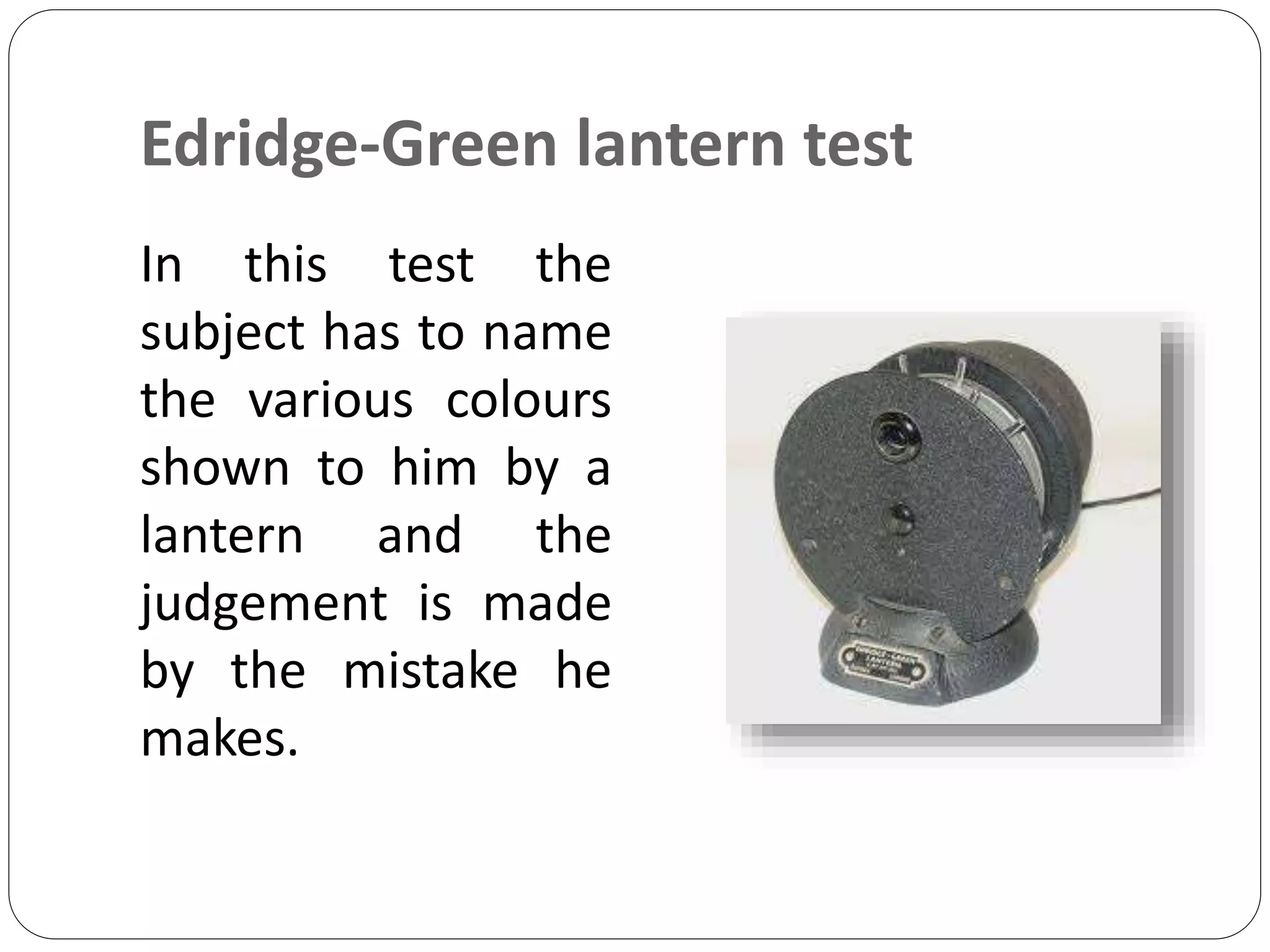
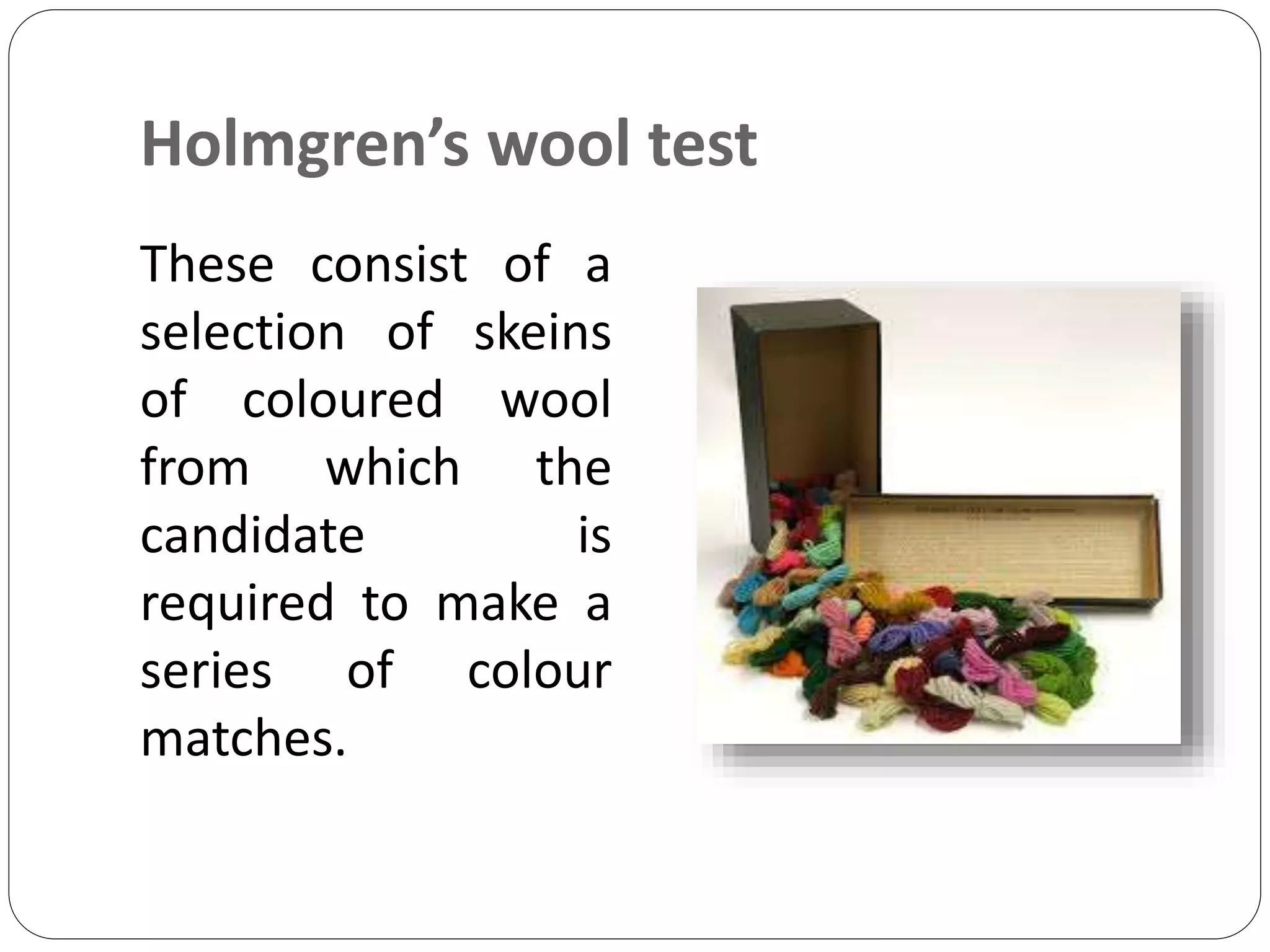
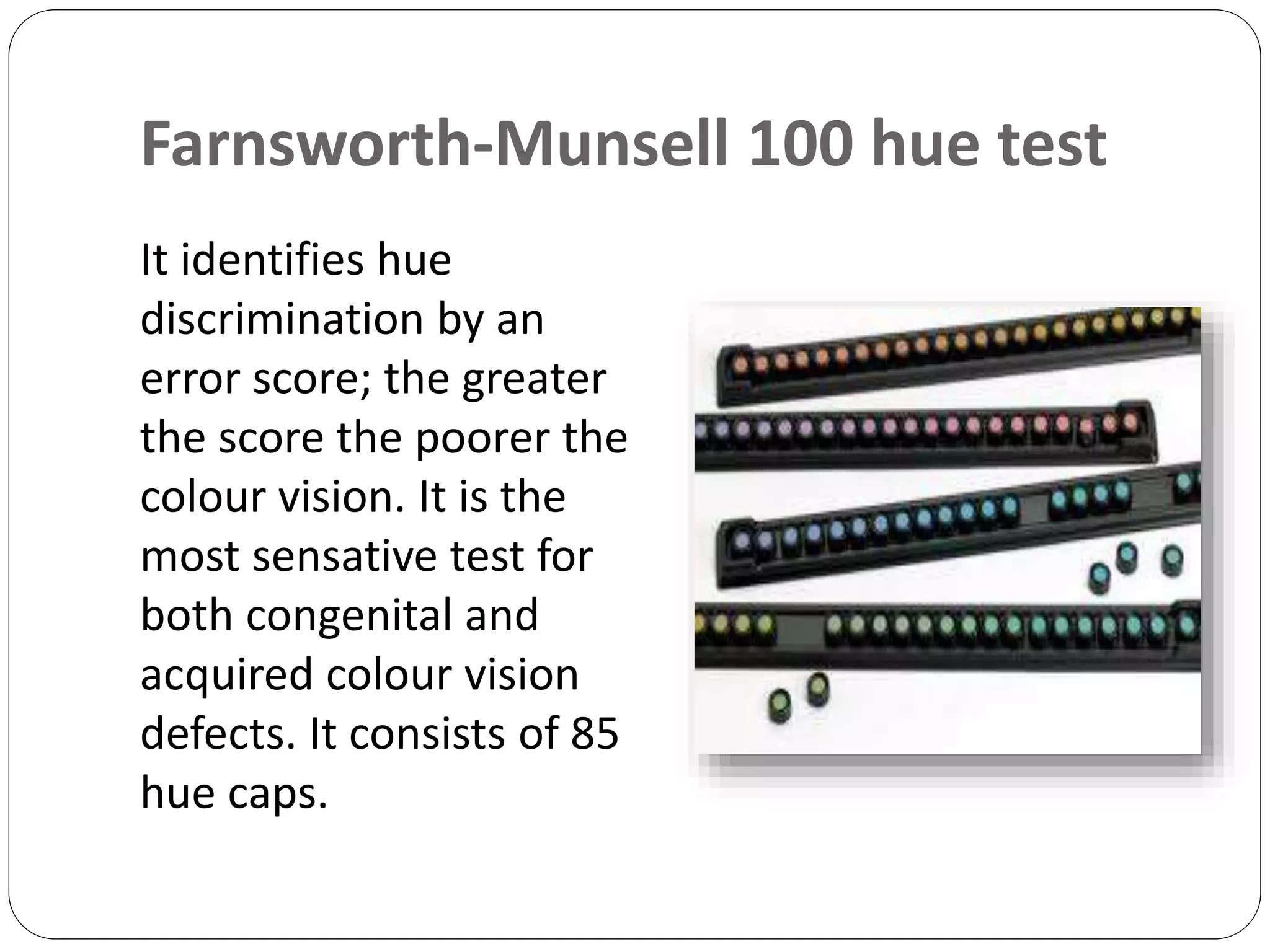
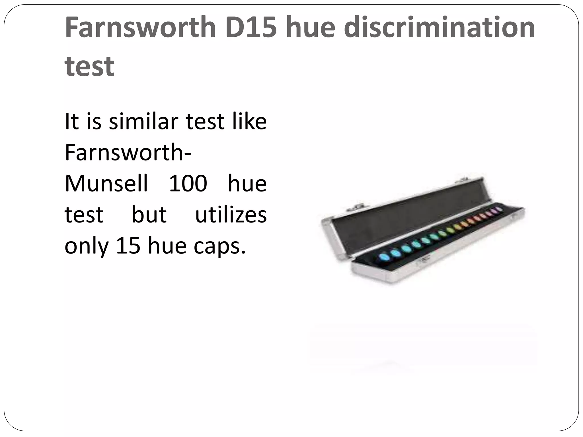
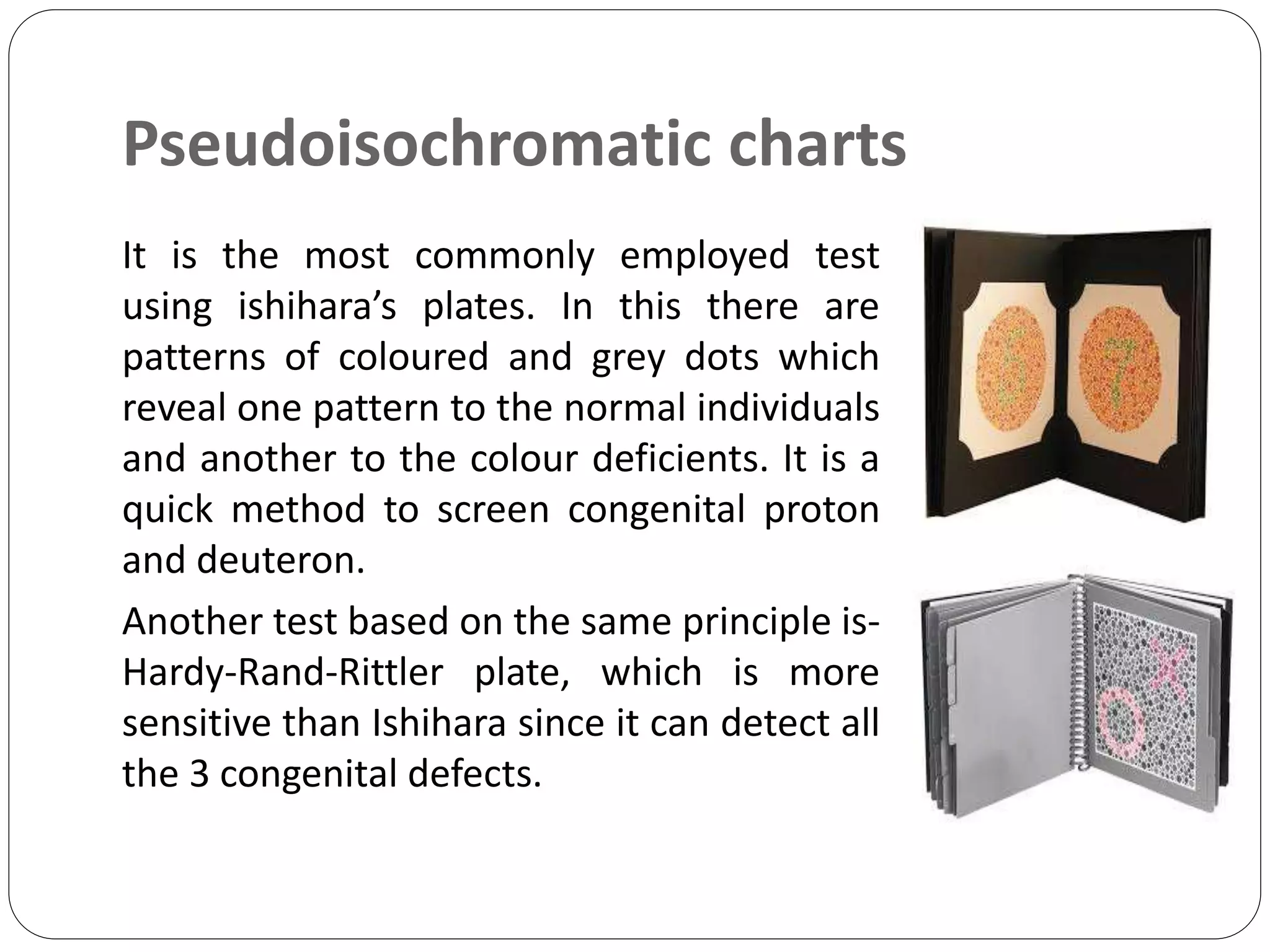
3. Introduction
Colour vision is the ability of the eye to
perceive light rays of different wavelengths
or different colours
It is a function of cones and thus better
appreciated in photopic vision
The visible wavelength of the
electromagnetic spectrum are between
400nm-780nm
4. Introduction
Colour doesn’t comes directly from the
objects we view, but rather, the brain
interprets wavelength bouncing off the
object we view
All colours are a result of admixture in
different proportion of the 3 primary
colours
5. Introduction
Theories of colour vision are-
⇨Trichromatic theory
⇨Opponent colour theory
⇨Additive colour theory
⇨Subtractive colour theory
6. Tests for colour vision
Test are design for:
(1)Screening defective vision from
normal
(2)Qualitative classification of colour
blindness, i.e. protons, deuteron and tritan
(3)Quantitative analysis of degree of
deficiency, i.e. mild, moderate or marked.
7. Tests for colour vision
Commonly employed colour vision tests are
as follows-
(i)Edridge-green lantern test
(ii)Holmgren’s wools test
(iii)Farnsworth-Munsell 100 hue test
(iv)Farnsworth D15 hue discrimination
test
8. Tests for colour vision
(v)Pseudoisochromatic charts
(vi)Nagel’s anomaloscope
9. Edridge-Green lantern test
In this test the
subject has to name
the various colours
shown to him by a
lantern and the
judgement is made
by the mistake he
makes.
10. Holmgren’s wool test
These consist of a
selection of skeins
of coloured wool
from which the
candidate is
required to make a
series of colour
matches.
11. Farnsworth-Munsell 100 hue test
It identifies hue
discrimination by an
error score; the greater
the score the poorer the
colour vision. It is the
most sensative test for
both congenital and
acquired colour vision
defects. It consists of 85
hue caps.
12. Farnsworth D15 hue discrimination
test
It is similar test like
Farnsworth-
Munsell 100 hue
test but utilizes
only 15 hue caps.
13. Pseudoisochromatic charts
It is the most commonly employed test
using ishihara’s plates. In this there are
patterns of coloured and grey dots which
reveal one pattern to the normal individuals
and another to the colour deficients. It is a
quick method to screen congenital proton
and deuteron.
Another test based on the same principle is-
Hardy-Rand-Rittler plate, which is more
sensitive than Ishihara since it can detect all
the 3 congenital defects.
14. Nagel’s anomaloscope
These are instruments in which
one looking down a telescope
a bright disc is seen dividing
into 2 halves by horizontal line.
One half is illuminated by
yellow light, and this has to be
matched by a mixture of red
and green in the other half. By
turning knobs, the relative
amounts of red and green in
the mixture and the brightness
can be varied.
15. References
Anatomy and Physiology of eye, 3th Edition,
by- A.K Khurana and Indu Khurana, Page no-
284-285.
Comprehensive Opthalmology, 7th Edition,
by- A.K Khurana, Page no-343
Parson’s Diseases of the Eye, 22nd Edition,
edited by Ramanjit Sihota and Radhika
Tandon, Page no- 107-109