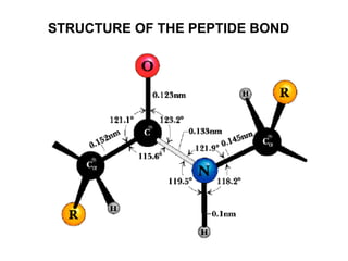
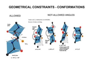
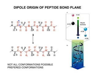
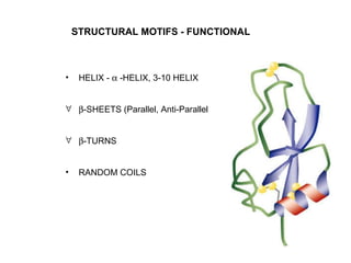
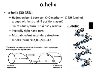
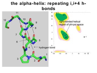
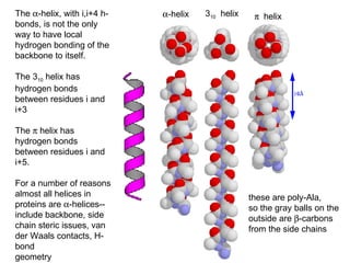
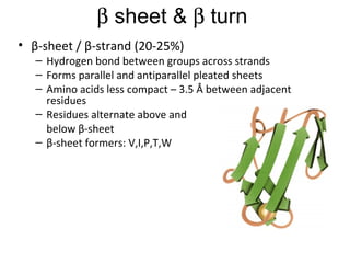
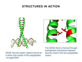
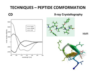
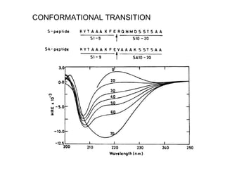
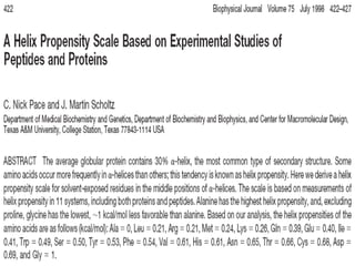
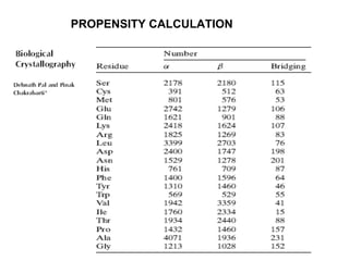
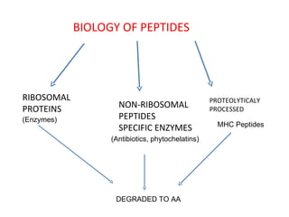
PEPTIDE STRUCTURE - FUNCTION
Rational Design of Peptides  -  Driving Force CRYSTALLOGRAPHY NMR – HIGH RES ABS, FLUORES, CD, IR - LOW SEQUENCING SEQUENCE  STRUCTURE
+ PEPTIDE BOND FORMATION AA1 AA2 H 2 0 DIPEPTIDE PROTEASES H 2 NC  HC OH O R HHNC  HC OH O R H 2 NC  HC O R 1 HNC  HC OH O R 2
PHYSICO-CHEMICAL PROPERTIES PHYSICAL PROPERTIES ADDITIVE LEGNTH, MASS NOT ADDI IVE Pka  => AA1+AA2  =====  DIPEPEPTIDE ENERGETICS, REACTIVITY ETC
STRUCTURE OF THE PEPTIDE BOND
GEOMETRICAL CONSTRAINTS - CONFORMATIONS ALLOWED NOT-ALLOWED ANGLES
DIPOLE ORIGIN OF PEPTIDE BOND PLANE NOT ALL CONFORMATIONS POSSIBLE PREFERED CONFORMATIONS
STRUCTURAL MOTIFS - FUNCTIONAL HELIX -    -HELIX, 3-10 HELIX  -SHEETS (Parallel, Anti-Parallel  -TURNS RANDOM COILS
   helix α-helix (30-35%) Hydrogen bond between C=O (carbonyl) & NH (amine) groups within strand (4 positions apart) 3.6 residues / turn, 1.5 Å rise / residue Typically right hand turn Most abundant secondary structure α-helix formers: A,R,L,M,E,Q,K
the alpha-helix: repeating i,i+4 h-bonds 2 1 3 4 5 7 8 9 6 10 11 12 right-handed helical region of phi-psi space hydrogen bond
The   -helix, with i,i+4 h-bonds, is not the only way to have local hydrogen bonding of the backbone to itself. The 3 10  helix has hydrogen bonds between residues i and i+3 The    helix has hydrogen bonds between residues i and i+5.  For a number of reasons almost all helices in proteins are   -helices--include backbone, side chain steric issues, van der Waals contacts, H-bond geometry  -helix 3 10   helix    helix these are poly-Ala, so the gray balls on the outside are   -carbons  from the side chains
   sheet &    turn β-sheet / β-strand (20-25%) Hydrogen bond between groups across strands Forms parallel and antiparallel pleated sheets Amino acids less compact – 3.5 Å between adjacent residues Residues alternate above and  below β-sheet β-sheet formers: V,I,P,T,W
 
β-turn Short turn (4 residues) Hydrogen bond between C=O &  NH groups within strand  (3 positions apart) Usually polar, found near surface β-turn formers: S,D,N,P,R TURNS
Others Loop (bridging region) Regions between α-helices and β-sheets On the surface, vary in length and 3D configurations Do not have regular periodic structures Loop formers: small polar residues Coil (40-50%) Generally speaking, anything besides α-helix, β-sheet, β-turn
Principal types of secondary structure found in proteins Repeating  (  )  values -63 o   -42 o  -57 o   -30 o  -119 o   +113 o  -139 o   +135 o      -helix (1  5)  (right-handed)    helix (1  4) Parallel   -sheet Antiparallel   -sheet
STRUCTURES IN ACTION GCN4 “leucine zipper” (green) bound as a dimer (two copies of the polypeptide) to target DNA The GCN4 dimer is formed through hydrophobic interactions between leucines (red) in the two polypeptide  chains Leu Leu
TECHNIQUES – PEPTIDE COMFORMATION CD X-ray Crystallography NMR
Do Small Peptides have Conformation Yes & No.  S-Peptide Ribonuclease A – Helical structure in solution Use of Helix Inducing Solvents – TFE and N-Propanol
Helix Induction and Propensity
CONFORMATIONAL TRANSITION
 
PROPENSITY CALCULATION
BIOLOGY OF PEPTIDES RIBOSOMAL PROTEINS NON-RIBOSOMAL PEPTIDES SPECIFIC ENZYMES PROTEOLYTICALY PROCESSED DEGRADED TO AA (Antibiotics, phytochelatins) (Enzymes) MHC Peptides
CHEMICAL METHOD –PEPTIDE SYNTHESIS STAGE 1:  ASSEMBLE AA ON POLYMER SUPPORT (R – PROTECTED) NON-REACTIVE STAGE 2:  CLEAVE THE SYNTHESIZED PEPTIDE a) CLEAVAGE OF CHAIN b) DE-PROTECT SIDE CHAIN STAGE 3: PURIFY CRUDE PEPTIDES – HPLC STAGE 4: STORAGE – LYOPHILIZE, SPEEDVAC, ETC STAGE 5: SEQUENCE, MALDI-TOF
SOLID-PHASE PEPTIDE SYNTHESIS (SPPS) STAGE 1: a) Attach N-terminal + Side Chain Protected to Polymer Support (Activation of C & Coupling to Support) b) Deprotection (N-term ) c) Coupling Next AA (Protected) d) Deprotection (N-term)  Continued ….
V 8 Protease “ Conformational Trap” Protease-mediated Protein Splicing – Nature’s Choice LYGSTSQE VASVKQAFDAVGVK NH-VASVKQAFDAVGVK-OH NH-LYGSTSQE-OH  “ Proteolysis” “ Reverse Proteolysis” LYGSTSQE VASVKQAFDAVGVK
TAAAKFE “ Conformational Trap” can act alone
Conformational Trap of product – ambient conditions, easy Isolation, and Purification of Products Applications 1. Ability to Incorporate Non-Natural aminoacids or synthesize Man-made peptides or proteins of therapeutic interest Semisynthetic Insulin, Hemoglobin, and IL-10 Sortases – Glycoprotein synthesis Laboratory reagents :- Protein with reporter groups, Kinases or Phosphotases with Pmp(phosphonomethylene phenylalanine )

Peptide+structure

  • 1.
  • 2.
    Rational Design ofPeptides - Driving Force CRYSTALLOGRAPHY NMR – HIGH RES ABS, FLUORES, CD, IR - LOW SEQUENCING SEQUENCE STRUCTURE
  • 3.
    + PEPTIDE BONDFORMATION AA1 AA2 H 2 0 DIPEPTIDE PROTEASES H 2 NC  HC OH O R HHNC  HC OH O R H 2 NC  HC O R 1 HNC  HC OH O R 2
  • 4.
    PHYSICO-CHEMICAL PROPERTIES PHYSICALPROPERTIES ADDITIVE LEGNTH, MASS NOT ADDI IVE Pka => AA1+AA2 ===== DIPEPEPTIDE ENERGETICS, REACTIVITY ETC
  • 5.
    STRUCTURE OF THEPEPTIDE BOND
  • 6.
    GEOMETRICAL CONSTRAINTS -CONFORMATIONS ALLOWED NOT-ALLOWED ANGLES
  • 7.
    DIPOLE ORIGIN OFPEPTIDE BOND PLANE NOT ALL CONFORMATIONS POSSIBLE PREFERED CONFORMATIONS
  • 8.
    STRUCTURAL MOTIFS -FUNCTIONAL HELIX -  -HELIX, 3-10 HELIX  -SHEETS (Parallel, Anti-Parallel  -TURNS RANDOM COILS
  • 9.
    helix α-helix (30-35%) Hydrogen bond between C=O (carbonyl) & NH (amine) groups within strand (4 positions apart) 3.6 residues / turn, 1.5 Å rise / residue Typically right hand turn Most abundant secondary structure α-helix formers: A,R,L,M,E,Q,K
  • 10.
    the alpha-helix: repeatingi,i+4 h-bonds 2 1 3 4 5 7 8 9 6 10 11 12 right-handed helical region of phi-psi space hydrogen bond
  • 11.
    The -helix, with i,i+4 h-bonds, is not the only way to have local hydrogen bonding of the backbone to itself. The 3 10 helix has hydrogen bonds between residues i and i+3 The  helix has hydrogen bonds between residues i and i+5. For a number of reasons almost all helices in proteins are  -helices--include backbone, side chain steric issues, van der Waals contacts, H-bond geometry  -helix 3 10 helix  helix these are poly-Ala, so the gray balls on the outside are  -carbons from the side chains
  • 12.
    sheet &  turn β-sheet / β-strand (20-25%) Hydrogen bond between groups across strands Forms parallel and antiparallel pleated sheets Amino acids less compact – 3.5 Å between adjacent residues Residues alternate above and below β-sheet β-sheet formers: V,I,P,T,W
  • 13.
  • 14.
    β-turn Short turn(4 residues) Hydrogen bond between C=O & NH groups within strand (3 positions apart) Usually polar, found near surface β-turn formers: S,D,N,P,R TURNS
  • 15.
    Others Loop (bridgingregion) Regions between α-helices and β-sheets On the surface, vary in length and 3D configurations Do not have regular periodic structures Loop formers: small polar residues Coil (40-50%) Generally speaking, anything besides α-helix, β-sheet, β-turn
  • 16.
    Principal types ofsecondary structure found in proteins Repeating (  ) values -63 o -42 o -57 o -30 o -119 o +113 o -139 o +135 o   -helix (1  5) (right-handed)    helix (1  4) Parallel  -sheet Antiparallel  -sheet
  • 17.
    STRUCTURES IN ACTIONGCN4 “leucine zipper” (green) bound as a dimer (two copies of the polypeptide) to target DNA The GCN4 dimer is formed through hydrophobic interactions between leucines (red) in the two polypeptide chains Leu Leu
  • 18.
    TECHNIQUES – PEPTIDECOMFORMATION CD X-ray Crystallography NMR
  • 19.
    Do Small Peptideshave Conformation Yes & No. S-Peptide Ribonuclease A – Helical structure in solution Use of Helix Inducing Solvents – TFE and N-Propanol
  • 20.
  • 21.
  • 22.
  • 23.
  • 24.
    BIOLOGY OF PEPTIDESRIBOSOMAL PROTEINS NON-RIBOSOMAL PEPTIDES SPECIFIC ENZYMES PROTEOLYTICALY PROCESSED DEGRADED TO AA (Antibiotics, phytochelatins) (Enzymes) MHC Peptides
  • 25.
    CHEMICAL METHOD –PEPTIDESYNTHESIS STAGE 1: ASSEMBLE AA ON POLYMER SUPPORT (R – PROTECTED) NON-REACTIVE STAGE 2: CLEAVE THE SYNTHESIZED PEPTIDE a) CLEAVAGE OF CHAIN b) DE-PROTECT SIDE CHAIN STAGE 3: PURIFY CRUDE PEPTIDES – HPLC STAGE 4: STORAGE – LYOPHILIZE, SPEEDVAC, ETC STAGE 5: SEQUENCE, MALDI-TOF
  • 26.
    SOLID-PHASE PEPTIDE SYNTHESIS(SPPS) STAGE 1: a) Attach N-terminal + Side Chain Protected to Polymer Support (Activation of C & Coupling to Support) b) Deprotection (N-term ) c) Coupling Next AA (Protected) d) Deprotection (N-term) Continued ….
  • 27.
    V 8 Protease“ Conformational Trap” Protease-mediated Protein Splicing – Nature’s Choice LYGSTSQE VASVKQAFDAVGVK NH-VASVKQAFDAVGVK-OH NH-LYGSTSQE-OH “ Proteolysis” “ Reverse Proteolysis” LYGSTSQE VASVKQAFDAVGVK
  • 28.
    TAAAKFE “ ConformationalTrap” can act alone
  • 29.
    Conformational Trap ofproduct – ambient conditions, easy Isolation, and Purification of Products Applications 1. Ability to Incorporate Non-Natural aminoacids or synthesize Man-made peptides or proteins of therapeutic interest Semisynthetic Insulin, Hemoglobin, and IL-10 Sortases – Glycoprotein synthesis Laboratory reagents :- Protein with reporter groups, Kinases or Phosphotases with Pmp(phosphonomethylene phenylalanine )