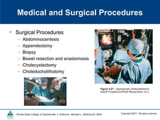
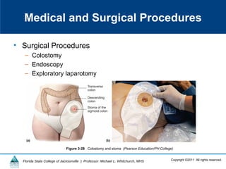
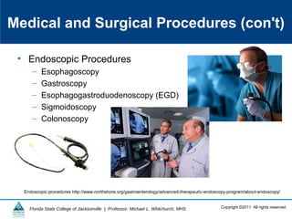
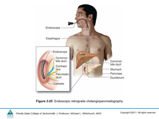
![Copyright ©2011 All rights reserved.Florida State College of Jacksonville | Professor: Michael L. Whitchurch, MHS
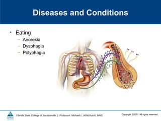
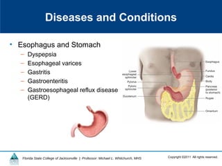
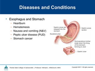
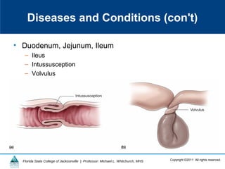
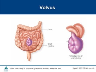
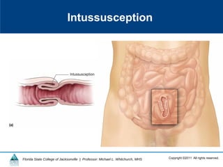
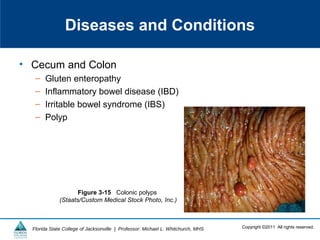
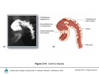
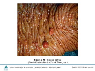
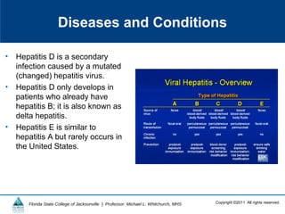
Diseases and Conditions
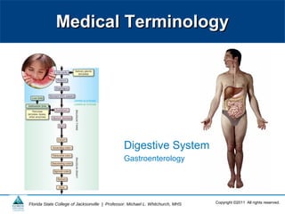
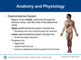
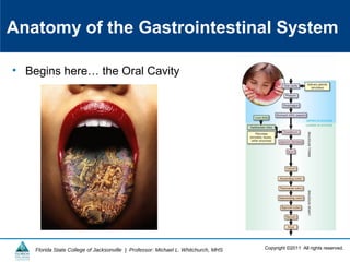
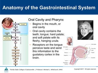
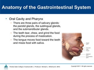
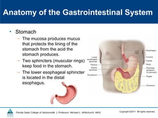
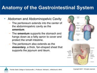
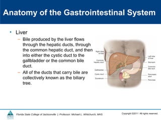
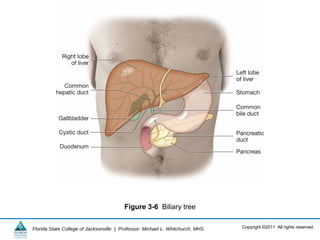
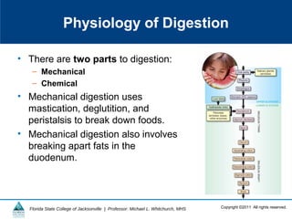
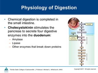
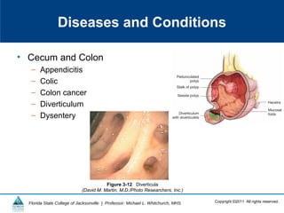
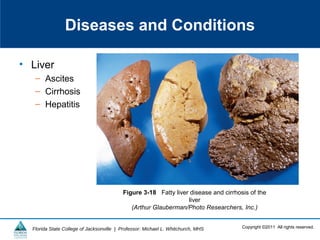
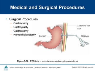
• Mouth and Lips
– Cheilitis
– Sialolithiasis
– Stomatitis
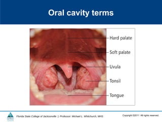
– Glossitis
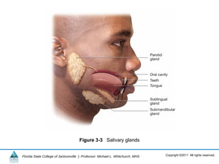
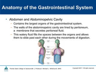
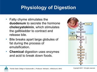
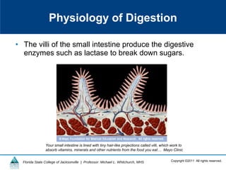
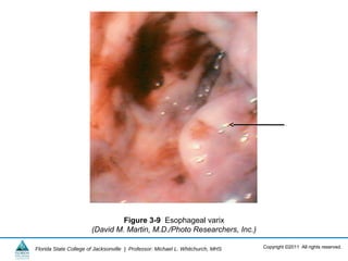
Figure 3-8 Glossitis
(Centers for Disease Control and Prevention [CDC]](https://image.slidesharecdn.com/ch03lnopen20campus20no-crs1-130520155220-phpapp02/85/Digestive-System-Terminology-44-320.jpg)
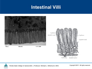
![Copyright ©2011 All rights reserved.Florida State College of Jacksonville | Professor: Michael L. Whitchurch, MHS
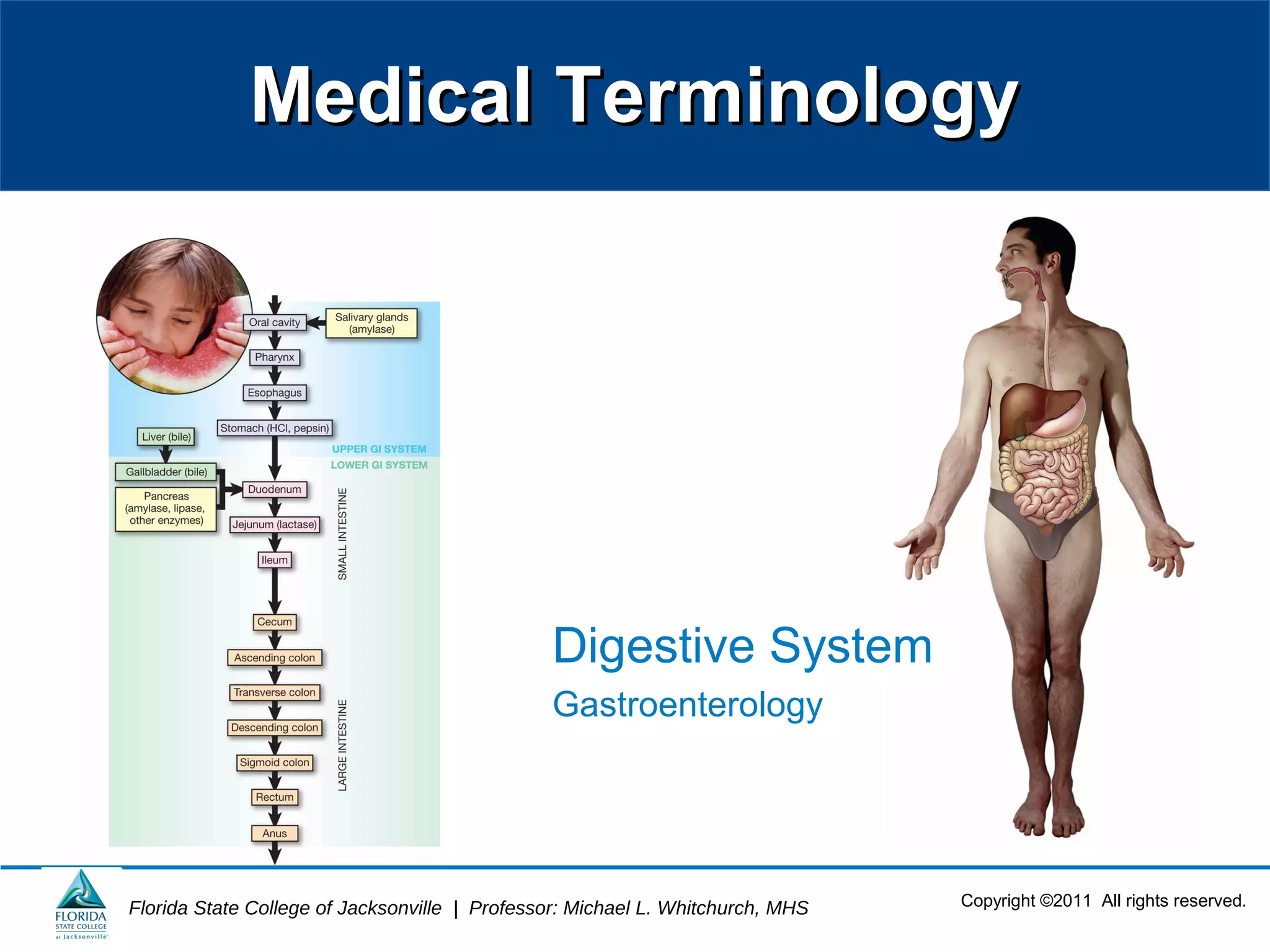
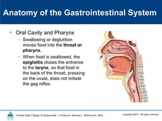
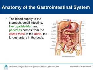
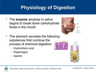
Figure 3-8 Glossitis
(Centers for Disease Control and Prevention [CDC])](https://image.slidesharecdn.com/ch03lnopen20campus20no-crs1-130520155220-phpapp02/85/Digestive-System-Terminology-45-320.jpg)


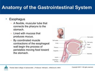
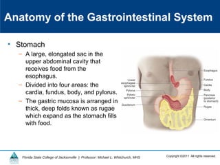
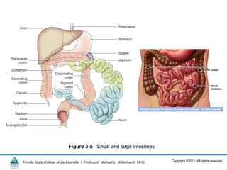
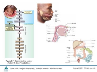
This document provides an overview of the gastrointestinal system and digestive process. It describes the anatomy and physiology of the gastrointestinal tract, from the mouth through the intestines. Key parts that are discussed include the oral cavity, esophagus, stomach, small intestine, large intestine, liver, gallbladder and pancreas. The document also covers the mechanical and chemical processes of digestion, absorption of nutrients, and elimination of waste. Common gastrointestinal diseases, conditions, and diagnostic procedures are listed but not described. Learning objectives focus on identifying structures, describing processes, and understanding medical terminology related to the gastrointestinal system.


![PERI-PROSTHETIC FRACTURE NAIL-PLATE CONSTRUCT [NPC].pptx](https://cdn.slidesharecdn.com/ss_thumbnails/drarunkumardrmohamedashrafperiprostheticfrasturenail-plateconstructnpc-260209164459-7e9d15a1-thumbnail.jpg?width=640&height=640&fit=bounds)


