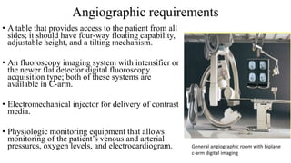
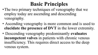
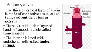
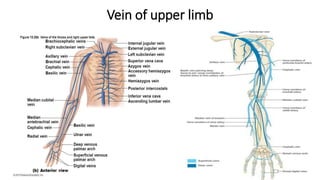
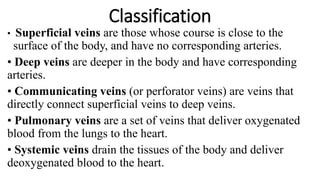
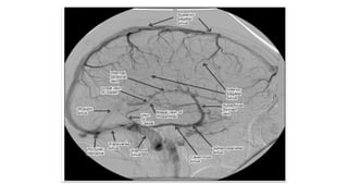
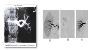
3. What is venography?
• Is the general term that describes the radiologic examination of vascular
structures within the body after the introduction of an iodinated contrast
medium or gas
• Radiological study of veins using ionization radiation (x-ray ) and the
contrast agents with the help of real time imaging
• Most accurate tests for deep vein thrombosis
• Most commonly done in legs for deep vein thrombosis
4. History
• Contrast-enhanced venography has been practiced for more than half a
century. In 1940, Bauer published details of normal venographic anatomy and
the venographic appearance of acute and chronic deep venous thrombosis
(DVT).
• Early attempts at venography were marked by complications related to the
equipment, contrast agents used at that time, and incomplete understanding of
venous hemodynamics.
• Since 1960, the procedure has become the leading diagnostic test for
DVT. The technique evolved during many years and became the “gold
standard” for the diagnosis of DVT.
• Early leg venography required venous cutdown for access to the deep venous
system, but this technique was modified by Welch and coworkers, who
recommended that contrast material be injected into a superficial vein in the
foot with a tourniquet applied above the ankle to prevent filling of the
superficial veins, which often obscured the deep system.
5. Basic Principles
•The two primary techniques of venography that we
employ today are ascending and descending
venography.
• Ascending venography is more common and is used to
elucidate the presence of DVT in the lower extremity.
• Descending venography predominantly evaluates
incompetent valves in patients with chronic venous
insufficiency. This requires direct access to the deep
venous system.
6. Veins• In the circulatory system, veins are blood vessels that
carry blood toward the heart.
• Most veins carry deoxygenated blood from the
tissues back to the heart; exceptions are the
pulmonary and umbilical veins.
• Veins differ from arteries in structure and function;
for example, arteries are more muscular than veins,
veins are often closer to the skin and contain valves
to help keep blood flowing toward the heart, while
arteries carry blood away from the heart.
7. Anatomy of veins
• The thick outermost layer of a vein
is made of connective tissue, called
tunica adventitia or tunica
externa.
• There is a middle thin layer of
bands of smooth muscle called
tunica media.
• The interior is lined with
endothelial cells called tunica
intima.
17. Classification
• Superficial veins are those whose course is close to the
surface of the body, and have no corresponding arteries.
• Deep veins are deeper in the body and have corresponding
arteries.
• Communicating veins (or perforator veins) are veins that
directly connect superficial veins to deep veins.
• Pulmonary veins are a set of veins that deliver oxygenated
blood from the lungs to the heart.
• Systemic veins drain the tissues of the body and deliver
deoxygenated blood to the heart.
18. Angiographic requirements
• A table that provides access to the patient from all
sides; it should have four-way floating capability,
adjustable height, and a tilting mechanism.
• An fluoroscopy imaging system with intensifier or
the newer flat detector digital fluoroscopy
acquisition type; both of these systems are
available in C-arm.
• Electromechanical injector for delivery of contrast
media.
• Physiologic monitoring equipment that allows
monitoring of the patient’s venous and arterial
pressures, oxygen levels, and electrocardiogram. General angiographic room with biplane
c-arm digital imaging
19. ANGIOGRAPHIC TRAY
• A sterile tray contains the basic equipment necessary for a
catheterization. Basic sterile items include the following:
1)Hemostats
2)Preparation sponges & antiseptic solution
3)Scalpel blade
4)Syringe and needle for local anesthetic
5)Basins and medicine cup
6)Sterile drapes and towels
7)Band-Aids(Fixing Tapes)
8)Sterile image intensifier cover
9)Needle(Butterfly, IV cannula) for vessel(vein) puncture, if
catheterization is not done
20. EMBOLISM
• Process of partial or complete obstruction
of some part of the cardiovascular system
by any mass carried in the circulation.
• Transported intravascular mass detached
from its site of origin is called an embolus.
• Emboli (90%) are thromboemboli.
21. THROMBOSIS
• THROMBOSIS is the formation of a
clotted mass of blood within the
cardiovascular system.
• The clotted mass is called as
THROMBUS.
22. •Venography can be divided into following sections:
1)Peripheral Venography
a)Lower limb venography
b)Upper limb Venography
c)Peripheral Varicography
2)Central Venography
a)Inferior Vena Cavography
b)Superior Vena Cavography
3)Selective visceral Venography
a)Renal venography
b)Hepatic Venography
c)Portal venography
23. Relative Contraindications
• Common Contraindications for venography include
• contrast media allergy,
• impaired renal function
• blood-clotting disorders
• Anticoagulant medication
• unstable cardiopulmonary or neurologic status.
• non-consent by patient to procedure
25. Lower limb venography
• It is the study of the veins of the lower limb by
the introduction of contrast medium
• The veins of the lower limb can be divided into
a superficial and a deep venous system.
• Superficial veins:
1)Great saphenous vein
2)Small saphenous vein
3)Superficial veins of the foot.
27. Patient preparation
• Some common procedure for patient preparation are:
• NPO for 4-6hrs prior to examination
• Check recent serum creatinine and urea level.
• Taking proper medical history so that appropriate
premedication can be used.
• Changing patient to an appropriate gown.
• Signing Informed consent
28. •Indications
1. Deep venous thrombosis
2. To demonstrate incompetent perforating veins
3. Oedema of unknown cause
4. Congenital abnormality of the venous system (rare).
•Contraindications
1.Local sepsis.
29. Contd…
• Contrast medium
1.LOCM 240mgI/ml.
• Equipment
1. Fluoroscopy unit with spot film device
2. Tilting radiography table.
• Patient preparation
1.Elevated leg overnight if oedema is severe.
30. Technique
1. The patient is supine and tilted 40° head up, to delay the transit time
of the contrast medium.
2. A tourniquet is applied tightly just above the ankle to occlude the
superficial venous system. It is important to remember that this may also
occlude the anterior tibial vein, and so its absence should not
automatically be interpreted as due to a venous thrombosis.
3. A 18-G butterfly needle (smaller if necessary) is inserted into a distal
vein on the dorsum of the foot. If the needle is too proximal, the contrast
medium may bypass the deep veins and so give the impression of a deep
venous occlusion.
4. 40 ml of contrast medium is injected by hand. The first series of spot
films is then taken.
31. Cont.
• 5. A further 20 ml of contrast are injected quickly whilst the patient
performs a Valsalva manoeuvre to delay the transit of contrast
medium into the proximal and pelvic veins. The patient is tilted
quickly into a slightly head down position and the Valsalva
manoeuvre is relaxed. Alternatively, if the patient is unable to
Valsalva, direct manual pressure over the femoral vein whilst the
table is being tilted into the head-down position will delay transit of
contrast medium proximally. Films are taken 2-3 s after releasing
pressure.
• 6. At the end of the procedure the needle should be flushed with
0.9% saline to avoid the risk of phlebitis due to stasis of contrast
medium.
32. • Films
(Collimated to include all veins)
1. AP of calf
2. Both obliques of calf (foot internally and externally rotated)
3. AP of popliteal, common femoral and iliac veins.
• Aftercare
1)The limb should be exercised.
• Complications
• Due to the contrast medium
1 As for the general complications of intravascular contrast media.
2 Thrombophlebitis.
3 Tissue necrosis due to extravasation of contrast medium. This is rare, but may occur in
patients with peripheral ischaemia.
4. Cardiac arrhythmia - more likely if the patient has pulmonary hypertension.
• Due to the technique
1 Haematoma
2 Pulmonary embolus - due to dislodged clot or air.
33. • Complications
Due to the contrast medium
1As for the general complications of intravascular contrast media.
2 Thrombophlebitis.
3 Tissue necrosis due to extravasation of contrast medium. This is
rare, but may occur in patients with peripheral ischaemia.
4 Cardiac arrhythmia - more likely if the patient has pulmonary
hypertension.
• Due to the technique
1 Haematoma
2 Pulmonary embolus - due to dislodged clot or air.
34.
35.
36. UPPER LIMB
• It is the study of the veins of the upper limb by the introduction of
contrast medium.
• Indications
1. Oedema
2. To demonstrate the site of a venous obstruction
3. SVC obstruction
• Contrast medium
1)LOCM 300mgI/ml.
• Equipment
1)Fluoroscopy unit with spot film device.
Preliminary film
37. •Technique(For intravenous venography )
1. The patient is supine.
2. an 18G butterfly needle is inserted into the median
cubital vein at the elbow. The cephalic vein is not used as it
bypass the axillary vein.
3. Spot films are taken of the region of interest during a
hand injection of 30 ml of contrast medium.
•Aftercare
1)None
•Complications
1)Due to the contrast medium.
38.
39. PERIPHERAL VARICOGRAPHY• Indications
1)To demonstrate distribution of varicose veins.
2)To demonstrate sites of communication with deep
venous system.
3) Assessment of recurrent varicosity.
• Contraindications
1)Local sepsis.
• Contrast medium
LOCM 240mgI/ml. Volume depends on extent and
volume of varicosities.
• Equipment
1) fuoroscopy unit with spot film device
2) Tilting fluoroscopy table.
40. Technique
1. The patient lies supine and tilted 40° head up to delay washout of contrast.
2. A 18G butterfly needle is inserted into a suitable varix below the knee.
3. 40-50 ml of contrast are injected by hand under fluoroscopic control.
4. A series of spot films is taken:
a. AP calf and 2 obliques
b. b. lateral knee - to assess the short saphenopopliteal junction.
5. If contrast filling above the knee is adequate, then further views of the thigh can
be taken to demonstrate the extent of long saphenous varicosity.
6. Due to the large volume of varicose veins, it may be necessary to re-site the
needle in a suitable varix above the knee to obtain adequate contrast filling of the
entire system.
41. Cont..
• 7. A further 40 ml of contrast are then injected and spot films taken.
• a. AP thigh and oblique - particular attention should be given to the potential sites of communication, e.g. mid-
thigh perforator b. AP and oblique of groin - views to demonstrate the saphenofemoral junction arc particularly
necessary in assessing recurrent varicosity even if there has been previous saphenofemoral ligation, as
recurrence at this site is common.
• 8. After injection and imaging is complete the veins should be flushed
with saline to prevent contrast stasis and the risk of phlebitis.
• 9. The needles are removed and pressure applied to ensure
haemostasis.
• Aftercare
The limb should be exercised gently to washout any remaining
contrast.
42. CENTRAL VENOGRAPHY(SUPERIOR VENA
CAVOGRAPHY)
• Indications
1. To demonstrate the site of a venous obstruction.
2. Congenital abnormality of the venous system,
e.g. left-sided superior vena cava.
• Contrast medium
LOCM 370mgI/ml, 60 ml.
• Equipment
-Rapid serial radiography unit.
2) Patient preparation
-Nil orally for 5 h prior to the procedure.
3)Preliminary films
-PA film of upper chest and lower neck.
43. Technique
1.The patient is supine.
2.18-G butterfly needles are inserted into the median antecubital vein of
both arms.
3.Hand injections of contrast medium 30 ml per side, are made
simultaneously, as rapidly as possible by two operators. The injection is
recorded by rapid serial radiography . The film sequence is commenced
after about two thirds of the contrast medium has been injected.
Note: If the study is to demonstrate a congenital abnormality, or on
the rare occasion that the opacification obtained by the above
method is too poor, a 5-F catheter with side holes, introduced by the
Seldinger technique, may be used.
44. Cont..
• Films
Rapid serial radiography is performed: one film per s for 10 s.
• Aftercare
None, unless a catheter is used.
• Complications
Due to the contrast medium
45. INFERIOR VENA CAVOGRAPHY
• Indications
1. To demonstrate the site of a venous obstruction, displacement or
infiltration.
2. Congenital abnormality of the venous system.
• Contrast medium
LOCM 370, 40 ml.
• Technique
1. With the patient supine, the catheter is inserted into the femoral
vein using the Seldinger technique. A Valsalva manoeuvre may
facilitate vene-puncture by dilating the veins.
2. An injection of 40 ml of contrast medium is made in 2 s by the
pump injector, and recorded by rapid serial radiography.
47. Cont..
• Aftercare
1.Pressure at venepuncture site.
2.Routine observations for 2 h.
• Complications
1.Due to the contrast medium
2.Due to the technique complications of catheter technique.
49. • Indications
1. To demonstrate prior to operation the anatomy of
the portal system in patients with portal
hypertension.
2. To check the patency of a portosystemic
anastomosis.
• Contrast medium
LOCM 370mgI/ml, 50 ml.
• Equipment
1. Rapid serial radiography unit.
2. Arterial catheter.
3. 10-cm needle (20-G) with stilette and outer
plastic sheath.
50. •Patient preparation
1. Admission to hospital. A surgeon should be informed in
case complications of procedure arise (for the trans-splenic
approach).
2. Clotting factors are checked.
3. Severe ascites is drained.
4. Nil orally for 5 h prior to the procedure.
5. Premedication, e.g. diazepam 10 mg orally.
51. Risk Factors and Complications
• Previous thrombosis
• Dilution of the contrast dye in the lower limb
• Difficulty accessing the veins due to:
1)Obesity
2)Severe swelling (edema)
3)Inflammation in the cells ( cellulitis )
53. Technique
• The patient is placed in the supine position and the spleen is
identified with ultrasound.
• Selected region anaesthetized.
• The patient is asked to hold breath in mid inspiration and the
needle is inserted inwards and upwards into the spleen about 3
quarters i.e 7.5 cm .
• 50 ml volume of contrast is given at 10 ml/sec
• 1 image per second is taken for 8 seconds
• Exposures made at the end of suspended expiration
54. Acute thrombus in the deep femoral and great
saphenous veins. The deep femoral vein (short
arrows) and the great saphenous vein (long
arrow) are shown. The femoral vein is occluded.
55. Subacute thrombus in the popliteal
vein. Arrows denote retracted
thrombus, indicative of subacute
thrombosis.
56. Chronic popliteal venous thrombosis. Note the
extensive recanalization of the popliteal vein
(open arrows) and relatively large caliber of
the great saphenous vein (solid arrow).
57. Chronic deep venous thrombosis.
Note the extensive collateral
channels and no visualization of
normal deep venous structures.
59. Venogram of the inferior vena cava (IVC)
before attempted retrieval of a
prophylactic IVC filter with trapped
thrombus
60. Deep Vein Thrombosis
• Primarily involves lower limbs
• Major source of fatal PE
• Risks
1) Restricted mobility
2)Surgery
3)Obesity
4) Pregnant
5) Birth Control pills
6) Long trips
61. Deep Vein Thrombosis of Iliac Vein
• On x-ray appears as a constant filling
defect
• Largely replaced by duplex color doppler
ultrasound
-Demonstrates the velocity of the
venous blood flow
•Doppler is 95% accurate and is the
preferred initial imaging modality.
• Demonstrates lack of compressibility of
the vein and this indicates the presence of a
thrombus.
64. Cerebral Venography
• Cerebral veins are usually visualized during the procedure of cerebral
angiography because the transit time from cerebral arteries two veins is
very less(about 2-4 sec)
• Abnormalities at the base of the skull can be visualized by Jugular
vein approach. Improper technique can results in a lack of intracranial
Dural sinus filling which may disguise as venous occlusion. This
problem is avoided by adequate neck compression along with proper
volume and rate of delivery of contrast
65.
66. CAPNOCAVOGRAPHY
• Visualization of inferior vena cava by injection of CO2 into the vein
• It is ideal in patients who are allergic to iodinated contrast media and
compromised renal function.
• The buoyancy of CO2 makes it necessary for the procedure to be
done in the left lateral position for optimal visualization of the IVC
• The main limitation of the technique is the possibility of neurotoxicity
67.
68. Renal venography
• It is usually performed to rule out thrombosis of the renal vein . It is
also catheterized for blood sampling , usually to measure the
production of renin, an enzyme produced by kidney when it lacks
adequate blood supply.
• The renal vein is most usually catheterized from a femoral vein
approach.
• 16 ml volume of contrast is given at 8 ml/sec.
• 2 images per second are taken for 4 seconds.
69.
70. Risk and Complications
• Angiographic, therefore venography procedures always
involve some level of risk for the patient. Common risks
and complications include the following:
A Due to technique:
• Bleeding at the puncture site
• Thrombus formation
• Embolus formation
• Hematoma
• Dissection of a vessel
• Infection of puncture site
• Tissue necrosis due to extravasation
71. Due to contrast media
• Contrast media reaction
• Thrombophlebitis
• Cardiac arrhythmia
72. Other modalities(Ultrasonography)
• Ultrasound is the most widely used imaging method for the venous system
• The advantages are that it is low cost and readily available
• Duplex scanning involves a combination of pulsed Doppler and real time for
direct visualization.
• Expansion and filling of the normal echo-free lumen can be identified but
slow moving blood may be misinterpreted for thrombus.
• The most reliable sign is compressibility.
• Direct pressure with the US probe over the vein will cause the normal vein to
collapse. If thrombus is present, this will not occur.
73.
74. Computed tomography
• Multidetector CT (MDCT) with standard IV
contrast and scan delay protocols for the
chest or abdomen/pelvis is very effective for
detection of compression or thrombosis of
major veins including the superior and
inferior vena cava, iliac and renal veins.
• Although it would be possible to perform
direct lower-limb CT venography after
infusion of contrast via a foot vein, this
technique has found little application and is
not used in clinical practice.
75. MAGNETIC RESONANCE IMAGING(MRI)
• MRI is well suited to imaging the venous system, but
because of cost and limited availability it is used
infrequently
• Peripheral MR venography (MRV) is currently used in
selected cases of:
-venous thrombosis in pregnant subjects and
-where fractured limbs are immobilized in casts.
• It is useful in evaluation of congenital abnormalities
of peripheral venous anatomy and venous
malformations.
• This is the best and most versatile imaging modality
for the brain.
76. References
A guide to radiological procedure ;Stephen Chapman, 6th edition
Merrill’s atlas of radiological procedures
Radiopedia
Shareslide/slideplayer
google
Editor's Notes
MAKE SOMETHING CLEAR,EXPLAIN LACKING ABILITY
1 IN NUMBER (UMBILICAL VEIN )
Left gastric vein
Right gastric vein
Superior pancreaticoduodenal
Cystic vein
Paraumbilical vein
anterior jugular vein
posterior external jugular vein
suprascapular vein
transverse cervical vein
receives greater part of blood from exterior of cranium & deep parts of face.
Inferior petrosal sinus
Pharyngeal vein
Common facial vein
Lingual vein
Superior thyroid vein
Middle thyroid vein
Occipital vein.
Collects blood from brain & superficial parts of face and neck.
vein of Rosenthal (great cerebral vein ) vein of trolard
connects the systems of superior vena cava and inferior vena cava and can provide an alternative path for blood to the right atrium when either of the venae cavae is blocked.[2][3]
A blood clot (thrombus) in a deep vein in the thigh or leg. The clot can break off as an embolus and make its way to the lung, where it can cause respiratory distress and respiratory failure.