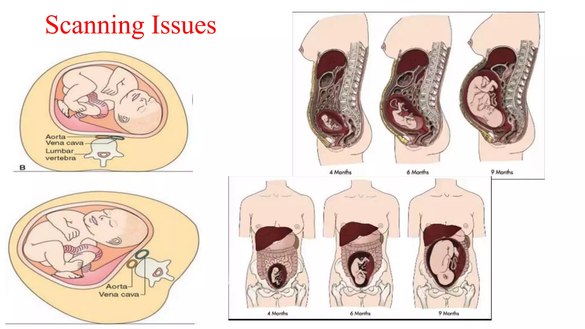
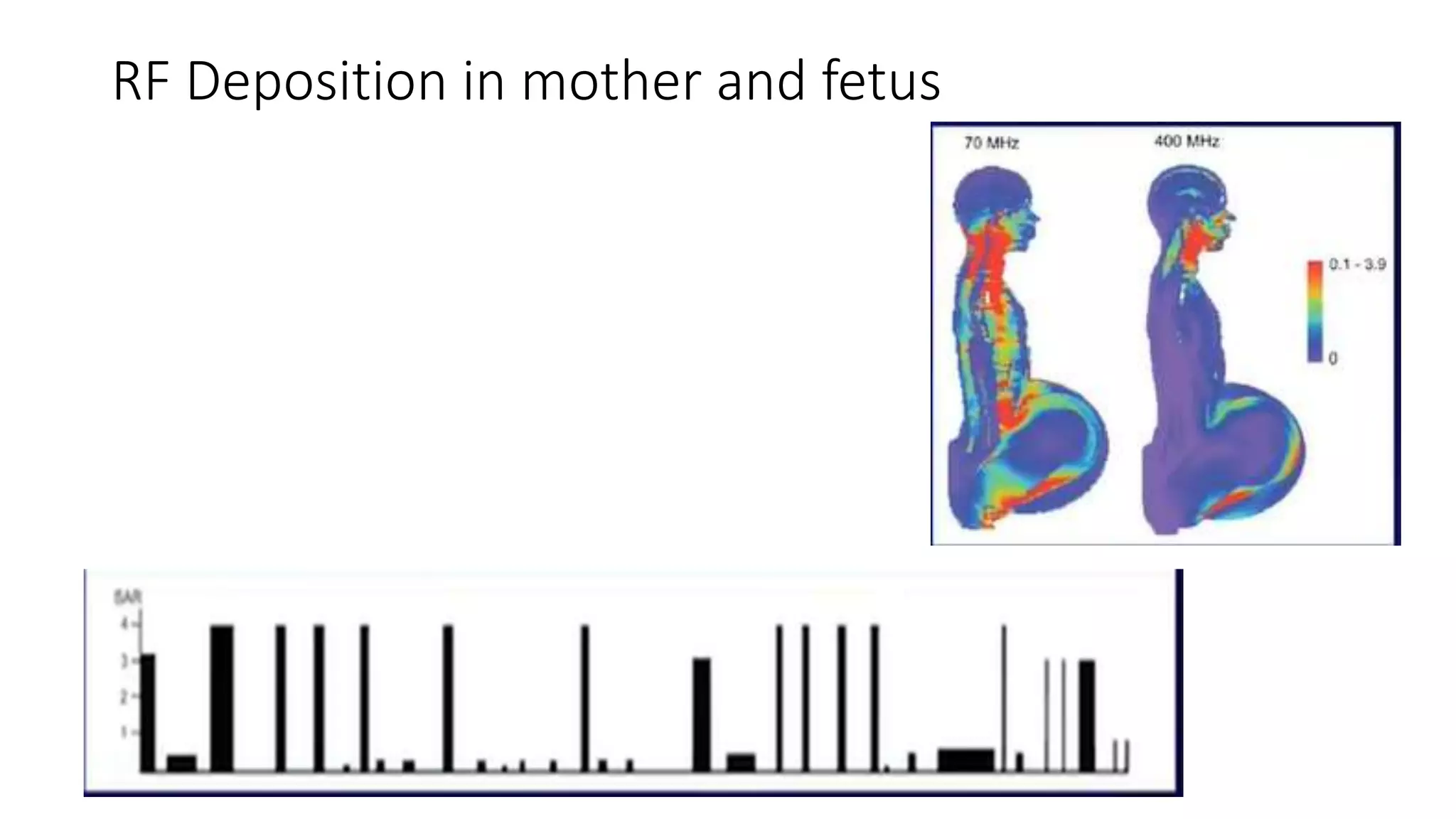
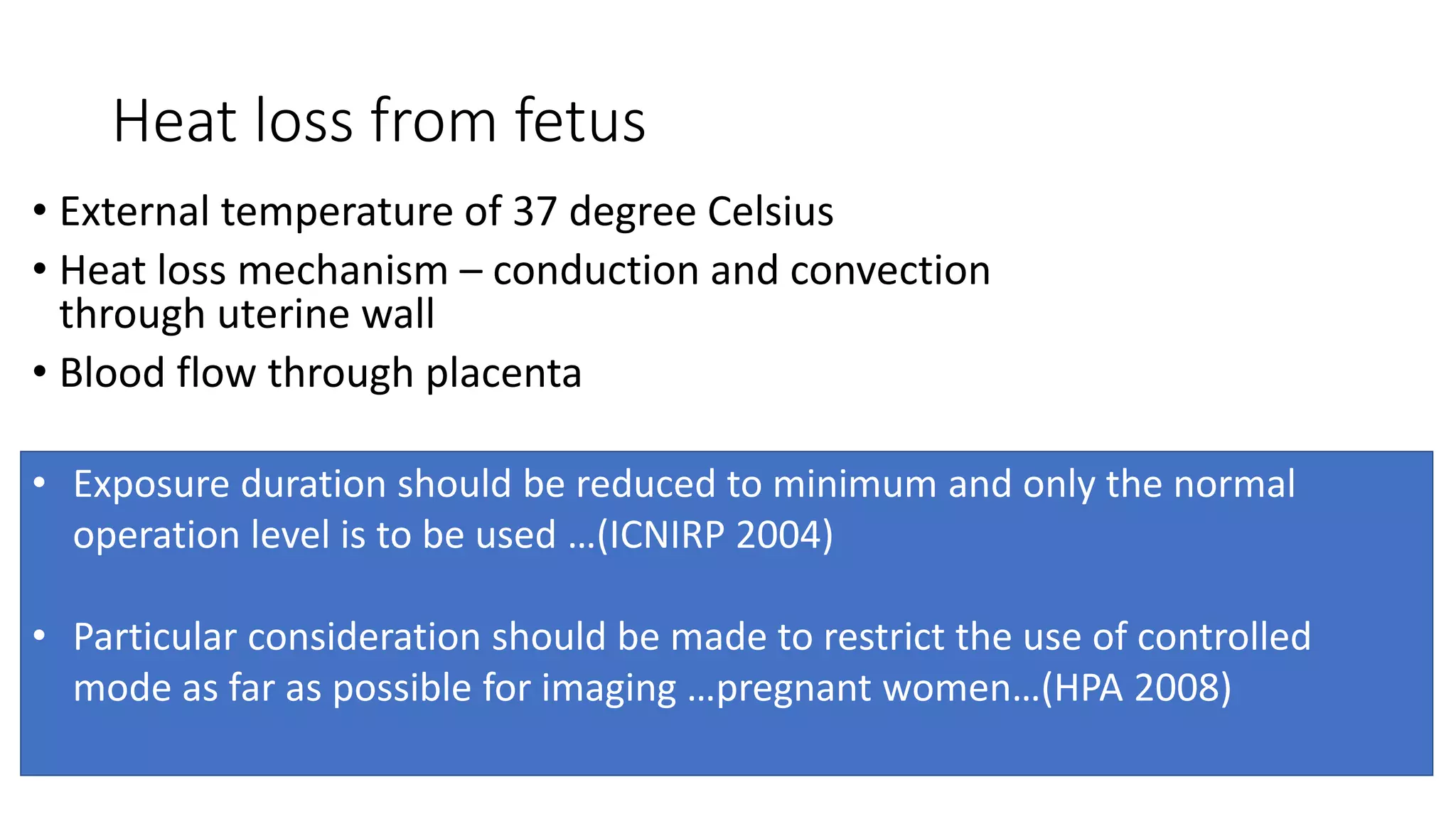
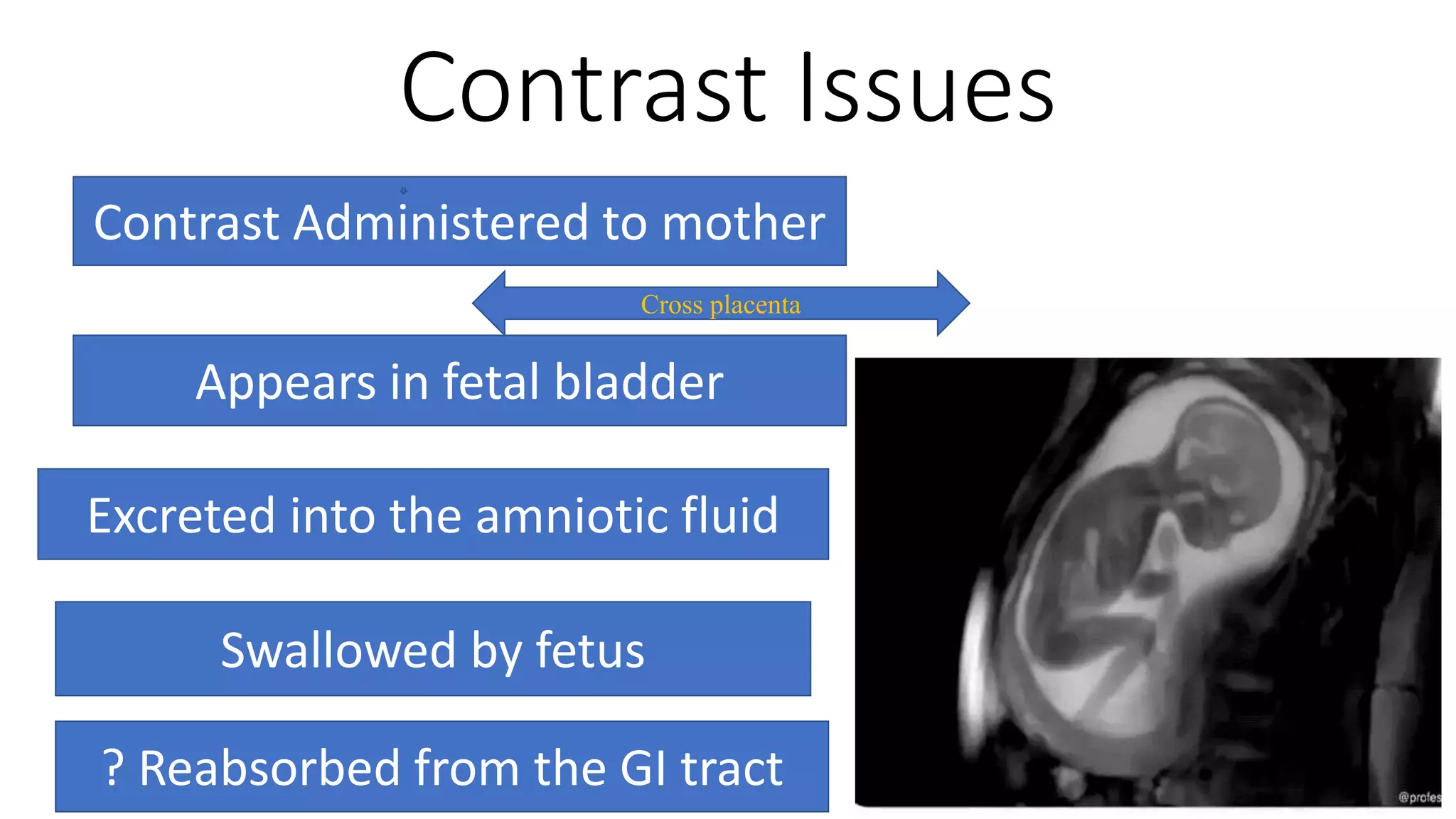
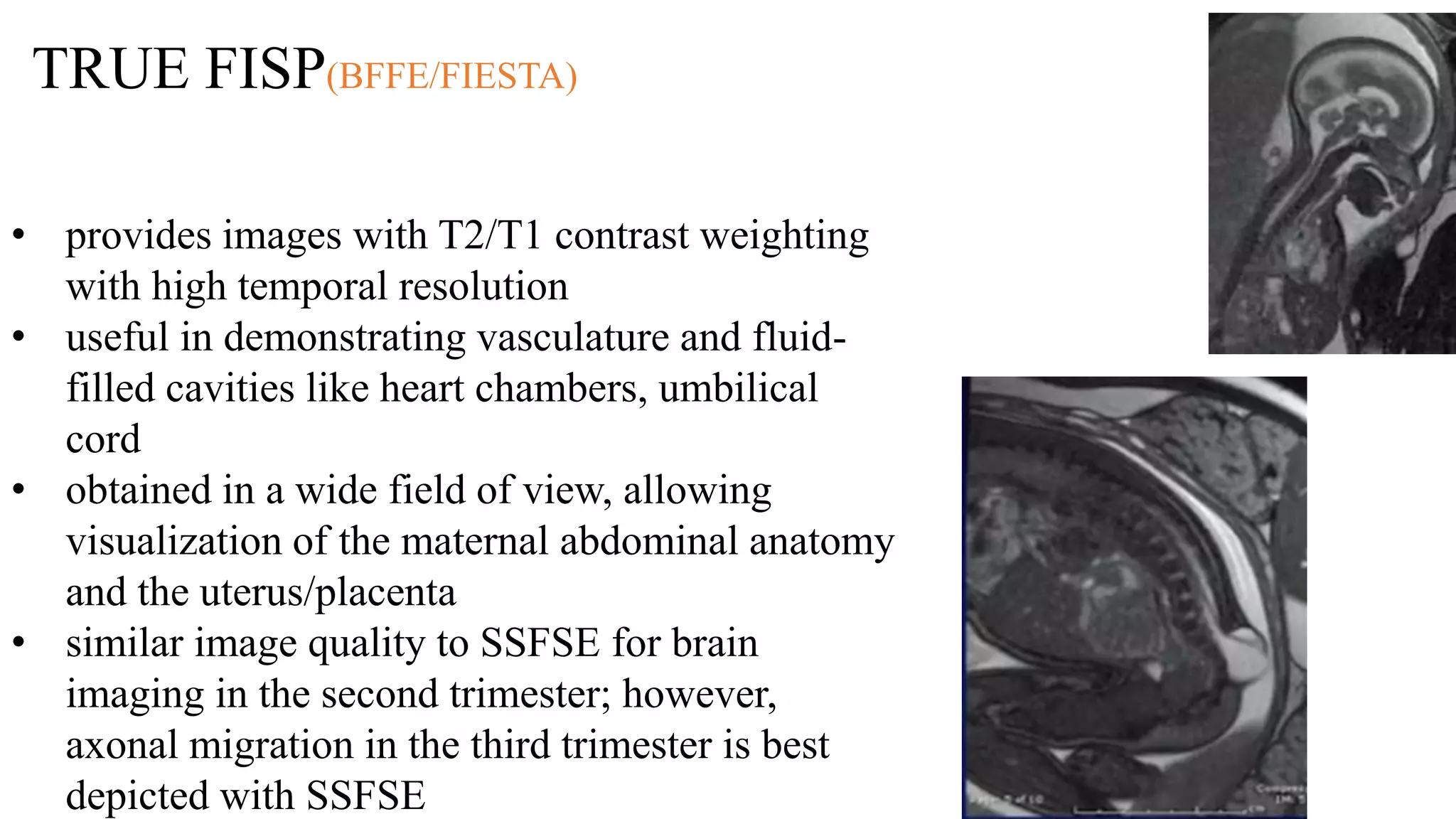
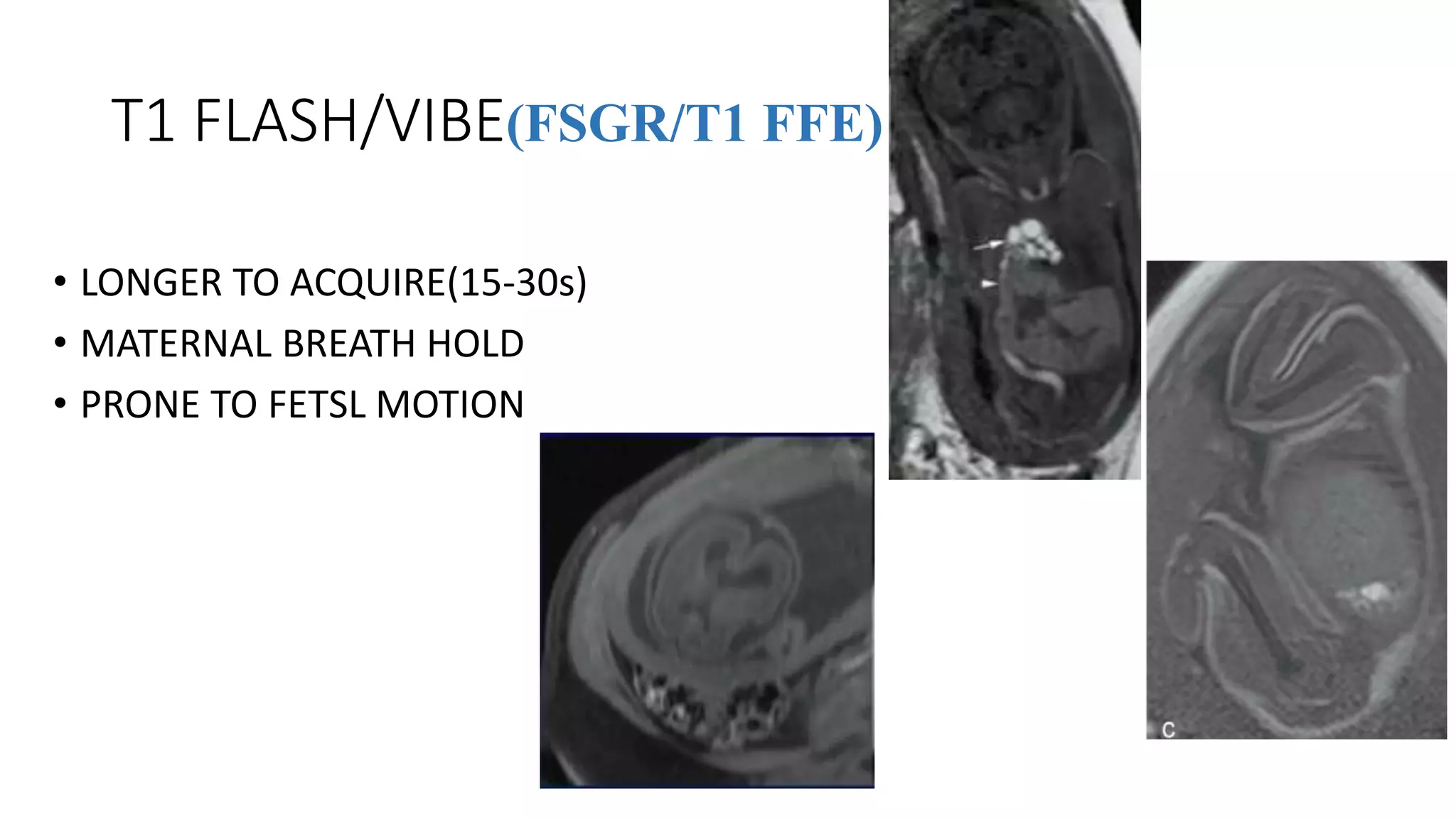
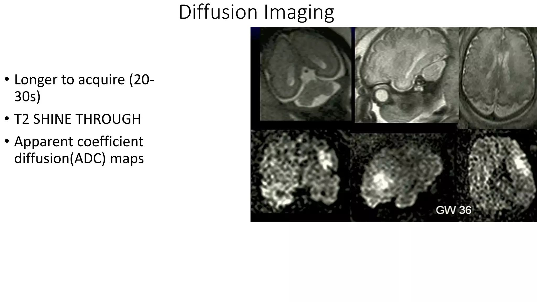
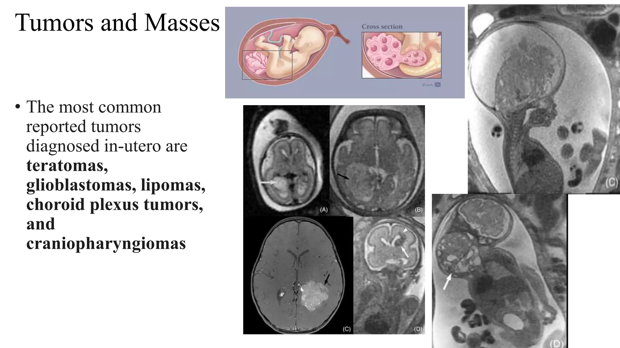
FETAL MRI
Presenter :Sujan Karki
B.Sc. MIT Final year
National Academy of Medical Sciences(NAMS)
Bir Hospital
Contents
• Scanning issues
• Safety of MRI –
• Specific Absorption Rate (Heat Deposition )
• Acoustic Issues
•Timing of MRI - 1st, 2nd and 3rd trimester
• Contrast Issue – Inability to use gadolinium
• Imaging Sequences
• Challenges in fetal imaging
• Fetal MRI indications
FETAL MRI : WHEN AND WHY ?
US MADE A DIAGNOSIS
US HAS ABNORMALITY
US IS LIMITED
Better understanding of anatomic features is
needed(surgical planning and prognosis )
MRI Multiplanar Capabilities
Comprehensive visualization
Further information is essential for diagnosis
MRI provides tissue characterization
A diagnosis is required
Oligohydramnios, Obesity, Multiple
fetuses, and late gestational age
Scanning Issues
Scanning Issues
• Long Scan time
• Repeat Examination
• Motion
1)Maternal Breathing
2)Fetal Motion
a) bulk motion
b) internal motion ( breathing and swallowing)
c) extremity motion
Sedation
• Maternal oral administration of 1 mg of flunitrazepam has been
recommended in Europe to reduce fetal movement.
• In some cases mother is sedated using benzodiazepam
• Conventionally pancuronium was injected on fetal muscles avoid the motion
Proper communication with the patient is
the most important sedation .
Fasting
• Four hour fasting or overnight fasting causes hypoglycemia which
reduces the fetal MRI
• Avoid caffeine and fizzy drinks which helps in the reduction of patient
motion
Coils and Coil Position
• 3T MRI – 32 Channel phased array
coil and a surface coil in abdomen
with 18 elements
• 1.5T uses 6 channel phased array
coil with surface coils in abdomen
with 6 elements
1. Phased array technique
combines multiple coil elements
to provide a large field of view
while maintaining signal.
2. Parallel imaging ????
PARALLEL IMAGING
Safety in MRI
• Specific absorption rate(heat deposition )
• Increases with field strength, RF power, transmitter-coil type and body size
• 1.5 to 3 T = 4 X SAR
• Multi echo, multi-slice pulse creates higher SAR
• IEC 60601-2-33(ed.3.1) sets limits pertaining to MR
• As 6/30/13,FDA required manufactures to comply wl IEC 60601
FDA : SAR LIMITS
Site Dose Time(min)= or> SAR(W/KG)
Whole body Average over 15 >4
Head Average over 10 >3.2
• Maximum limits
• FDA do not define operating mode
• In 2013 ,FDA adopted the IEC limits for MRI manufacturers
• IEC: Normal and First level operating mode
IEC/FDA: SAR
Operating mode Whole Body
SAR(W/kg)
Head SAR(W/kg)
Normal 2 3.2
First Level Controlled
(match FDA Limits)
4 3.2
Normal operating mode : no outputs cause physiologic stress
Suitable For: Any patient with impaired heat regulation
Pregnant/neonate patients
First level controlled Mode: under medical supervision
RF Deposition in mother and fetus
Temperature Rise Limits
Operating Mode Max Rise of Core Temp
Normal 0.5
First Level Controlled 1
Temperature rise is ultimately what we care about
Heat loss from mother
• External temp about 20 degree Celsius
• Heat loss mainly through skin
peripheral vasodilation
sweating
• To avoid heating
Good bore ventilation
No blankets
Minimal clothing
Heat loss from fetus
• External temperature of 37 degree Celsius
• Heat loss mechanism – conduction and convection
through uterine wall
• Blood flow through placenta
• Exposure duration should be reduced to minimum and only the normal
operation level is to be used …(ICNIRP 2004)
• Particular consideration should be made to restrict the use of controlled
mode as far as possible for imaging …pregnant women…(HPA 2008)
Exposure to RF
• Edward et.al (2013) : performed on animal models and found CNS is most
vulnerable
• 2 degree rise over 2hrs may cause neural tube defects and cranio-facial defects
• Any pulse sequence developed must not cause a body temperature rise above
0.5 degree Celsius for normal mode , 1 degree Celsius for controlled mode and
greater than 1 degree Celsius in experimental mode .
• Manufacturer estimates the SAR value for each pulse sequence
• Hand et.al –typically body coil with 1.5 T and 3T
fetus – 40-70% of maximum mom SAR
Acoustic Noise
• About 30% reduction in dB intensity provided by fetal surroundings
• Protocols involving rapid gradients creates higher noises
• EPI produces loudest sound reaches up to 120 dB
• Bakers et.al conducted a study on 18 patients and 16 passed a hearing test so
Acoustic damage from MRI is appeared to be theoretical concern rather
than a real concern
Timing of MRI – 1st ,2nd and 3rd trimester
• Yip et.al conducted a study in chicks embryo at 1.5 T
MRI
• At different developmental stages
• Increased abnormalities and mortality rates in six days
embryos after exposure
• ACR white paper --- pregnant patient can be accepted
to undergo MRI at any stage of pregnancy
considering the risk-benefit ratio .
Contrast Issues
Contrast Administered to mother
Appears in fetal bladder
Excreted into the amniotic fluid
Swallowed by fetus
? Reabsorbed from the GI tract
Cross placenta
Patient preparation
• Maternal 4 hrs. fasting prior exam – reduces bowl peristalsis and prevent postprandial
fetal motion
• Patient should empty bladder just before examination
• Written consent is advisable
• Explain the mother about procedure and indication and assure her that she is the part of
examination
• Check the indications and if possible ask radiologist to stay in console room ??????
Patient positioning
• Feet first supine
• Left lateral decubitus
Scout
images
• Planned orthogonal to maternal pelvis
• Large FOV
• Overview and Orientation
• Fetal lie in relation with mother …to confirm which side is which
• Always use last sequence to plan for new sequence
T2 Weighted/HASTE(single shot FSE/TSE)
• Single Shot ? ??? Breath hold
• 3 orthogonal plane
• Alter TE ???
• 140 for excellent grey-white discrimination
• Can reduce TE with increase in GE age of >30
• TE of 90ms for body imaging
• use of T2 HASTE, the foundation of fetal
imaging, produced no significant temperature
increase in the fetal brain or amniotic fluid of a
pig model.
• And in a pregnant model, in normal mode at 1.5
T and 3.0T, the calculated temperature increase
and SAR limits were found to be within a safe
range
TRUE FISP(BFFE/FIESTA)
• provides images with T2/T1 contrast weighting
with high temporal resolution
• useful in demonstrating vasculature and fluid-
filled cavities like heart chambers, umbilical
cord
• obtained in a wide field of view, allowing
visualization of the maternal abdominal anatomy
and the uterus/placenta
• similar image quality to SSFSE for brain
imaging in the second trimester; however,
axonal migration in the third trimester is best
depicted with SSFSE
Fetogropahy
• Thick slab MRCP sequence
• 3D visualization/Global
Visualization
• Heavily T2 weighted
• 20-50 mm slice thickness
T1 FLASH/VIBE(FSGR/T1 FFE)
• LONGER TO ACQUIRE(15-30s)
• MATERNAL BREATH HOLD
• PRONE TO FETSL MOTION
EPI
• FAST ACQUISTION ( 4s for whole head )
• HIGH SENSITIVITY FOR HEMMOHRAGE
• Helps for the evaluation for
developments
• CAN EPI BE USED FOR SKELETAL
EVLUATION ????
Spin echo based EPI and GRE Based EPI
Diffusion Imaging
• Longer to acquire (20-
30s)
• T2 SHINE THROUGH
• Apparent coefficient
diffusion(ADC) maps
Dynamic Sequences
• Swallowing
• Bulk fetal motion
•Fetal Cardiac
Assessment
• Fetal akinesia
•Movement disorders
Challenges to imaging and quality
Fetal Motion
During slice acquisition
Sleep-wake phase
Multiple pregnancies
Maternal comfort
Time constrains
Maternal breathing and movements
Dependance on diaphragmatic excursion
Effect dependent on fetal position
Limit to comfort time
FETAL MOTION
Maternal Breathing and Movements
Multiple Pregnancies
MOVEMENTS BETWEEN THE SEQUENCES
RADIOLOGIST PRESENCE
DURING THE ACQUISTION ??
Indications of fetal MRI
• CNS (38%)
• Lung/Thoracic (34%) Congenital Diaphragmatic Hernia most common
• In 29.3% post referral diagnosis changed completely
• In 28% additional findings were discovered
• In 42% referral diagnosis was concordant with post referral diagnosis.
28% extra pathological findings by fetal MRI
Indications and reasons of performing fetal MRI at
1.5T and 3T
Indications Reasons
Indication for 3 T
Brain Intraparenchymal Resolution
Bones Sensitivity for susceptibility effects
Gestational Age >18 weeks,
cartilage(joints), abdominal organs
Resolution
Angiography Background Suppression
Indication at 1.5 T
Polyhydramnios Less sensitive to moving fluids`
Maternal sensitivity to heat Less warming
Large maternal habitus Less sensitive to artefacts
Ventriculomegaly, Callosal Agenesis, Chiari malformation
Tumors and Masses
• The most common
reported tumors
diagnosed in-utero are
teratomas,
glioblastomas, lipomas,
choroid plexus tumors,
and
craniopharyngiomas
Diaphragmatic Hernia
• Congenital diaphragmatic hernia is a
developmental defect in the
posterolateral diaphragm with herniation
of abdominal viscera into the thorax.
• The cause is unknown, but one third of
cases are associated with chromosomal
or additional anatomic abnormalities and
have a mortality rate of 76%
• The sonographic diagnosis of congenital
diaphragmatic hernia and the evaluation
of liver position can be difficult because
lung and liver are of similar echogenicity
Biometric measurements
Monochorionic twin pregnancy complications
Esophageal Atresia
• Esophageal atresia is
a birth defect in
which part of a
baby's
esophagus (the tube
that connects the
mouth to the
stomach) does not
develop properly.
Placenta, Umbilical Cord and fetal Presentation
Evaluation of the placenta is a part
of the second and third trimester
examination
Multiplanar imaging allows clear and
detailed placental position and size
The umbilical cord and its insertion
can be imaged
Meningocele ,Myelomeningocele
Omphalocoele
• a condition in which
there is a skin or
membrane covered
herniation of
abdominal contents
into the base of the
umbilical cord.
Placenta Previa & Placenta Accreta
Other maternal indications
• Appendicitis -100% sensitivity and 94% specificity
• Intestinal Bowl Diseases- 91% sensitivity and 71%
specificity.
• Diverticulitis- 86-94% sensitivity and 88-92% specificity .
• Pulmonary Embolism
Limitations of fetal MRI
• reduced signal to noise ratio (especially prior to 18 weeks gestation)
• maternal weight/size exceeds table recommendations/MRI caliber
• claustrophobia
• implanted ferromagnetic devices
References
• T2-Weighted Fast MR Imaging with True FISP Versus HASTE
Comparative Efficacy in the Evaluation of Normal Fetal Brain Maturation
Hsiao-Wen Chung1 , Cheng-Yu Chen2, Robert A. Zimmerman3
• Fetal magnetic resonance imaging: jumping from 1.5 to 3 tesla
(preliminary experience) Teresa Victoria & Diego Jaramillo & Timothy Paul
Leslie Roberts.
• MRI Fetal Imaging –A Literature Review Lisa Roorda, BSc., M.R.T. (R.)
Red River College MRI Technologist Student, Winnipeg, Manitoba.
• Techniques, terminology, and indications for MRI in pregnancy Ray O.
Bahado-Singh, MD

Fetal mri

  • 1.
    FETAL MRI Presenter :SujanKarki B.Sc. MIT Final year National Academy of Medical Sciences(NAMS) Bir Hospital
  • 2.
    Contents • Scanning issues •Safety of MRI – • Specific Absorption Rate (Heat Deposition ) • Acoustic Issues •Timing of MRI - 1st, 2nd and 3rd trimester • Contrast Issue – Inability to use gadolinium • Imaging Sequences • Challenges in fetal imaging • Fetal MRI indications
  • 3.
    FETAL MRI :WHEN AND WHY ? US MADE A DIAGNOSIS US HAS ABNORMALITY US IS LIMITED Better understanding of anatomic features is needed(surgical planning and prognosis ) MRI Multiplanar Capabilities Comprehensive visualization Further information is essential for diagnosis MRI provides tissue characterization A diagnosis is required Oligohydramnios, Obesity, Multiple fetuses, and late gestational age
  • 5.
  • 6.
    Scanning Issues • LongScan time • Repeat Examination • Motion 1)Maternal Breathing 2)Fetal Motion a) bulk motion b) internal motion ( breathing and swallowing) c) extremity motion
  • 7.
    Sedation • Maternal oraladministration of 1 mg of flunitrazepam has been recommended in Europe to reduce fetal movement. • In some cases mother is sedated using benzodiazepam • Conventionally pancuronium was injected on fetal muscles avoid the motion Proper communication with the patient is the most important sedation .
  • 8.
    Fasting • Four hourfasting or overnight fasting causes hypoglycemia which reduces the fetal MRI • Avoid caffeine and fizzy drinks which helps in the reduction of patient motion
  • 9.
    Coils and CoilPosition • 3T MRI – 32 Channel phased array coil and a surface coil in abdomen with 18 elements • 1.5T uses 6 channel phased array coil with surface coils in abdomen with 6 elements 1. Phased array technique combines multiple coil elements to provide a large field of view while maintaining signal. 2. Parallel imaging ????
  • 10.
  • 11.
    Safety in MRI •Specific absorption rate(heat deposition ) • Increases with field strength, RF power, transmitter-coil type and body size • 1.5 to 3 T = 4 X SAR • Multi echo, multi-slice pulse creates higher SAR • IEC 60601-2-33(ed.3.1) sets limits pertaining to MR • As 6/30/13,FDA required manufactures to comply wl IEC 60601
  • 12.
    FDA : SARLIMITS Site Dose Time(min)= or> SAR(W/KG) Whole body Average over 15 >4 Head Average over 10 >3.2 • Maximum limits • FDA do not define operating mode • In 2013 ,FDA adopted the IEC limits for MRI manufacturers • IEC: Normal and First level operating mode
  • 13.
    IEC/FDA: SAR Operating modeWhole Body SAR(W/kg) Head SAR(W/kg) Normal 2 3.2 First Level Controlled (match FDA Limits) 4 3.2 Normal operating mode : no outputs cause physiologic stress Suitable For: Any patient with impaired heat regulation Pregnant/neonate patients First level controlled Mode: under medical supervision
  • 14.
    RF Deposition inmother and fetus
  • 15.
    Temperature Rise Limits OperatingMode Max Rise of Core Temp Normal 0.5 First Level Controlled 1 Temperature rise is ultimately what we care about
  • 16.
    Heat loss frommother • External temp about 20 degree Celsius • Heat loss mainly through skin peripheral vasodilation sweating • To avoid heating Good bore ventilation No blankets Minimal clothing
  • 17.
    Heat loss fromfetus • External temperature of 37 degree Celsius • Heat loss mechanism – conduction and convection through uterine wall • Blood flow through placenta • Exposure duration should be reduced to minimum and only the normal operation level is to be used …(ICNIRP 2004) • Particular consideration should be made to restrict the use of controlled mode as far as possible for imaging …pregnant women…(HPA 2008)
  • 18.
    Exposure to RF •Edward et.al (2013) : performed on animal models and found CNS is most vulnerable • 2 degree rise over 2hrs may cause neural tube defects and cranio-facial defects • Any pulse sequence developed must not cause a body temperature rise above 0.5 degree Celsius for normal mode , 1 degree Celsius for controlled mode and greater than 1 degree Celsius in experimental mode . • Manufacturer estimates the SAR value for each pulse sequence • Hand et.al –typically body coil with 1.5 T and 3T fetus – 40-70% of maximum mom SAR
  • 19.
    Acoustic Noise • About30% reduction in dB intensity provided by fetal surroundings • Protocols involving rapid gradients creates higher noises • EPI produces loudest sound reaches up to 120 dB • Bakers et.al conducted a study on 18 patients and 16 passed a hearing test so Acoustic damage from MRI is appeared to be theoretical concern rather than a real concern
  • 20.
    Timing of MRI– 1st ,2nd and 3rd trimester • Yip et.al conducted a study in chicks embryo at 1.5 T MRI • At different developmental stages • Increased abnormalities and mortality rates in six days embryos after exposure • ACR white paper --- pregnant patient can be accepted to undergo MRI at any stage of pregnancy considering the risk-benefit ratio .
  • 21.
    Contrast Issues Contrast Administeredto mother Appears in fetal bladder Excreted into the amniotic fluid Swallowed by fetus ? Reabsorbed from the GI tract Cross placenta
  • 22.
    Patient preparation • Maternal4 hrs. fasting prior exam – reduces bowl peristalsis and prevent postprandial fetal motion • Patient should empty bladder just before examination • Written consent is advisable • Explain the mother about procedure and indication and assure her that she is the part of examination • Check the indications and if possible ask radiologist to stay in console room ??????
  • 23.
    Patient positioning • Feetfirst supine • Left lateral decubitus
  • 24.
    Scout images • Planned orthogonalto maternal pelvis • Large FOV • Overview and Orientation • Fetal lie in relation with mother …to confirm which side is which • Always use last sequence to plan for new sequence
  • 25.
    T2 Weighted/HASTE(single shotFSE/TSE) • Single Shot ? ??? Breath hold • 3 orthogonal plane • Alter TE ??? • 140 for excellent grey-white discrimination • Can reduce TE with increase in GE age of >30 • TE of 90ms for body imaging • use of T2 HASTE, the foundation of fetal imaging, produced no significant temperature increase in the fetal brain or amniotic fluid of a pig model. • And in a pregnant model, in normal mode at 1.5 T and 3.0T, the calculated temperature increase and SAR limits were found to be within a safe range
  • 26.
    TRUE FISP(BFFE/FIESTA) • providesimages with T2/T1 contrast weighting with high temporal resolution • useful in demonstrating vasculature and fluid- filled cavities like heart chambers, umbilical cord • obtained in a wide field of view, allowing visualization of the maternal abdominal anatomy and the uterus/placenta • similar image quality to SSFSE for brain imaging in the second trimester; however, axonal migration in the third trimester is best depicted with SSFSE
  • 27.
    Fetogropahy • Thick slabMRCP sequence • 3D visualization/Global Visualization • Heavily T2 weighted • 20-50 mm slice thickness
  • 28.
    T1 FLASH/VIBE(FSGR/T1 FFE) •LONGER TO ACQUIRE(15-30s) • MATERNAL BREATH HOLD • PRONE TO FETSL MOTION
  • 29.
    EPI • FAST ACQUISTION( 4s for whole head ) • HIGH SENSITIVITY FOR HEMMOHRAGE • Helps for the evaluation for developments • CAN EPI BE USED FOR SKELETAL EVLUATION ????
  • 30.
    Spin echo basedEPI and GRE Based EPI
  • 31.
    Diffusion Imaging • Longerto acquire (20- 30s) • T2 SHINE THROUGH • Apparent coefficient diffusion(ADC) maps
  • 32.
    Dynamic Sequences • Swallowing •Bulk fetal motion •Fetal Cardiac Assessment • Fetal akinesia •Movement disorders
  • 33.
    Challenges to imagingand quality Fetal Motion During slice acquisition Sleep-wake phase Multiple pregnancies Maternal comfort Time constrains Maternal breathing and movements Dependance on diaphragmatic excursion Effect dependent on fetal position Limit to comfort time
  • 34.
  • 35.
  • 36.
  • 37.
    MOVEMENTS BETWEEN THESEQUENCES RADIOLOGIST PRESENCE DURING THE ACQUISTION ??
  • 38.
    Indications of fetalMRI • CNS (38%) • Lung/Thoracic (34%) Congenital Diaphragmatic Hernia most common • In 29.3% post referral diagnosis changed completely • In 28% additional findings were discovered • In 42% referral diagnosis was concordant with post referral diagnosis.
  • 39.
    28% extra pathologicalfindings by fetal MRI
  • 40.
    Indications and reasonsof performing fetal MRI at 1.5T and 3T Indications Reasons Indication for 3 T Brain Intraparenchymal Resolution Bones Sensitivity for susceptibility effects Gestational Age >18 weeks, cartilage(joints), abdominal organs Resolution Angiography Background Suppression Indication at 1.5 T Polyhydramnios Less sensitive to moving fluids` Maternal sensitivity to heat Less warming Large maternal habitus Less sensitive to artefacts
  • 41.
  • 42.
    Tumors and Masses •The most common reported tumors diagnosed in-utero are teratomas, glioblastomas, lipomas, choroid plexus tumors, and craniopharyngiomas
  • 43.
    Diaphragmatic Hernia • Congenitaldiaphragmatic hernia is a developmental defect in the posterolateral diaphragm with herniation of abdominal viscera into the thorax. • The cause is unknown, but one third of cases are associated with chromosomal or additional anatomic abnormalities and have a mortality rate of 76% • The sonographic diagnosis of congenital diaphragmatic hernia and the evaluation of liver position can be difficult because lung and liver are of similar echogenicity
  • 44.
  • 45.
  • 46.
    Esophageal Atresia • Esophagealatresia is a birth defect in which part of a baby's esophagus (the tube that connects the mouth to the stomach) does not develop properly.
  • 47.
    Placenta, Umbilical Cordand fetal Presentation Evaluation of the placenta is a part of the second and third trimester examination Multiplanar imaging allows clear and detailed placental position and size The umbilical cord and its insertion can be imaged
  • 48.
  • 49.
    Omphalocoele • a conditionin which there is a skin or membrane covered herniation of abdominal contents into the base of the umbilical cord.
  • 50.
    Placenta Previa &Placenta Accreta
  • 51.
    Other maternal indications •Appendicitis -100% sensitivity and 94% specificity • Intestinal Bowl Diseases- 91% sensitivity and 71% specificity. • Diverticulitis- 86-94% sensitivity and 88-92% specificity . • Pulmonary Embolism
  • 52.
    Limitations of fetalMRI • reduced signal to noise ratio (especially prior to 18 weeks gestation) • maternal weight/size exceeds table recommendations/MRI caliber • claustrophobia • implanted ferromagnetic devices
  • 53.
    References • T2-Weighted FastMR Imaging with True FISP Versus HASTE Comparative Efficacy in the Evaluation of Normal Fetal Brain Maturation Hsiao-Wen Chung1 , Cheng-Yu Chen2, Robert A. Zimmerman3 • Fetal magnetic resonance imaging: jumping from 1.5 to 3 tesla (preliminary experience) Teresa Victoria & Diego Jaramillo & Timothy Paul Leslie Roberts. • MRI Fetal Imaging –A Literature Review Lisa Roorda, BSc., M.R.T. (R.) Red River College MRI Technologist Student, Winnipeg, Manitoba. • Techniques, terminology, and indications for MRI in pregnancy Ray O. Bahado-Singh, MD

Editor's Notes

  • #12 International Electrotechnical Commission;
  • #22 gadopentetate dimeglumine 
  • #43 Sacrococcygeal terartoma faciocervical teratoma