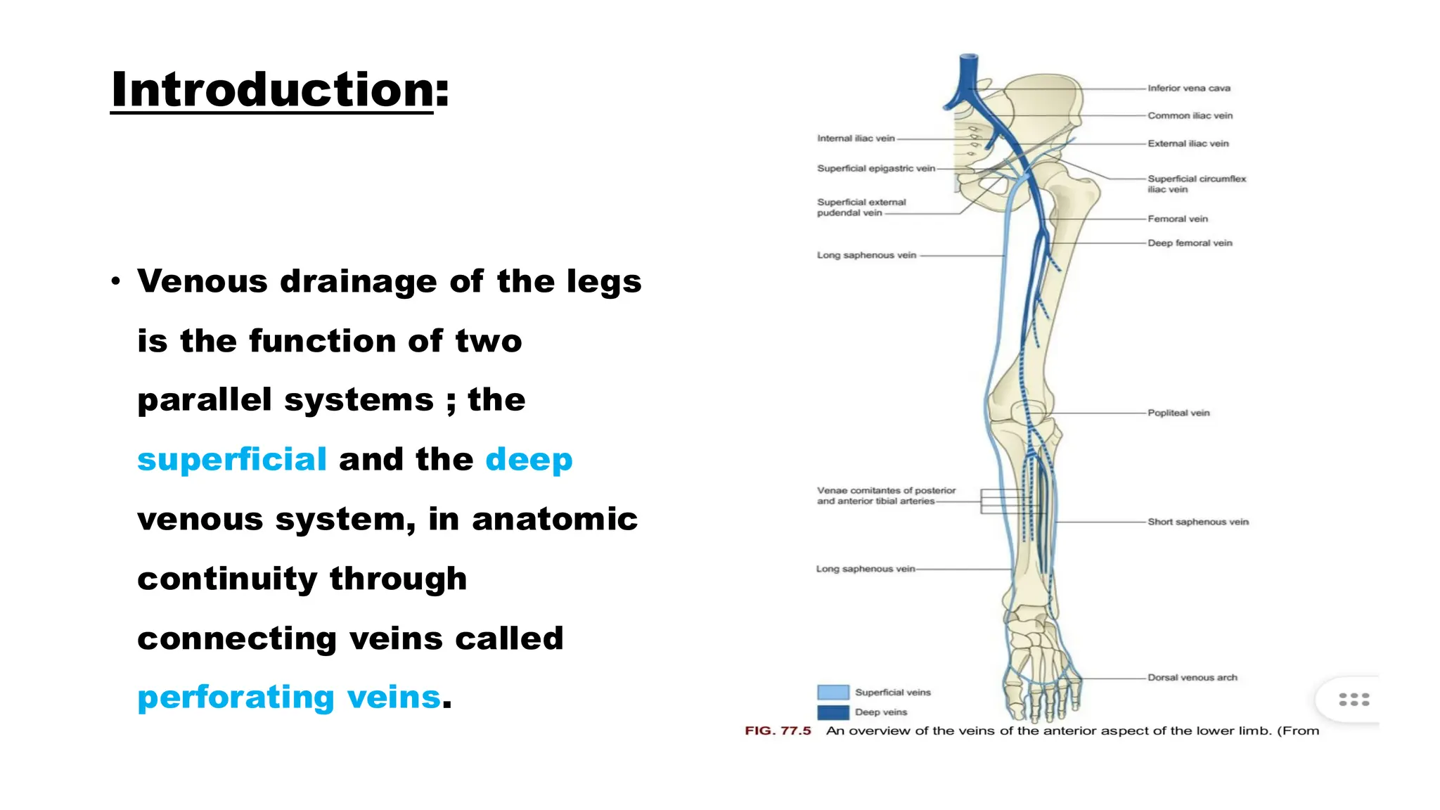
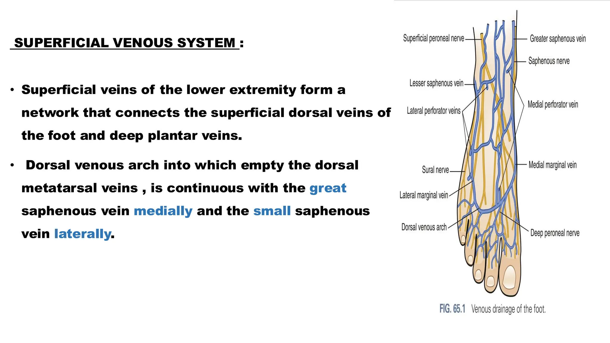
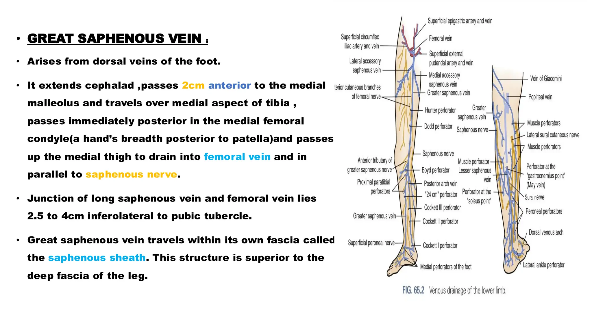
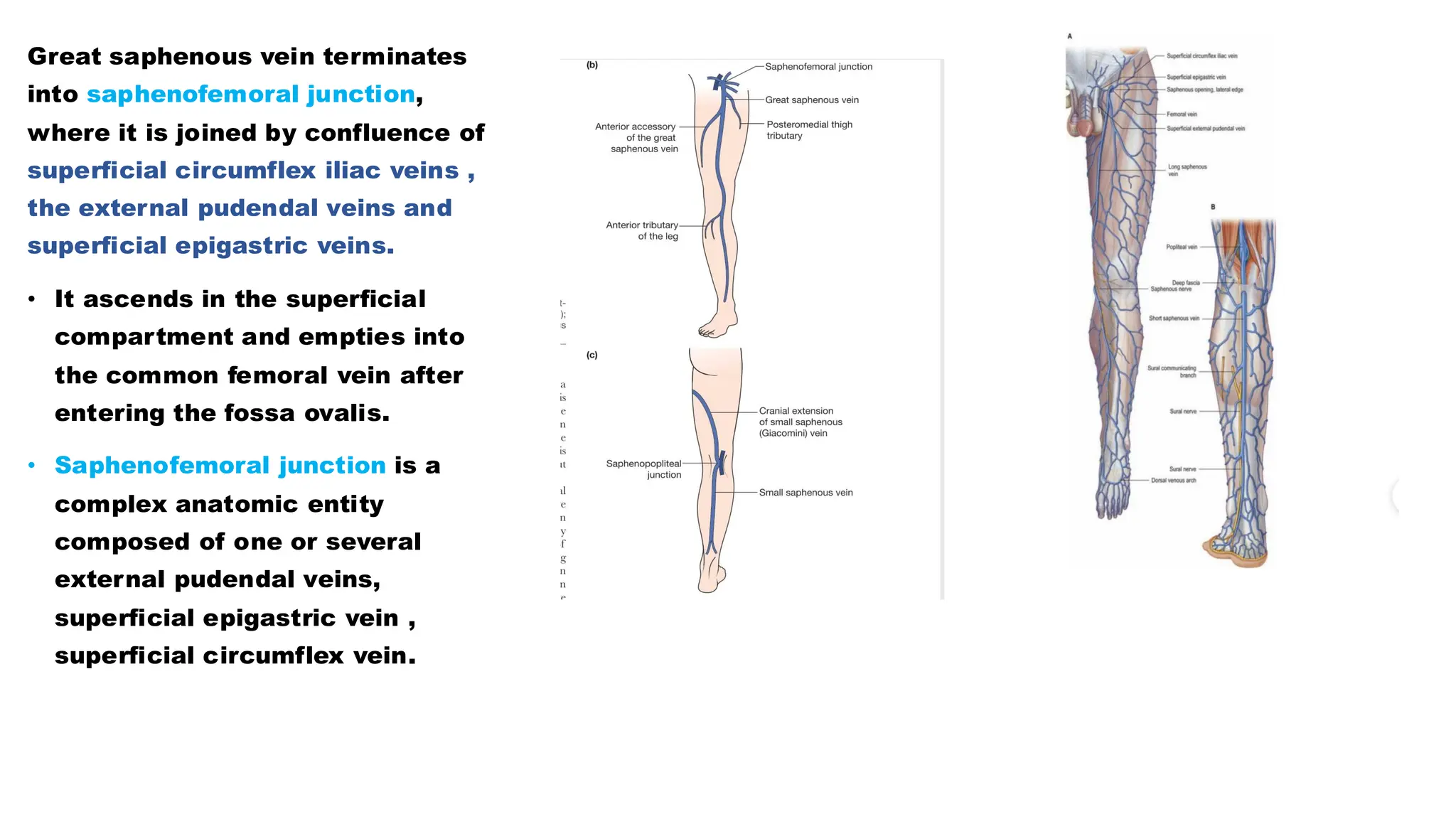
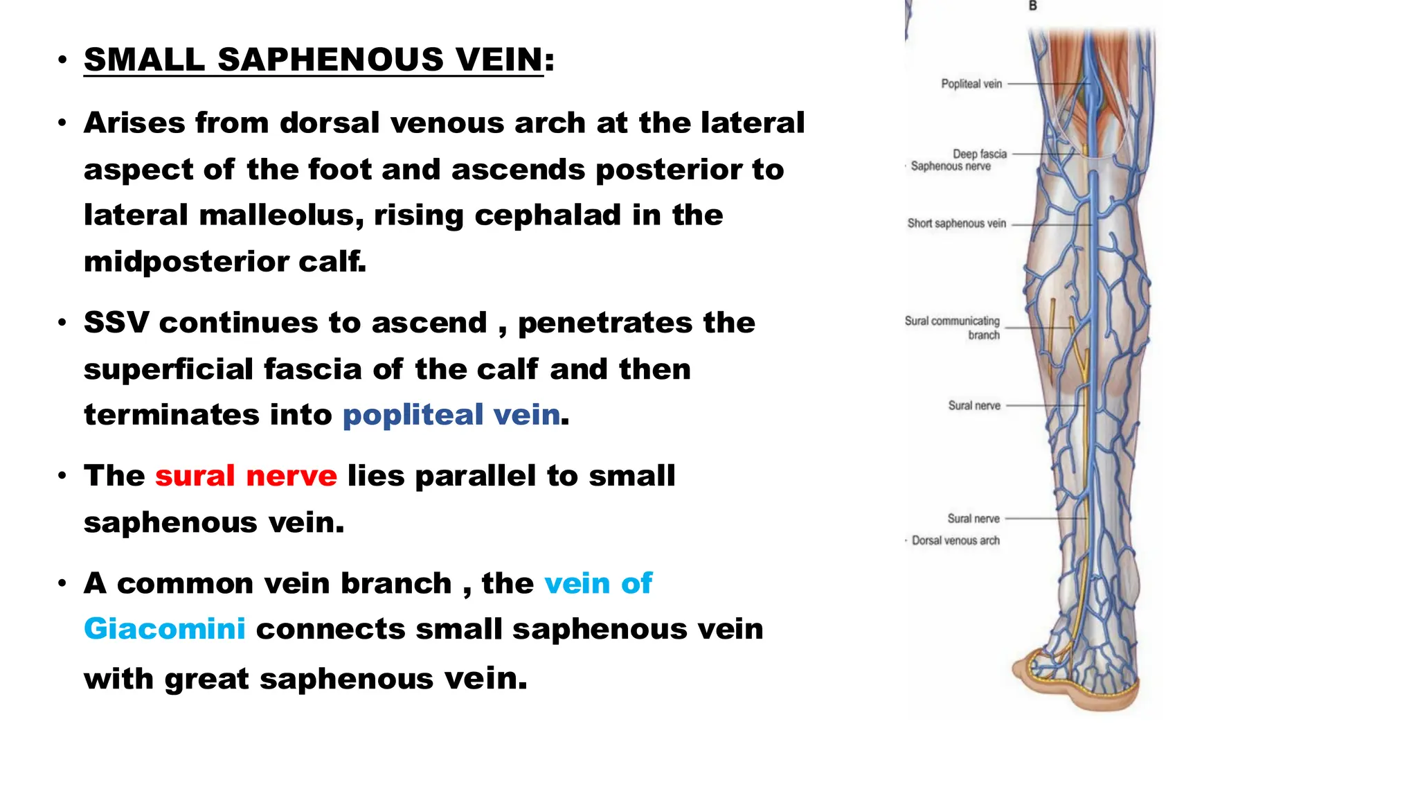
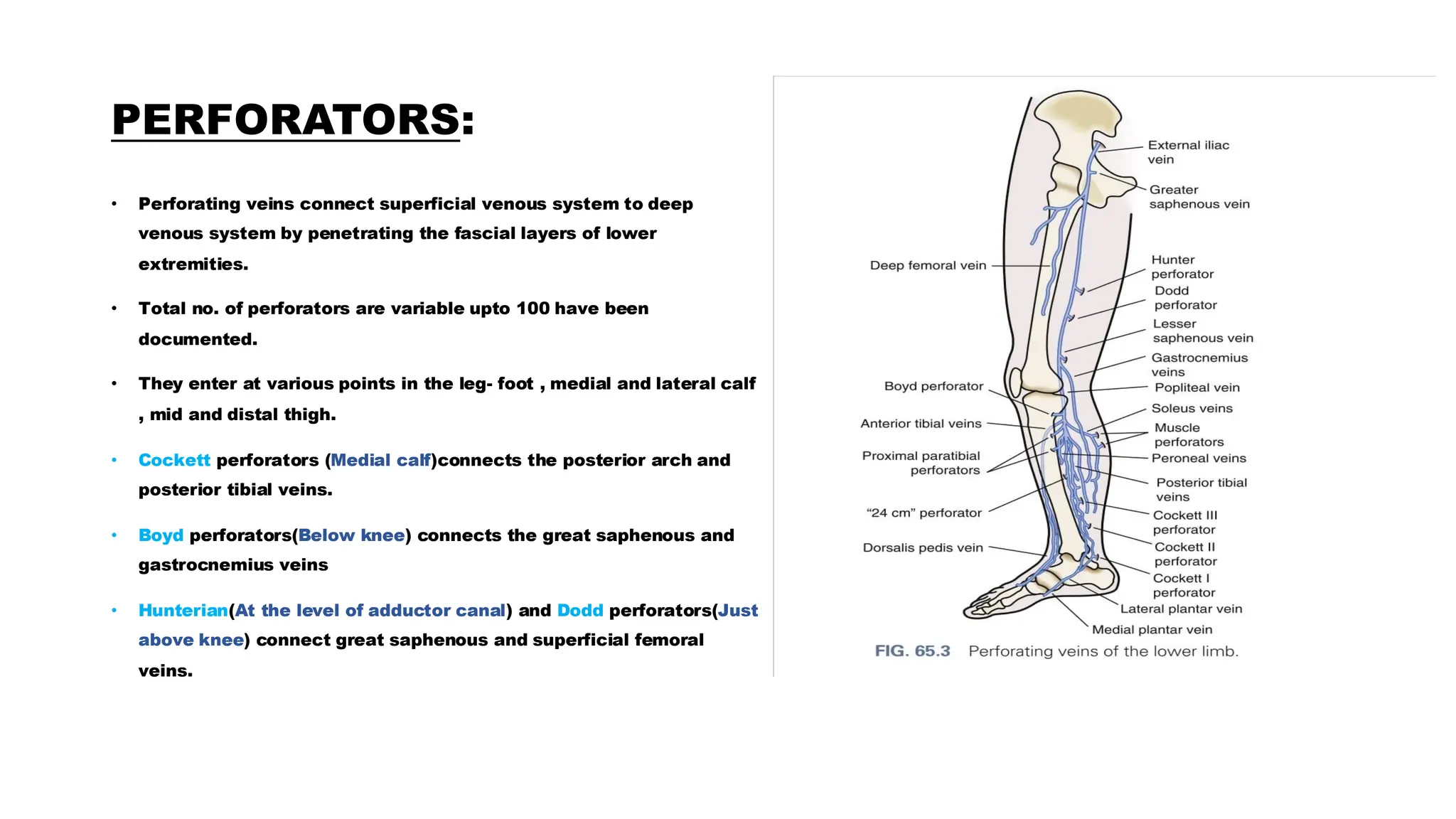
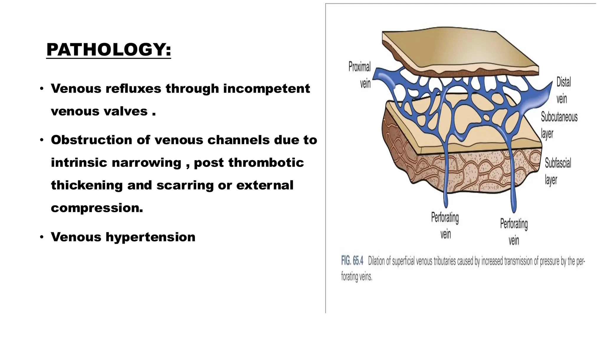
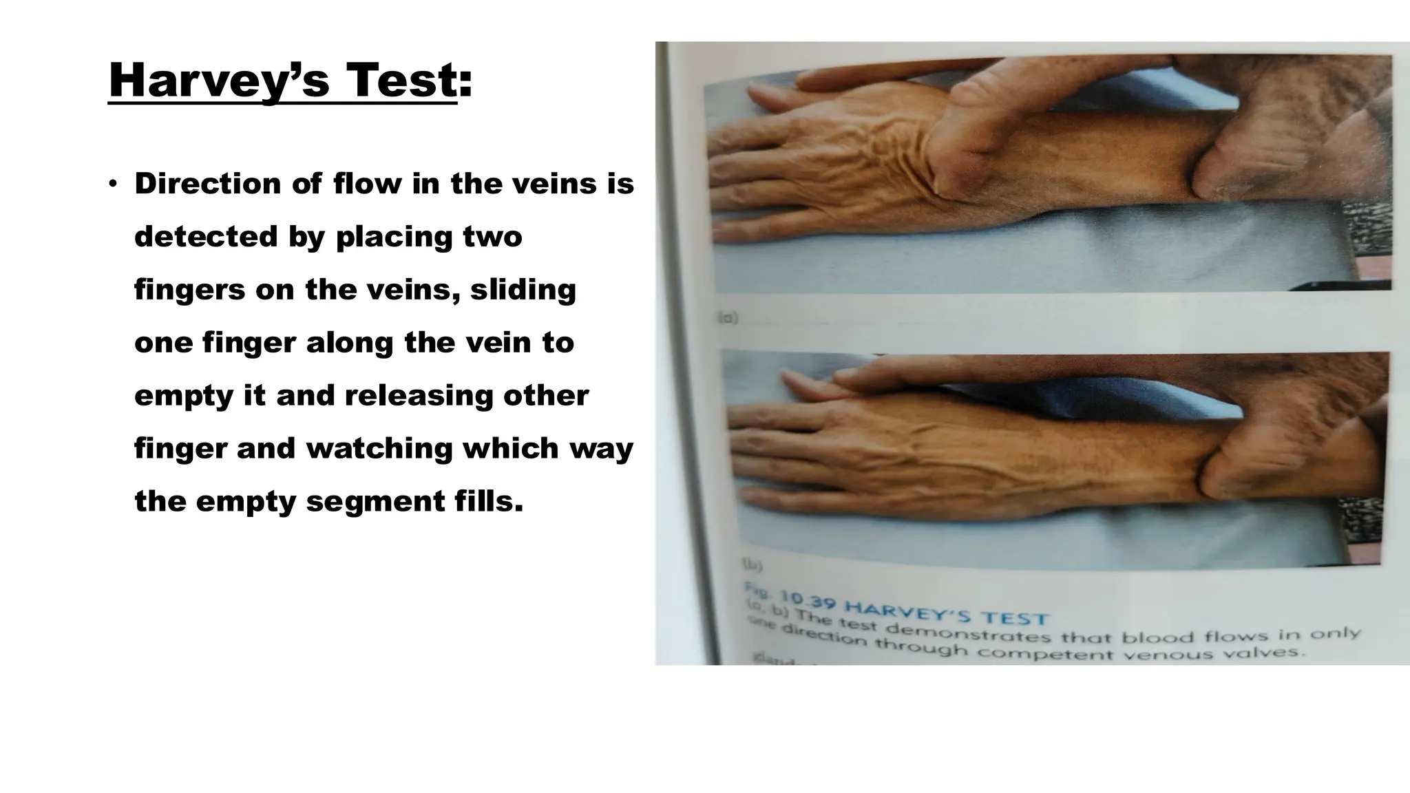
Varicose vein
Presenter: Dr. Suhaib Bashir
Moderator: Prof Dr. Mushtaq Chalkoo
Introduction:
• Venous drainage of the legs
is the function of two
parallel systems ; the
superficial and the deep
venous system, in anatomic
continuity through
connecting veins called
perforating veins.
SUPERFICIAL VENOUS SYSTEM :
• Superficial veins of the lower extremity form a
network that connects the superficial dorsal veins of
the foot and deep plantar veins.
• Dorsal venous arch into which empty the dorsal
metatarsal veins , is continuous with the great
saphenous vein medially and the small saphenous
vein laterally.
• GREAT SAPHENOUS VEIN :
• Arises from dorsal veins of the foot.
• It extends cephalad ,passes 2cm anterior to the medial
malleolus and travels over medial aspect of tibia ,
passes immediately posterior in the medial femoral
condyle(a hand’s breadth posterior to patella)and passes
up the medial thigh to drain into femoral vein and in
parallel to saphenous nerve.
• Junction of long saphenous vein and femoral vein lies
2.5 to 4cm inferolateral to pubic tubercle.
• Great saphenous vein travels within its own fascia called
the saphenous sheath. This structure is superior to the
deep fascia of the leg.
Great saphenous vein terminates
into saphenofemoral junction,
where it is joined by confluence of
superficial circumflex iliac veins ,
the external pudendal veins and
superficial epigastric veins.
• It ascends in the superficial
compartment and empties into
the common femoral vein after
entering the fossa ovalis.
• Saphenofemoral junction is a
complex anatomic entity
composed of one or several
external pudendal veins,
superficial epigastric vein ,
superficial circumflex vein.
• SMALL SAPHENOUS VEIN:
• Arises from dorsal venous arch at the lateral
aspect of the foot and ascends posterior to
lateral malleolus, rising cephalad in the
midposterior calf.
• SSV continues to ascend , penetrates the
superficial fascia of the calf and then
terminates into popliteal vein.
• The sural nerve lies parallel to small
saphenous vein.
• A common vein branch , the vein of
Giacomini connects small saphenous vein
with great saphenous vein.
DEEP VENOUS SYSTEM:
Plantar digital veins in the foot empty into a
network of metatarsal vein that compose deep
plantar venous arch.
Continues into medial and lateral plantar veins
which then drains into posterior tibial veins .
Dorsalis pedis veins on the dorsum of foot form the
paired anterior tibial veins at the ankle.
Paired posterior tibial veins , to and flanking the
posterior tibial artery, run under the fascia of the
deep posterior compartment. These veins enter the
soleus and join the popliteal vein , after joining with
paired peroneal and anterior tibial veins.
Popliteal vein enters a window in the adductor
magnus at which point termed as femoral vein .
PERFORATORS:
• Perforating veins connect superficial venous system to deep
venous system by penetrating the fascial layers of lower
extremities.
• Total no. of perforators are variable upto 100 have been
documented.
• They enter at various points in the leg- foot , medial and lateral calf
, mid and distal thigh.
• Cockett perforators (Medial calf)connects the posterior arch and
posterior tibial veins.
• Boyd perforators(Below knee) connects the great saphenous and
gastrocnemius veins
• Hunterian(At the level of adductor canal) and Dodd perforators(Just
above knee) connect great saphenous and superficial femoral
veins.
Compartments of venous
system of lower limb:
Normal Venous Histology and Function :
• Venous wall is composed of three layers, intima, media and adventitia.
• Vein walls have less smooth muscle and elastin than their arterial
counterparts.
• Venous intima: Endothelial cell layer resting on basement membrane.
Intima enfolds forming bicuspid valves whose function is to assure
venous return to heart.
• Media: Smooth muscle cells and elastin connective tissue.
• Adventitia : Adrenergic fibers (cutaneous veins)
FUNCTIONS:
• Venous valves prevent retrograde flow.
• Its their failure or valvular incompetence that leads to reflux .
• Veins can withstand large volume shifts with comparatively small
changes in pressure because veins do not have significant amounts of
elastin.
RISK FACTORS :
• Advancing age
• Female gender
• Multiparity
• Hereditary
• History of trauma to extremity
• Prolonged standing
PATHOLOGY:
• Venous refluxes through incompetent
venous valves .
• Obstruction of venous channels due to
intrinsic narrowing , post thrombotic
thickening and scarring or external
compression.
• Venous hypertension
VARICOSE VEIN:
• DEFINITION :
• Subcutaneous
dilated
,elongated and
tortuous veins
with diameter
3mm or more.
Veins:
Spider veins Ankle Blow-outs Saphena Varices
Causes of Varicose veins:
EPIDEMIOLOGY:
• Prevalence : 30 – 50 %
• Gender : Females > Males
• Age : 55-64 years
• Body mass index and height : higher prevalence
• Pregnancy (Increases risk)
• Family History
• Occupation and lifestyle ( Prolonged Standing)
SYMPTOMS:
• Pain (Aching, heaviness, throbbing ,burning lower limb) :exacerbated in
afternoons , after prolonged standing.
• Venous neuropathy (Cutaneous burning)
• Pruritis
• Venous claudication ( venous outflow obstruction, secondary venous
insufficiency)
PHYSICAL EXAMINATION:
• Assessment of arterial
circulation
• Venous assessment in
standing and supine
positions.
• Visual inspection (
Telangiectasias, reticular
veins. Varicose veins)
• Hyperpigmentation (
Lipodermatosclerosis ,
Hypertrophic skin ,
Atrophie Blanche )
Diagnostic evaluation of Venous dysfunction :
• Brodie – Trendelenburg Test :
• This test is performed to determine
the incompetency of sapheno-
femoral valve and other
communicating systems.
• Tourniquet test :
• Variant of Trendelenburg test.
Principles of tourniquet tests:
Doppler flow detector studies:
• uniphasic signal on
squeezing, with no sound on
relaxation indicates
competent valves with
forward flow.
• Biphasic signal, with
prolonged retrograde flow
on releasing the
compression , indicates
reflux and valvular
incompetence.
Perthes’ test:
• Affected lower extremity is
wrapped with elastic bandage.
With elastic bandage on patient
is instructed to move around and
exercise. Severe crampy pain is
complained of if there is deep
venous thrombosis.
Schwartz test:
• If a tap is made on long
saphenous vein in the
lower part of the leg an
impulse can be felt at the
saphenous opening .
• Pratt’ s test( To know
position of leg perforators)
• Morrisey’s cough impulse
test
• Fegan’s method to identify
site of perforators.
Harvey’s Test:
• Direction of flow in the veins is
detected by placing two
fingers on the veins, sliding
one finger along the vein to
empty it and releasing other
finger and watching which way
the empty segment fills.
INVESTIGATIONS:
• Duplex imaging
• Ultrasound doppler
• Air displacement plethysmography
• MRVI( Magnetic resonance venous
imaging)
MANAGEMENT:
• Compression
• Open Surgery
• Saphenofemoral ligation and great saphenous stripping
• Endothermal ablation
• Laser ablation
• Radiofrequency ablation (RFA)
• Ultrasound guided foam sclerotherapy (UGFS)
• Endovenous Glue
Open surgery:
• Saphenofemoral ligation and great saphenous stripping
• Saphenopopliteal junction and small saphenous stripping
• Phlebectomy
• Perforator Ligation
Laser ablation:
• Laser energy (wavelength 1470mm)is
transmitted down the fibre leading to
absorption of radiation and production of
thermal energy with around 60-80 J/cm.
Radiofrequency ablation:
• RFA uses same treatment principle as that of EVLA, except that an
electromagnetic current is used to create thermal energy.
• ClosureFast device(Medtronic) has a wire coil on the end of a treatment
catheter.
• Generator passes through a coil reaching 120 degree centigrade and
maintained for treatment cycle of 20 seconds.
• Coils are withdrawn for a set length and another treatment cycle is commenced.
Endothermal Ablation:
Treatment device is inserted into the incompetent axial vein percutaneously
Vein is surrounded by tumescent local anaesthetic solution
Compresses the vein onto the treatment device, emptying of blood
Hydro-dissects tissues (nerves)
Thermal device that destroys the structure of vein leading to permenant
occlusion
Advantages of RFA over EVLA:
• Marginal reduction in pain and bruising.
• EVLA requires specific safety protocols including designs and function
of room , specific training for operator and theatre team.
• EVLA requires understanding of power settings and pullback speeds.
• Allows better communication with the patient.
Advantages of EVLA over RFA:
• Veins of very large diameter > 15mm provides better efficacy rates.
• Less expensive
Non-endothermal ,non –tumescent ablation:
• Ultrasound guided foam sclerotherapy:
• Sclerosing Agent : Sodium tetradecyl sulphate, hypertonic saline ,
polidocanol
• 1:3 or 1:4 ratio mixture of sclerosant and air drawn into 1ml syringe, and
then oscillated vigorously oscillated between 2 syringes about 10-20
times.
• About 1-2 ml of foam should be injected.
• Max. volume should not exceed 10-12ml .
ASVAL(Ambulatory selective varices ablation under
local anesthesia )
• Surgical treatment of epifascial veins while sparing refluxing saphenous
vein.
• Principle:
• Reflux spreads from epifascial to saphenous vein in an antegrade
fashion.
Bibliography:
• Sabiston Textbook of surgery – 21st edition
• Bailey and Love’s Short practice of surgery -28th edition
• A Manual on clinical surgery by S Das -16th edition
• Fischer’s Mastery of Surgery 8th edition
• Browse’s Introduction to Symptoms and Signs of Surgical Disease.(5th
edition)
• CMDT Surgery -15th edition- Gerard M . Doherty
THANK YOU

Anatomy of Varicose veins and its management

  • 1.
    Varicose vein Presenter: Dr.Suhaib Bashir Moderator: Prof Dr. Mushtaq Chalkoo
  • 2.
    Introduction: • Venous drainageof the legs is the function of two parallel systems ; the superficial and the deep venous system, in anatomic continuity through connecting veins called perforating veins.
  • 3.
    SUPERFICIAL VENOUS SYSTEM: • Superficial veins of the lower extremity form a network that connects the superficial dorsal veins of the foot and deep plantar veins. • Dorsal venous arch into which empty the dorsal metatarsal veins , is continuous with the great saphenous vein medially and the small saphenous vein laterally.
  • 4.
    • GREAT SAPHENOUSVEIN : • Arises from dorsal veins of the foot. • It extends cephalad ,passes 2cm anterior to the medial malleolus and travels over medial aspect of tibia , passes immediately posterior in the medial femoral condyle(a hand’s breadth posterior to patella)and passes up the medial thigh to drain into femoral vein and in parallel to saphenous nerve. • Junction of long saphenous vein and femoral vein lies 2.5 to 4cm inferolateral to pubic tubercle. • Great saphenous vein travels within its own fascia called the saphenous sheath. This structure is superior to the deep fascia of the leg.
  • 5.
    Great saphenous veinterminates into saphenofemoral junction, where it is joined by confluence of superficial circumflex iliac veins , the external pudendal veins and superficial epigastric veins. • It ascends in the superficial compartment and empties into the common femoral vein after entering the fossa ovalis. • Saphenofemoral junction is a complex anatomic entity composed of one or several external pudendal veins, superficial epigastric vein , superficial circumflex vein.
  • 6.
    • SMALL SAPHENOUSVEIN: • Arises from dorsal venous arch at the lateral aspect of the foot and ascends posterior to lateral malleolus, rising cephalad in the midposterior calf. • SSV continues to ascend , penetrates the superficial fascia of the calf and then terminates into popliteal vein. • The sural nerve lies parallel to small saphenous vein. • A common vein branch , the vein of Giacomini connects small saphenous vein with great saphenous vein.
  • 7.
    DEEP VENOUS SYSTEM: Plantardigital veins in the foot empty into a network of metatarsal vein that compose deep plantar venous arch. Continues into medial and lateral plantar veins which then drains into posterior tibial veins . Dorsalis pedis veins on the dorsum of foot form the paired anterior tibial veins at the ankle. Paired posterior tibial veins , to and flanking the posterior tibial artery, run under the fascia of the deep posterior compartment. These veins enter the soleus and join the popliteal vein , after joining with paired peroneal and anterior tibial veins. Popliteal vein enters a window in the adductor magnus at which point termed as femoral vein .
  • 8.
    PERFORATORS: • Perforating veinsconnect superficial venous system to deep venous system by penetrating the fascial layers of lower extremities. • Total no. of perforators are variable upto 100 have been documented. • They enter at various points in the leg- foot , medial and lateral calf , mid and distal thigh. • Cockett perforators (Medial calf)connects the posterior arch and posterior tibial veins. • Boyd perforators(Below knee) connects the great saphenous and gastrocnemius veins • Hunterian(At the level of adductor canal) and Dodd perforators(Just above knee) connect great saphenous and superficial femoral veins.
  • 9.
  • 10.
    Normal Venous Histologyand Function : • Venous wall is composed of three layers, intima, media and adventitia. • Vein walls have less smooth muscle and elastin than their arterial counterparts. • Venous intima: Endothelial cell layer resting on basement membrane. Intima enfolds forming bicuspid valves whose function is to assure venous return to heart. • Media: Smooth muscle cells and elastin connective tissue. • Adventitia : Adrenergic fibers (cutaneous veins)
  • 11.
    FUNCTIONS: • Venous valvesprevent retrograde flow. • Its their failure or valvular incompetence that leads to reflux . • Veins can withstand large volume shifts with comparatively small changes in pressure because veins do not have significant amounts of elastin.
  • 12.
    RISK FACTORS : •Advancing age • Female gender • Multiparity • Hereditary • History of trauma to extremity • Prolonged standing
  • 13.
    PATHOLOGY: • Venous refluxesthrough incompetent venous valves . • Obstruction of venous channels due to intrinsic narrowing , post thrombotic thickening and scarring or external compression. • Venous hypertension
  • 14.
    VARICOSE VEIN: • DEFINITION: • Subcutaneous dilated ,elongated and tortuous veins with diameter 3mm or more.
  • 15.
    Veins: Spider veins AnkleBlow-outs Saphena Varices
  • 16.
  • 17.
    EPIDEMIOLOGY: • Prevalence :30 – 50 % • Gender : Females > Males • Age : 55-64 years • Body mass index and height : higher prevalence • Pregnancy (Increases risk) • Family History • Occupation and lifestyle ( Prolonged Standing)
  • 18.
    SYMPTOMS: • Pain (Aching,heaviness, throbbing ,burning lower limb) :exacerbated in afternoons , after prolonged standing. • Venous neuropathy (Cutaneous burning) • Pruritis • Venous claudication ( venous outflow obstruction, secondary venous insufficiency)
  • 19.
    PHYSICAL EXAMINATION: • Assessmentof arterial circulation • Venous assessment in standing and supine positions. • Visual inspection ( Telangiectasias, reticular veins. Varicose veins) • Hyperpigmentation ( Lipodermatosclerosis , Hypertrophic skin , Atrophie Blanche )
  • 20.
    Diagnostic evaluation ofVenous dysfunction : • Brodie – Trendelenburg Test : • This test is performed to determine the incompetency of sapheno- femoral valve and other communicating systems. • Tourniquet test : • Variant of Trendelenburg test.
  • 21.
  • 22.
    Doppler flow detectorstudies: • uniphasic signal on squeezing, with no sound on relaxation indicates competent valves with forward flow. • Biphasic signal, with prolonged retrograde flow on releasing the compression , indicates reflux and valvular incompetence.
  • 23.
    Perthes’ test: • Affectedlower extremity is wrapped with elastic bandage. With elastic bandage on patient is instructed to move around and exercise. Severe crampy pain is complained of if there is deep venous thrombosis.
  • 24.
    Schwartz test: • Ifa tap is made on long saphenous vein in the lower part of the leg an impulse can be felt at the saphenous opening . • Pratt’ s test( To know position of leg perforators) • Morrisey’s cough impulse test • Fegan’s method to identify site of perforators.
  • 25.
    Harvey’s Test: • Directionof flow in the veins is detected by placing two fingers on the veins, sliding one finger along the vein to empty it and releasing other finger and watching which way the empty segment fills.
  • 26.
    INVESTIGATIONS: • Duplex imaging •Ultrasound doppler • Air displacement plethysmography • MRVI( Magnetic resonance venous imaging)
  • 27.
    MANAGEMENT: • Compression • OpenSurgery • Saphenofemoral ligation and great saphenous stripping • Endothermal ablation • Laser ablation • Radiofrequency ablation (RFA) • Ultrasound guided foam sclerotherapy (UGFS) • Endovenous Glue
  • 28.
    Open surgery: • Saphenofemoralligation and great saphenous stripping • Saphenopopliteal junction and small saphenous stripping • Phlebectomy • Perforator Ligation
  • 30.
    Laser ablation: • Laserenergy (wavelength 1470mm)is transmitted down the fibre leading to absorption of radiation and production of thermal energy with around 60-80 J/cm.
  • 31.
    Radiofrequency ablation: • RFAuses same treatment principle as that of EVLA, except that an electromagnetic current is used to create thermal energy. • ClosureFast device(Medtronic) has a wire coil on the end of a treatment catheter. • Generator passes through a coil reaching 120 degree centigrade and maintained for treatment cycle of 20 seconds. • Coils are withdrawn for a set length and another treatment cycle is commenced.
  • 32.
    Endothermal Ablation: Treatment deviceis inserted into the incompetent axial vein percutaneously Vein is surrounded by tumescent local anaesthetic solution Compresses the vein onto the treatment device, emptying of blood Hydro-dissects tissues (nerves) Thermal device that destroys the structure of vein leading to permenant occlusion
  • 33.
    Advantages of RFAover EVLA: • Marginal reduction in pain and bruising. • EVLA requires specific safety protocols including designs and function of room , specific training for operator and theatre team. • EVLA requires understanding of power settings and pullback speeds. • Allows better communication with the patient.
  • 34.
    Advantages of EVLAover RFA: • Veins of very large diameter > 15mm provides better efficacy rates. • Less expensive
  • 35.
    Non-endothermal ,non –tumescentablation: • Ultrasound guided foam sclerotherapy: • Sclerosing Agent : Sodium tetradecyl sulphate, hypertonic saline , polidocanol • 1:3 or 1:4 ratio mixture of sclerosant and air drawn into 1ml syringe, and then oscillated vigorously oscillated between 2 syringes about 10-20 times. • About 1-2 ml of foam should be injected. • Max. volume should not exceed 10-12ml .
  • 38.
    ASVAL(Ambulatory selective varicesablation under local anesthesia ) • Surgical treatment of epifascial veins while sparing refluxing saphenous vein. • Principle: • Reflux spreads from epifascial to saphenous vein in an antegrade fashion.
  • 39.
    Bibliography: • Sabiston Textbookof surgery – 21st edition • Bailey and Love’s Short practice of surgery -28th edition • A Manual on clinical surgery by S Das -16th edition • Fischer’s Mastery of Surgery 8th edition • Browse’s Introduction to Symptoms and Signs of Surgical Disease.(5th edition) • CMDT Surgery -15th edition- Gerard M . Doherty
  • 40.