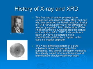
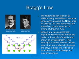
X-ray diffraction
Outline
 History and background information of X-
ray diffraction.
 Theory behind the equipment
 Challenges of applying traditional XRD to
nano-technology
 Summary
History of X-ray and XRD
 Wilhelm Conrad Röntgen discovered
X-Rays in 1895.
 1901 Nobel prize in Physics
Wilhelm Conrad Röntgen (1845-1923)
A modern radiograph of a hand
Bertha Röntgen’s Hand 8 Nov, 1895
History of X-ray and XRD
 Radiographs like the ones in the
last slide are simply shadowgrams.
 The X-rays either pass straight
through or are stopped by the
object. The diagram on the upper
left illustrates the principle and
shows a perfect shadow.
 In reality, a large fraction of the X-
rays are not simply absorbed or
transmitted by the object but are
scattered. The diagram on the
bottom left illustrates this effect and
illustrates the fuzzy edge of the
object that is produced in the image
by the scattered X-rays.
 The first kind of scatter process to be
recognised was discovered by Max von Laue
who was awarded the Nobel prize for physics
in 1914 "for his discovery of the diffraction of
X-rays by crystals". His collaborators Walter
Friedrich and Paul Knipping took the picture
on the bottom left in 1912. It shows how a
beam of X-rays is scattered into a
characteristic pattern by a crystal. In this
case it is copper sulphate.
 The X-ray diffraction pattern of a pure
substance is like a fingerprint of the
substance. The powder diffraction method is
thus ideally suited for characterization and
identification of polycrystalline phases.
History of X-ray and XRD
Max von Laue (1897-1960)
Bragg’s Law
 The father and son team of Sir
William Henry and William Lawrence
Bragg were awarded the Nobel prize
for physics "for their services in the
analysis of crystal structure by
means of Xrays“ in 1915.
 Bragg's law was an extremely
important discovery and formed the
basis for the whole of what is now
known as crystallography. This
technique is one of the most widely
used structural analysis techniques
and plays a major role in fields as
diverse as structural biology and
materials science.
William Lawrence
Bragg (1890-1971)
Sir William Henry
Bragg (1862-1942)
Equipment
Bruker D8 Analytical X-ray Systems
Goniometer
2-Theta:Theta Setup
Goniometer
Theta:Theta Setup
Fundamentals
 How it works?
 An electron in an alternating electromagnetic field will oscillate with the
same frequency as the field. When an X-ray beam hits an atom, the
electrons around the atom start to oscillate with the same frequency as
the incoming beam. In almost all directions we will have destructive
interference, that is, the combining waves are out of phase and there is
no resultant energy leaving the solid sample. However the atoms in a
crystal are arranged in a regular pattern, and in a very few directions we
will have constructive interference. The waves will be in phase and
there will be well defined X-ray beams leaving the sample at various
directions. Hence, a diffracted beam may be described as a beam
composed of a large number of scattered rays mutually reinforcing one
another.
 Crystal systems, space groups, reciprocal lattice, Miller indices…
 Planes going through areas with high electron density will reflect
strongly, planes with low electron density will give weak intensities.
Sample preparation
 Single crystal X-ray diffraction
 The single crystal sample is a perfect crystal (all unit cells
aligned in a perfect extended pattern) with a cross section of
about 0.3 mm.
 The single crystal diffractometer and associated computer
package is used mainly to elucidate the molecular structure of
novel compounds.
 Powder (polycrystalline) X-ray diffraction
 It is important to have a sample with a smooth plane
surface. If possible, we normally grind the sample
down to particles of about 0.002 mm to 0.005 mm
cross section. The ideal sample is homogeneous and
the crystallites are randomly distributed. The sample
is pressed into a sample holder so that we have a
smooth flat surface.
Data collection and analysis
 Collecting data: computer and software
 Analysis:
 ICDD database – Identification
 Structure refinement – GSAS
 Quantitative phase analysis – GSAS
 Novel structure – single crystal
Applications
 Identification
 Polymer crystallinity
 Residual stress
 Texture analysis
Challenge of applying to
nanotechnology
 Traditional X-ray powder-diffraction techniques rely on
the long-range order in crystals to produce sharp "Bragg
peaks" in a diffraction pattern. By examining these Bragg
peaks, which result from X-ray scattering, scientists can
determine the material's atomic structure.
 But nanocrystals lack long-range order and often
incorporate a large number of defects. As a result, their
diffraction patterns are much more diffuse with few, if
any, Bragg peaks.
 "This poses a real challenge to the traditional techniques
for structure determination," -- Valeri Petkov of Michigan
State.
Breakthroughs for diffraction
 RTMS detection technology and the
implementation of mono-capillary have reduced
measurement times and minimum amounts of
material required considerably.
 Many X-ray diffraction techniques are at disposal
of the nanoscientist now for the structural
characterization of the nanomaterials, such as
high-resolution diffraction, reflectometry, small-
angle X-ray scattering and line profile analysis.
Summary
 X-ray diffraction provides a powerful tool to
study the structure and composition of the
materials which is a key requirement for
understanding materials properties
 An X-ray diffraction system should not be
missing in a modern laboratory for
research on nano- and advanced
materials. – www.panalytical.com
Some useful links
 GSAS:
 http://www.ncnr.nist.gov/xtal/software/gsas.html
 ICDD:
 http://www.icdd.com/
 CCP14
 http://ccp14.sims.nrc.ca/
 Diffraction tutorials
 http://www.uni-
wuerzburg.de/mineralogie/crystal/teaching/basic.html
 Paper addressed the problem
 Phase Transitions, 2003, Vol 76, Nos. 1-2, pp. 171-185

99995067.ppt

  • 1.
  • 2.
    Outline  History andbackground information of X- ray diffraction.  Theory behind the equipment  Challenges of applying traditional XRD to nano-technology  Summary
  • 3.
    History of X-rayand XRD  Wilhelm Conrad Röntgen discovered X-Rays in 1895.  1901 Nobel prize in Physics Wilhelm Conrad Röntgen (1845-1923) A modern radiograph of a hand Bertha Röntgen’s Hand 8 Nov, 1895
  • 4.
    History of X-rayand XRD  Radiographs like the ones in the last slide are simply shadowgrams.  The X-rays either pass straight through or are stopped by the object. The diagram on the upper left illustrates the principle and shows a perfect shadow.  In reality, a large fraction of the X- rays are not simply absorbed or transmitted by the object but are scattered. The diagram on the bottom left illustrates this effect and illustrates the fuzzy edge of the object that is produced in the image by the scattered X-rays.
  • 5.
     The firstkind of scatter process to be recognised was discovered by Max von Laue who was awarded the Nobel prize for physics in 1914 "for his discovery of the diffraction of X-rays by crystals". His collaborators Walter Friedrich and Paul Knipping took the picture on the bottom left in 1912. It shows how a beam of X-rays is scattered into a characteristic pattern by a crystal. In this case it is copper sulphate.  The X-ray diffraction pattern of a pure substance is like a fingerprint of the substance. The powder diffraction method is thus ideally suited for characterization and identification of polycrystalline phases. History of X-ray and XRD Max von Laue (1897-1960)
  • 6.
    Bragg’s Law  Thefather and son team of Sir William Henry and William Lawrence Bragg were awarded the Nobel prize for physics "for their services in the analysis of crystal structure by means of Xrays“ in 1915.  Bragg's law was an extremely important discovery and formed the basis for the whole of what is now known as crystallography. This technique is one of the most widely used structural analysis techniques and plays a major role in fields as diverse as structural biology and materials science. William Lawrence Bragg (1890-1971) Sir William Henry Bragg (1862-1942)
  • 7.
  • 8.
  • 9.
  • 10.
    Fundamentals  How itworks?  An electron in an alternating electromagnetic field will oscillate with the same frequency as the field. When an X-ray beam hits an atom, the electrons around the atom start to oscillate with the same frequency as the incoming beam. In almost all directions we will have destructive interference, that is, the combining waves are out of phase and there is no resultant energy leaving the solid sample. However the atoms in a crystal are arranged in a regular pattern, and in a very few directions we will have constructive interference. The waves will be in phase and there will be well defined X-ray beams leaving the sample at various directions. Hence, a diffracted beam may be described as a beam composed of a large number of scattered rays mutually reinforcing one another.  Crystal systems, space groups, reciprocal lattice, Miller indices…  Planes going through areas with high electron density will reflect strongly, planes with low electron density will give weak intensities.
  • 11.
    Sample preparation  Singlecrystal X-ray diffraction  The single crystal sample is a perfect crystal (all unit cells aligned in a perfect extended pattern) with a cross section of about 0.3 mm.  The single crystal diffractometer and associated computer package is used mainly to elucidate the molecular structure of novel compounds.  Powder (polycrystalline) X-ray diffraction  It is important to have a sample with a smooth plane surface. If possible, we normally grind the sample down to particles of about 0.002 mm to 0.005 mm cross section. The ideal sample is homogeneous and the crystallites are randomly distributed. The sample is pressed into a sample holder so that we have a smooth flat surface.
  • 12.
    Data collection andanalysis  Collecting data: computer and software  Analysis:  ICDD database – Identification  Structure refinement – GSAS  Quantitative phase analysis – GSAS  Novel structure – single crystal
  • 13.
    Applications  Identification  Polymercrystallinity  Residual stress  Texture analysis
  • 14.
    Challenge of applyingto nanotechnology  Traditional X-ray powder-diffraction techniques rely on the long-range order in crystals to produce sharp "Bragg peaks" in a diffraction pattern. By examining these Bragg peaks, which result from X-ray scattering, scientists can determine the material's atomic structure.  But nanocrystals lack long-range order and often incorporate a large number of defects. As a result, their diffraction patterns are much more diffuse with few, if any, Bragg peaks.  "This poses a real challenge to the traditional techniques for structure determination," -- Valeri Petkov of Michigan State.
  • 15.
    Breakthroughs for diffraction RTMS detection technology and the implementation of mono-capillary have reduced measurement times and minimum amounts of material required considerably.  Many X-ray diffraction techniques are at disposal of the nanoscientist now for the structural characterization of the nanomaterials, such as high-resolution diffraction, reflectometry, small- angle X-ray scattering and line profile analysis.
  • 16.
    Summary  X-ray diffractionprovides a powerful tool to study the structure and composition of the materials which is a key requirement for understanding materials properties  An X-ray diffraction system should not be missing in a modern laboratory for research on nano- and advanced materials. – www.panalytical.com
  • 17.
    Some useful links GSAS:  http://www.ncnr.nist.gov/xtal/software/gsas.html  ICDD:  http://www.icdd.com/  CCP14  http://ccp14.sims.nrc.ca/  Diffraction tutorials  http://www.uni- wuerzburg.de/mineralogie/crystal/teaching/basic.html  Paper addressed the problem  Phase Transitions, 2003, Vol 76, Nos. 1-2, pp. 171-185