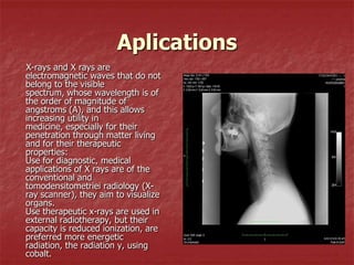
X-RAYS


         Berchesug Alexandra
           Margarit Alina
           Paun Iulia
           Soava Lavinia
           Clasa a XI –a C
Generate
 (or Röntgen) are issued in
tubes called Röntgen, are
electrons,are accelerated in
intense electric fields so that
they penetrate inside
electron shells of atoms anode
or gas in the tube and pull out
electrons from
layers near the core. After
braking these electrons from
subsequent transitions
electrons from levels with low
energies (layers K, L), X-rays
are obtained
Detection
     For X rays are used:
     gas-filled detectors
     (used in digital
     radiography to measure
     energy and position of
     individual X-ray photon
     counting speed
     radiological)
     type proportional
     counter,
     -calorimeters,
     -microchannel,
     -area photodetector,
     superconducting tunnel
     junction-detectors, etc…
The detector is a large, flat, with
one or more layers to absorb
radiation energy into electric
charge X. Convert using an
integrated surface electric charge
autoscanarea active matrix
image.
X radiation absorbed by a
phosphorescent screen release
light creates an image on the
surface. Lateral scattering of light
is limited by diffusion.
Digital detectors are capable of
running all current radiological
modalities, including radiography
and fluoroscopy
Element diameter is comparable
to the thickness of the screen
image. The screen is thicker (to
increase the absorption quantum
efficiency) decreases the image
resolution. Using fluorescent
screen, increase the image
resolution
Proprieties
-Vacuum it travels at the speed of
light;
-impress photographic plates;
- Are not diverted for electric and
magnetic fields;
- Are invisible, ie unlike light, does
not impress human eye
-Ionising gases that pass. Number
of urge intensity ion radiation
produced show. On this property
are made based operation of
radiation detectors.
- Have fiziological
action, destroying organic cells
and is generally harmful to
humans. This property are made
based on their use in the
treatment of cancerous tumors to
destroy diseased tissue.
- Substances produced
fluorescence (light emission)
Aplications
X-rays and X rays are
electromagnetic waves that do not
belong to the visible
spectrum, whose wavelength is of
the order of magnitude of
angstroms (A), and this allows
increasing utility in
medicine, especially for their
penetration through matter living
and for their therapeutic
properties:
Use for diagnostic, medical
applications of X rays are of the
conventional and
tomodensitometriei radiology (X-
ray scanner), they aim to visualize
organs.
Use therapeutic x-rays are used in
external radiotherapy, but their
capacity is reduced ionization, are
preferred more energetic
radiation, the radiation y, using
cobalt.

X rays

  • 1.
    X-RAYS Berchesug Alexandra Margarit Alina Paun Iulia Soava Lavinia Clasa a XI –a C
  • 2.
    Generate (or Röntgen)are issued in tubes called Röntgen, are electrons,are accelerated in intense electric fields so that they penetrate inside electron shells of atoms anode or gas in the tube and pull out electrons from layers near the core. After braking these electrons from subsequent transitions electrons from levels with low energies (layers K, L), X-rays are obtained
  • 3.
    Detection For X rays are used: gas-filled detectors (used in digital radiography to measure energy and position of individual X-ray photon counting speed radiological) type proportional counter, -calorimeters, -microchannel, -area photodetector, superconducting tunnel junction-detectors, etc…
  • 4.
    The detector isa large, flat, with one or more layers to absorb radiation energy into electric charge X. Convert using an integrated surface electric charge autoscanarea active matrix image. X radiation absorbed by a phosphorescent screen release light creates an image on the surface. Lateral scattering of light is limited by diffusion. Digital detectors are capable of running all current radiological modalities, including radiography and fluoroscopy Element diameter is comparable to the thickness of the screen image. The screen is thicker (to increase the absorption quantum efficiency) decreases the image resolution. Using fluorescent screen, increase the image resolution
  • 5.
    Proprieties -Vacuum it travelsat the speed of light; -impress photographic plates; - Are not diverted for electric and magnetic fields; - Are invisible, ie unlike light, does not impress human eye -Ionising gases that pass. Number of urge intensity ion radiation produced show. On this property are made based operation of radiation detectors. - Have fiziological action, destroying organic cells and is generally harmful to humans. This property are made based on their use in the treatment of cancerous tumors to destroy diseased tissue. - Substances produced fluorescence (light emission)
  • 6.
    Aplications X-rays and Xrays are electromagnetic waves that do not belong to the visible spectrum, whose wavelength is of the order of magnitude of angstroms (A), and this allows increasing utility in medicine, especially for their penetration through matter living and for their therapeutic properties: Use for diagnostic, medical applications of X rays are of the conventional and tomodensitometriei radiology (X- ray scanner), they aim to visualize organs. Use therapeutic x-rays are used in external radiotherapy, but their capacity is reduced ionization, are preferred more energetic radiation, the radiation y, using cobalt.