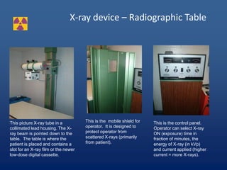
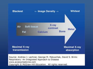
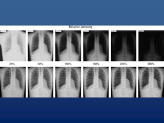
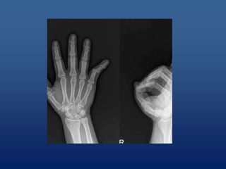
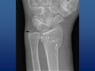
Dr. Aisha Arif
X-rays Fundamentals and uses
X-ray equipment
Early X-Ray Tube (1899): This tube is a
specimen of the first type of gas x-ray tube to
incorporate a water-cooled anode. The hollow
anode was supplied with water by gravity feed
from a supply held in the side bulb. This type
of tube was introduced by Mueller about 1899.
• Radiation is energy in the form of waves or particles.
Radiation which is high enough in energy to cause ionization is
called ionizing radiation. It includes particles and rays given
off by radioactive material and high-voltage equipment.
Ionizing radiation includes x-rays, gamma-rays, beta particles,
alpha particles, and neutrons.
• Without the use of monitoring equipment, humans
are not able to "find" ionizing radiation. In contrast
to heat, light, odors and noise, humans are not able
to see, feel, taste, smell, or hear ionizing radiation.
What is radiation?
What are X-rays?
• X rays are the ionizing electromagnetic radiation emitted
from a highly evacuated high-voltage tube. Inner orbital
electrons in the target anode are stimulated to emit x-
radiation via bombardment by a stream of electrons from
a heated cathode. X-rays, like gamma rays, are
penetrating and carry enough energy to ionize atoms in
their path. Nearly identical to gamma rays, x-rays require
shielding to reduce their intensity and minimize the
danger of tissue damage to personnel. Mishaps with x-
rays can cause severe radiation burns and deep tissue
damage and can lead to various cancers.
X-rays
X-rays were discovered in 1895 when Wilhelm
Conrad Roentgen observed that a screen
coated with a barium salt fluoresced when
placed near a cathode ray tube. Roentgen
concluded that a form of penetrating radiation
was being emitted by the cathode ray tube
and called the unknown rays, X-rays.
X-ray tube
An x-ray tube requires a source of electrons, a means to accelerate the
electrons, and a target to stop the high-speed electrons.
Diagnostic X-rays
• Two main types of diagnostic X-ray devices:
• Radiograph – a picture with film or image is
sent direct to computer screen. These are
quick ‘shots’.
• Fluoroscopic – a real time “moving”
inspection on inside functions. These longer
exposures yield high doses.
Diagnostic X-rays
• Diagnostic radiology is the branch of medicine
that involves taking and reading X-rays. The
physicians that prescribe the shots and the
technologists that operate the machines are
specifically trained and licensed to perform
these tasks. They also stay current through
continuing education. Institutions are always
striving to get quality images with the
minimum patient exposure.
X-Ray Effects
• The effects of x-ray exposure depends upon:
• Duration/Exposure time - How fast the dose is
delivered.
• Energy - How much energy was in the x-ray
– Low Energy (<50 KeV) - damage only to skin or outer part
of body
– High Energy - damage to internal organs
• Total Dose - The magnitude of the dose
RADIATION PROTECTION – TIME
The dose of radiation a worker receives is
directly proportional to the amount of time
spent in a radiation field. Thus, reducing the
time by one-half will reduce the radiation
dose received by one-half. Operators should
always continuously monitor, work quickly and
spend as little time as possible next to any
high-field X-ray equipment.
Radiation Protection - Distance
Radiation exposure decreases rapidly as the distance between
the worker and the X-ray device increases. The amount of
radiation at a given distance from the source varies inversely
with the square of the distance. For example, doubling the
distance from an x-ray tube will reduce the dose to one-fourth
of its original value, and increasing the distance by a factor of
three will reduce the dose to one-ninth of its original value.
Maintaining a safe distance, therefore, represents one the
simplest and most effective methods for reducing radiation
exposure to workers. Using the principle of distance is
especially important when working around open beam high-
field analytical x-ray equipment.
Radiation Protection - SHIELDING
Radiation exposure to personnel can also be reduced by
placing material between a worker and the x-ray tube. The
energy of the x-ray is reduced by subatomic interactions in the
shielding material. Dense materials such as lead, are the
most effective shielding, but any metal is useful. Even
concrete, water and natural earth are used in shielding. The
energy of scattered radiation from x-ray use is often so low
that clear plastic is useful.
Shielding is often incorporated into the equipment, such as
the metal lining surrounding the x-ray tube. It may also consist
of permanent barriers such as concrete and lead walls, leaded
glass, and plastic movable screens in the case of analytical x-
ray equipment.
X-ray device – Radiographic Table
This picture X-ray tube in a
collimated lead housing. The X-
ray beam is pointed down to the
table. The table is where the
patient is placed and contains a
slot for an X-ray film or the newer
low-dose digital cassette.
This is the mobile shield for
operator. It is designed to
protect operator from
scattered X-rays (primarily
from patient).
This is the control panel.
Operator can select X-ray
ON (exposure) time in
fraction of minutes, the
energy of X-ray (in kVp)
and current applied (higher
current = more X-rays).
Uses of x-rays
• Identifying fractures (cracks) and breaks or
infections in bones and teeth1
• Diagnosing cavities and evaluating
structures in the mouth and jaw
• Picking up on signs of joint changes that
indicate arthritis using a special type of X-
ray image called an arthrogram
• Revealing tumors on bones
•
Uses of x-rays
• Measuring bone density as a means of
diagnosing osteoporosis
• Finding evidence of pneumonia,
tuberculosis, or lung cancer (chest X-rays)
• Examining breast tissue for signs of
cancer using a special X-ray technique
called mammography1
Uses of x-rays
• Looking for signs of heart failure or
changes in blood flow to the lungs and
heart
• Revealing problems in the digestive tract
such as kidney stones, sometimes using a
contrast medium called barium
• Locating swallowed items such as a coin
or tiny toy
What are the absolute
contraindications for a plain
radiograph/X-ray?
• None. A plain X-ray is a low-dose
examination that is cheap and readily
available. The small risk must be weighed
up against the benefit (see Adverse effects
below).
What are the relative
contraindications for a plain
radiograph/X-ray?
• Pregnancy
• Weight of patient (X-ray tables have
weight limits).
Cost and risk vs benefit for plain
radiographs
• Cheap study among all radiology tests.
• Enough to inform about major emergency
conditions.
• Guides the treatment plan.
• Follow up easy.
• Only risk is accumulative dose of radiation
after repeat studie.

xrays basics.ppt

  • 1.
    Dr. Aisha Arif X-raysFundamentals and uses
  • 2.
    X-ray equipment Early X-RayTube (1899): This tube is a specimen of the first type of gas x-ray tube to incorporate a water-cooled anode. The hollow anode was supplied with water by gravity feed from a supply held in the side bulb. This type of tube was introduced by Mueller about 1899.
  • 3.
    • Radiation isenergy in the form of waves or particles. Radiation which is high enough in energy to cause ionization is called ionizing radiation. It includes particles and rays given off by radioactive material and high-voltage equipment. Ionizing radiation includes x-rays, gamma-rays, beta particles, alpha particles, and neutrons. • Without the use of monitoring equipment, humans are not able to "find" ionizing radiation. In contrast to heat, light, odors and noise, humans are not able to see, feel, taste, smell, or hear ionizing radiation. What is radiation?
  • 4.
    What are X-rays? •X rays are the ionizing electromagnetic radiation emitted from a highly evacuated high-voltage tube. Inner orbital electrons in the target anode are stimulated to emit x- radiation via bombardment by a stream of electrons from a heated cathode. X-rays, like gamma rays, are penetrating and carry enough energy to ionize atoms in their path. Nearly identical to gamma rays, x-rays require shielding to reduce their intensity and minimize the danger of tissue damage to personnel. Mishaps with x- rays can cause severe radiation burns and deep tissue damage and can lead to various cancers.
  • 5.
    X-rays X-rays were discoveredin 1895 when Wilhelm Conrad Roentgen observed that a screen coated with a barium salt fluoresced when placed near a cathode ray tube. Roentgen concluded that a form of penetrating radiation was being emitted by the cathode ray tube and called the unknown rays, X-rays.
  • 6.
    X-ray tube An x-raytube requires a source of electrons, a means to accelerate the electrons, and a target to stop the high-speed electrons.
  • 7.
    Diagnostic X-rays • Twomain types of diagnostic X-ray devices: • Radiograph – a picture with film or image is sent direct to computer screen. These are quick ‘shots’. • Fluoroscopic – a real time “moving” inspection on inside functions. These longer exposures yield high doses.
  • 8.
    Diagnostic X-rays • Diagnosticradiology is the branch of medicine that involves taking and reading X-rays. The physicians that prescribe the shots and the technologists that operate the machines are specifically trained and licensed to perform these tasks. They also stay current through continuing education. Institutions are always striving to get quality images with the minimum patient exposure.
  • 9.
    X-Ray Effects • Theeffects of x-ray exposure depends upon: • Duration/Exposure time - How fast the dose is delivered. • Energy - How much energy was in the x-ray – Low Energy (<50 KeV) - damage only to skin or outer part of body – High Energy - damage to internal organs • Total Dose - The magnitude of the dose
  • 10.
    RADIATION PROTECTION –TIME The dose of radiation a worker receives is directly proportional to the amount of time spent in a radiation field. Thus, reducing the time by one-half will reduce the radiation dose received by one-half. Operators should always continuously monitor, work quickly and spend as little time as possible next to any high-field X-ray equipment.
  • 11.
    Radiation Protection -Distance Radiation exposure decreases rapidly as the distance between the worker and the X-ray device increases. The amount of radiation at a given distance from the source varies inversely with the square of the distance. For example, doubling the distance from an x-ray tube will reduce the dose to one-fourth of its original value, and increasing the distance by a factor of three will reduce the dose to one-ninth of its original value. Maintaining a safe distance, therefore, represents one the simplest and most effective methods for reducing radiation exposure to workers. Using the principle of distance is especially important when working around open beam high- field analytical x-ray equipment.
  • 12.
    Radiation Protection -SHIELDING Radiation exposure to personnel can also be reduced by placing material between a worker and the x-ray tube. The energy of the x-ray is reduced by subatomic interactions in the shielding material. Dense materials such as lead, are the most effective shielding, but any metal is useful. Even concrete, water and natural earth are used in shielding. The energy of scattered radiation from x-ray use is often so low that clear plastic is useful. Shielding is often incorporated into the equipment, such as the metal lining surrounding the x-ray tube. It may also consist of permanent barriers such as concrete and lead walls, leaded glass, and plastic movable screens in the case of analytical x- ray equipment.
  • 13.
    X-ray device –Radiographic Table This picture X-ray tube in a collimated lead housing. The X- ray beam is pointed down to the table. The table is where the patient is placed and contains a slot for an X-ray film or the newer low-dose digital cassette. This is the mobile shield for operator. It is designed to protect operator from scattered X-rays (primarily from patient). This is the control panel. Operator can select X-ray ON (exposure) time in fraction of minutes, the energy of X-ray (in kVp) and current applied (higher current = more X-rays).
  • 19.
    Uses of x-rays •Identifying fractures (cracks) and breaks or infections in bones and teeth1 • Diagnosing cavities and evaluating structures in the mouth and jaw • Picking up on signs of joint changes that indicate arthritis using a special type of X- ray image called an arthrogram • Revealing tumors on bones •
  • 20.
    Uses of x-rays •Measuring bone density as a means of diagnosing osteoporosis • Finding evidence of pneumonia, tuberculosis, or lung cancer (chest X-rays) • Examining breast tissue for signs of cancer using a special X-ray technique called mammography1
  • 21.
    Uses of x-rays •Looking for signs of heart failure or changes in blood flow to the lungs and heart • Revealing problems in the digestive tract such as kidney stones, sometimes using a contrast medium called barium • Locating swallowed items such as a coin or tiny toy
  • 22.
    What are theabsolute contraindications for a plain radiograph/X-ray? • None. A plain X-ray is a low-dose examination that is cheap and readily available. The small risk must be weighed up against the benefit (see Adverse effects below).
  • 23.
    What are therelative contraindications for a plain radiograph/X-ray? • Pregnancy • Weight of patient (X-ray tables have weight limits).
  • 24.
    Cost and riskvs benefit for plain radiographs • Cheap study among all radiology tests. • Enough to inform about major emergency conditions. • Guides the treatment plan. • Follow up easy. • Only risk is accumulative dose of radiation after repeat studie.