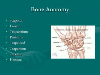
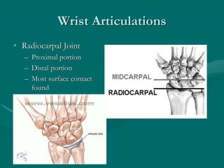
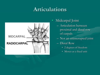
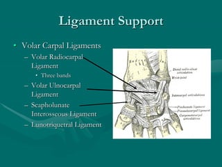
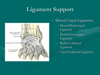
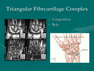
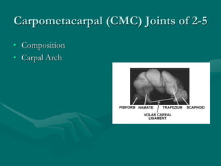
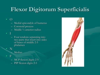
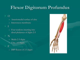
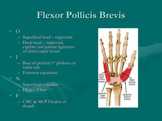
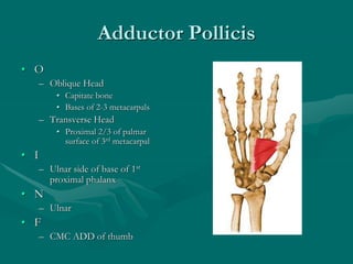
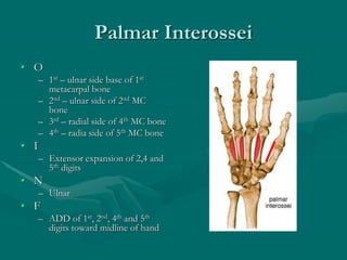
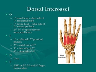
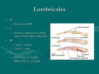
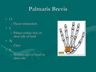
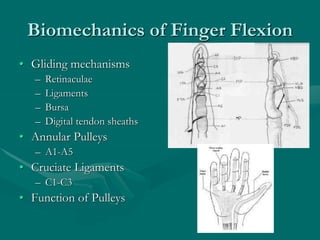
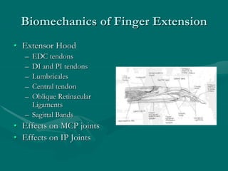
Wrist and Hand Anatomy
Bone Anatomy
• Scapoid
• Lunate
• Triquetrium
• Pisiform
• Trapeziod
• Trapezium
• Capitate
• Hamate
Wrist Articulations
• Radiocarpal Joint
– Proximal portion
– Distal portion
– Most surface contact
found
Articulations
• Midcarpal Joint
– Articulation between
proximal and distal row
of carpals
– Not an uninterupted joint
– Distal Row
• 2 degrees of freedom
• Moves as a fixed unit
Ligament Support
• Volar Carpal Ligaments
– Volar Radiocarpal
Ligament
• Three bands
– Volar Ulnocarpal
Ligament
– Scapholunate
Interosseous Ligament
– Lunotriquetral Ligament
Ligament Support
• Dorsal Carpal Ligaments
– Dorsal Radiocarpal
Ligament
– Dorsal Intercarpal
Ligament
– Radial Collateral
Ligament
– Ulnar Collateral Ligament
Triangular Fibrcartilage Complex
• Composition
• Role
Hand
Carpometacarpal (CMC) Joints of 2-5
• Composition
• Carpal Arch
Carpometacarpal (CMC) Joints of 2-5
• Composition
• Carpal Arch
• Ligament Support
– Transverse Carpal
Ligament
– Dorsal and Palmar CMC
Ligaments
– Dorsal and Palmar
Metacarpal Ligaments
– Metacarpal Interosseous
Ligaments
Carpometacarpal (CMC) Joints of 2-5
• Composition
• Carpal Arch
• Ligament Support
– Transverse Carpal Ligament
– Dorsal and Palmar CMC
Ligaments
– Dorsal and Palmar Metacarpal
Ligaments
– Metacarpal Interosseous
Ligaments
• Movement of CMC Joints
Metacarpophalangeal (MCP) Joints
of 2-5 Fingers
• Ligament Support
– Capsule
– Volar Plate
– Collateral Ligaments
– Motions
Interphalangeal Joints of 2-5 Fingers
• Hinge Joints
• Motions
Thumb
CMC of Thumb
• Saddle Joint
• Ligament Support
– Capsule
– Intermetacarpal Ligament
MCP of Thumb
• Ligament Support
• IP Joint of Thumb
Extrinsic Hand Muscles
Extensor Indicis
• O
– Dorsal surface lower ½ body
of ulna
– Interosseus membrane
• I
– Ulnar side of index finger’s
EDC tendon
• N
– Radial (posterior interosseus)
• F
– MCP and IP Ext of 2nd digit
Extensor Pollicis Longus
• O
– Posterior 1/3 ulna
– Interosseus membrane
• I
– Posterior surface of base of
thumb distal phalanx
• N
– Radial (posterior interosseus)
• F
– CMC, MCP and IP Ext of 1st
digit
Extensor Pollicis Brevis
• O
– Dorsal 2/3 of radius
• I
– Dorsal surface of base of
proximal 1st phalanx
• N
– Radial (posterior interosseus)
• F
– CMC & MCP Ext of thumb
– CMC ABD of thumb
Abductor Pollicis Longus
• O
– Posterior distal 2/3 of ulna
– Posterior middle 1/3 of radius
– Interosseus membrane
• I
– Radial side of base of 1st
metacarpal
• N
– Radial (posterior interosseus)
• F
– CMC ABD & Ext of thumb
Flexor Pollicis Longus
• O
– Anterior middle ½ of radius
– Interosseus membrane
• I
– Palmar surface of base of distal
1st phalanx
• N
– Median (anterior interosseus)
• F
– IP Flexion of thumb
Extensor Digiti Minimi
• O
– Lateral epicondyle of
humerus
• I
– Extensor expansion of 5th
digit
• N
– Radial (posteior
interosseus)
• F
– MCP and IP extension of
5th digit
Flexor Digitorum Superficialis
• O
– Medial epicondyle of humerus
– Coronoid process
– Middle ½ anterior radius
• I
– Four tendons separating into
two parts that insert into sides
of bases of middle 2-5
phalanxes
• N
– Median
• F
– MCP flexion digits 2-5
– PIP flexion digits 2-5
Flexor Digitorum Profundus
• O
– Anteriomedial surface of ulna
– Interosseus membrane
• I
– Four tendons inserting into
distal phalanxes of digits 2-5
• N
– Media 2-3 digits
– Ulna 4-5 digits
• F
– DIP flexion of 2-5 digits
Intrinsic Hand Muscles
Thenar Eminance
Abductor Pollicis Brevis
• O
– Scaphoid tuberosity
– Trapezium ridge
– Transverse carpal ligament
• I
– Lateral base f proximal 1st
phalanx
• N
– Median
• F
– CMC & MCP ABD of thumb
Flexor Pollicis Brevis
• O
– Superficial head – trapezium
– Deep head – trapezoid,
capitate and palmar ligaments
of distal carpal bones
• I
– Base of prximal 1st phalanx on
radial side
– Extensor expansion
• N
– Superficial – median
– Deep – Ulnar
• F
– CMC & MCP Flexion of
thumb
Opponens Pollicis
• O
– Trapezium
– Transverse Carpal
Ligament
• I
– Radial side of 1st
metacarpal shaft
• N
– Median
• F
– Opposition
Intrinsic Hand Muscles
Hypothenar Eminence
Abductor Digiti Minimi
• O
– Pisiform
• I
– Ulnar side base of 5th
proximal phalanx
• N
– Ulnar
• F
– MCP ABD of 5th digit
Opponen Digiti Minimi
• O
– Hook of hamate
– Transverse carpal ligament
• I
– Ulnar border of entire 5th
metacarpal bone
• N
– Ulnar
• F
– MCP flexion & rotation of 5th
digit
Flexor Digiti Minimi
• O
– Hamate bone
– Transverse carpal
ligament
• I
– Ulnar side of proximal 5th
phalanx
• N
– Ulnar
• F
– MCP Flexion of 5th digit
Other Intrinsic Hand
Muscles
Adductor Pollicis
• O
– Oblique Head
• Capitate bone
• Bases of 2-3 metacarpals
– Transverse Head
• Proximal 2/3 of palmar
surface of 3rd metacarpal
• I
– Ulnar side of base of 1st
proximal phalanx
• N
– Ulnar
• F
– CMC ADD of thumb
Palmar Interossei
• O
– 1st – ulnar side base of 1st
metacarpal bone
– 2nd – ulnar side of 2nd MC
bone
– 3rd – radial side of 4th MC bone
– 4th – radia side of 5th MC bone
• I
– Extensor expansion of 2,4 and
5th digits
• N
– Ulnar
• F
– ADD of 1st, 2nd, 4th and 5th
digits toward midline of hand
Dorsal Interossei
• O
– 1st lateral head – ulnar side of
1st metacarpal bone
– 1st medial head – radial side of
2nd metacarpal bone
– 2nd, 3rd, 4th space between
metacarpal bones
• I
– 1st – radial side 2nd proximal
phalanx
– 2nd – radial side of 3rd
– 3rd – ilnar side of 3rd
– 4th – ulnar side of 4th
• N
– Ulnar
• F
– ABD of 2nd, 3rd, and 5th finger
from midline
Lumbricales
• O
– Tendons of FDP
• I
– Extensor expansion on dorsal
aspect of each digits radial side
• N
– 1 and 2 – median
– 3 and 4 – ulnar
• F
– MCP flexion 2-5 digits
– DIP & PIP ext 2-5 digits
Palmaris Brevis
• O
– Flexor retinaculum
• I
– Palmar surface skin on
ulnar side of hand
• N
– Ulnar
• F
– Wrinkles skin of hand on
ulnar side
Biomechanics of Hand
Biomechanics of Finger Flexion
• Gliding mechanisms
– Retinaculae
– Ligaments
– Bursa
– Digital tendon sheaths
• Annular Pulleys
– A1-A5
• Cruciate Ligaments
– C1-C3
• Function of Pulleys
Biomechanics of Finger Extension
• Extensor Hood
– EDC tendons
– DI and PI tendons
– Lumbricales
– Central tendon
– Oblique Retinacular
Ligaments
– Sagittal Bands
• Effects on MCP joints
• Effects on IP Joints

WristandHandAnatomy (1).ppt

  • 1.
  • 2.
    Bone Anatomy • Scapoid •Lunate • Triquetrium • Pisiform • Trapeziod • Trapezium • Capitate • Hamate
  • 3.
    Wrist Articulations • RadiocarpalJoint – Proximal portion – Distal portion – Most surface contact found
  • 4.
    Articulations • Midcarpal Joint –Articulation between proximal and distal row of carpals – Not an uninterupted joint – Distal Row • 2 degrees of freedom • Moves as a fixed unit
  • 5.
    Ligament Support • VolarCarpal Ligaments – Volar Radiocarpal Ligament • Three bands – Volar Ulnocarpal Ligament – Scapholunate Interosseous Ligament – Lunotriquetral Ligament
  • 6.
    Ligament Support • DorsalCarpal Ligaments – Dorsal Radiocarpal Ligament – Dorsal Intercarpal Ligament – Radial Collateral Ligament – Ulnar Collateral Ligament
  • 7.
  • 8.
  • 9.
    Carpometacarpal (CMC) Jointsof 2-5 • Composition • Carpal Arch
  • 10.
    Carpometacarpal (CMC) Jointsof 2-5 • Composition • Carpal Arch • Ligament Support – Transverse Carpal Ligament – Dorsal and Palmar CMC Ligaments – Dorsal and Palmar Metacarpal Ligaments – Metacarpal Interosseous Ligaments
  • 11.
    Carpometacarpal (CMC) Jointsof 2-5 • Composition • Carpal Arch • Ligament Support – Transverse Carpal Ligament – Dorsal and Palmar CMC Ligaments – Dorsal and Palmar Metacarpal Ligaments – Metacarpal Interosseous Ligaments • Movement of CMC Joints
  • 12.
    Metacarpophalangeal (MCP) Joints of2-5 Fingers • Ligament Support – Capsule – Volar Plate – Collateral Ligaments – Motions
  • 13.
    Interphalangeal Joints of2-5 Fingers • Hinge Joints • Motions
  • 14.
  • 15.
    CMC of Thumb •Saddle Joint • Ligament Support – Capsule – Intermetacarpal Ligament
  • 16.
    MCP of Thumb •Ligament Support • IP Joint of Thumb
  • 17.
  • 19.
    Extensor Indicis • O –Dorsal surface lower ½ body of ulna – Interosseus membrane • I – Ulnar side of index finger’s EDC tendon • N – Radial (posterior interosseus) • F – MCP and IP Ext of 2nd digit
  • 20.
    Extensor Pollicis Longus •O – Posterior 1/3 ulna – Interosseus membrane • I – Posterior surface of base of thumb distal phalanx • N – Radial (posterior interosseus) • F – CMC, MCP and IP Ext of 1st digit
  • 21.
    Extensor Pollicis Brevis •O – Dorsal 2/3 of radius • I – Dorsal surface of base of proximal 1st phalanx • N – Radial (posterior interosseus) • F – CMC & MCP Ext of thumb – CMC ABD of thumb
  • 22.
    Abductor Pollicis Longus •O – Posterior distal 2/3 of ulna – Posterior middle 1/3 of radius – Interosseus membrane • I – Radial side of base of 1st metacarpal • N – Radial (posterior interosseus) • F – CMC ABD & Ext of thumb
  • 23.
    Flexor Pollicis Longus •O – Anterior middle ½ of radius – Interosseus membrane • I – Palmar surface of base of distal 1st phalanx • N – Median (anterior interosseus) • F – IP Flexion of thumb
  • 24.
    Extensor Digiti Minimi •O – Lateral epicondyle of humerus • I – Extensor expansion of 5th digit • N – Radial (posteior interosseus) • F – MCP and IP extension of 5th digit
  • 25.
    Flexor Digitorum Superficialis •O – Medial epicondyle of humerus – Coronoid process – Middle ½ anterior radius • I – Four tendons separating into two parts that insert into sides of bases of middle 2-5 phalanxes • N – Median • F – MCP flexion digits 2-5 – PIP flexion digits 2-5
  • 26.
    Flexor Digitorum Profundus •O – Anteriomedial surface of ulna – Interosseus membrane • I – Four tendons inserting into distal phalanxes of digits 2-5 • N – Media 2-3 digits – Ulna 4-5 digits • F – DIP flexion of 2-5 digits
  • 27.
  • 28.
    Abductor Pollicis Brevis •O – Scaphoid tuberosity – Trapezium ridge – Transverse carpal ligament • I – Lateral base f proximal 1st phalanx • N – Median • F – CMC & MCP ABD of thumb
  • 29.
    Flexor Pollicis Brevis •O – Superficial head – trapezium – Deep head – trapezoid, capitate and palmar ligaments of distal carpal bones • I – Base of prximal 1st phalanx on radial side – Extensor expansion • N – Superficial – median – Deep – Ulnar • F – CMC & MCP Flexion of thumb
  • 30.
    Opponens Pollicis • O –Trapezium – Transverse Carpal Ligament • I – Radial side of 1st metacarpal shaft • N – Median • F – Opposition
  • 31.
  • 32.
    Abductor Digiti Minimi •O – Pisiform • I – Ulnar side base of 5th proximal phalanx • N – Ulnar • F – MCP ABD of 5th digit
  • 33.
    Opponen Digiti Minimi •O – Hook of hamate – Transverse carpal ligament • I – Ulnar border of entire 5th metacarpal bone • N – Ulnar • F – MCP flexion & rotation of 5th digit
  • 34.
    Flexor Digiti Minimi •O – Hamate bone – Transverse carpal ligament • I – Ulnar side of proximal 5th phalanx • N – Ulnar • F – MCP Flexion of 5th digit
  • 35.
  • 36.
    Adductor Pollicis • O –Oblique Head • Capitate bone • Bases of 2-3 metacarpals – Transverse Head • Proximal 2/3 of palmar surface of 3rd metacarpal • I – Ulnar side of base of 1st proximal phalanx • N – Ulnar • F – CMC ADD of thumb
  • 37.
    Palmar Interossei • O –1st – ulnar side base of 1st metacarpal bone – 2nd – ulnar side of 2nd MC bone – 3rd – radial side of 4th MC bone – 4th – radia side of 5th MC bone • I – Extensor expansion of 2,4 and 5th digits • N – Ulnar • F – ADD of 1st, 2nd, 4th and 5th digits toward midline of hand
  • 38.
    Dorsal Interossei • O –1st lateral head – ulnar side of 1st metacarpal bone – 1st medial head – radial side of 2nd metacarpal bone – 2nd, 3rd, 4th space between metacarpal bones • I – 1st – radial side 2nd proximal phalanx – 2nd – radial side of 3rd – 3rd – ilnar side of 3rd – 4th – ulnar side of 4th • N – Ulnar • F – ABD of 2nd, 3rd, and 5th finger from midline
  • 39.
    Lumbricales • O – Tendonsof FDP • I – Extensor expansion on dorsal aspect of each digits radial side • N – 1 and 2 – median – 3 and 4 – ulnar • F – MCP flexion 2-5 digits – DIP & PIP ext 2-5 digits
  • 40.
    Palmaris Brevis • O –Flexor retinaculum • I – Palmar surface skin on ulnar side of hand • N – Ulnar • F – Wrinkles skin of hand on ulnar side
  • 41.
  • 42.
    Biomechanics of FingerFlexion • Gliding mechanisms – Retinaculae – Ligaments – Bursa – Digital tendon sheaths • Annular Pulleys – A1-A5 • Cruciate Ligaments – C1-C3 • Function of Pulleys
  • 43.
    Biomechanics of FingerExtension • Extensor Hood – EDC tendons – DI and PI tendons – Lumbricales – Central tendon – Oblique Retinacular Ligaments – Sagittal Bands • Effects on MCP joints • Effects on IP Joints