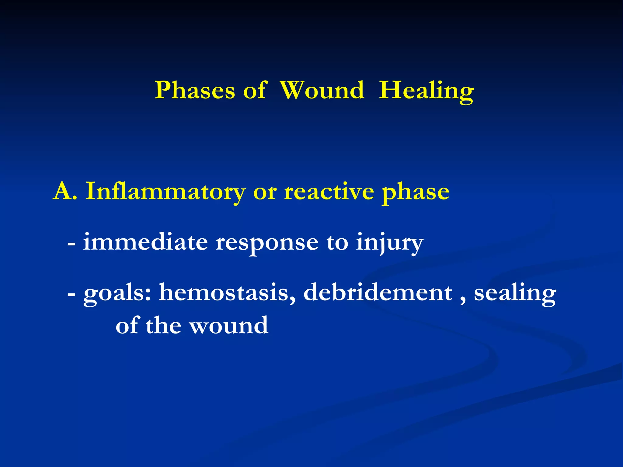
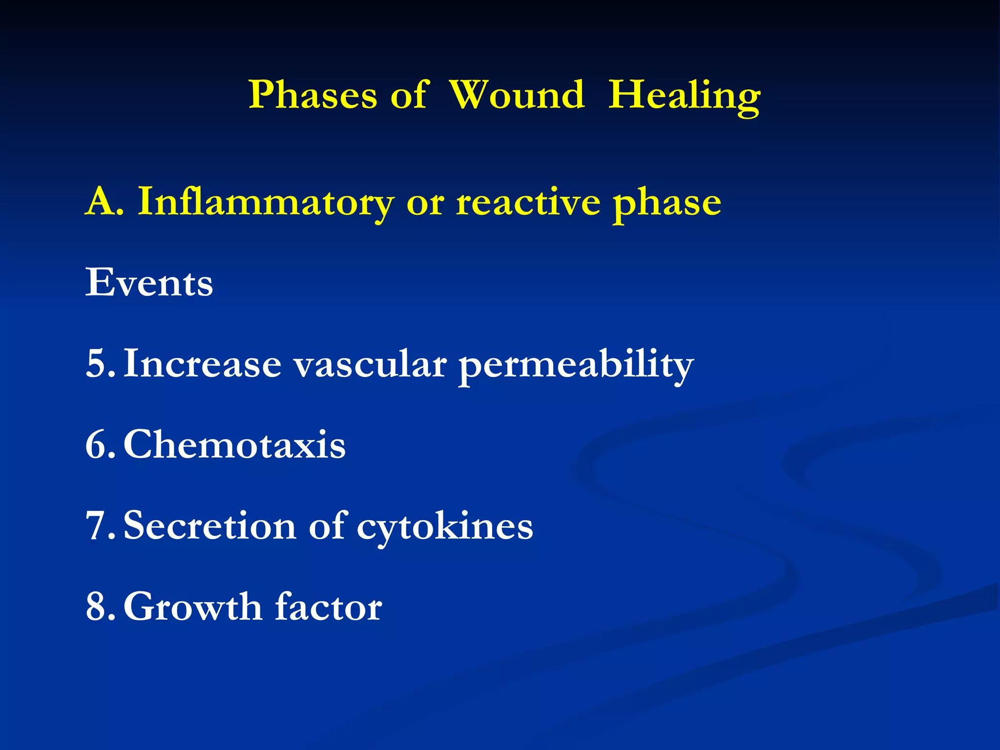
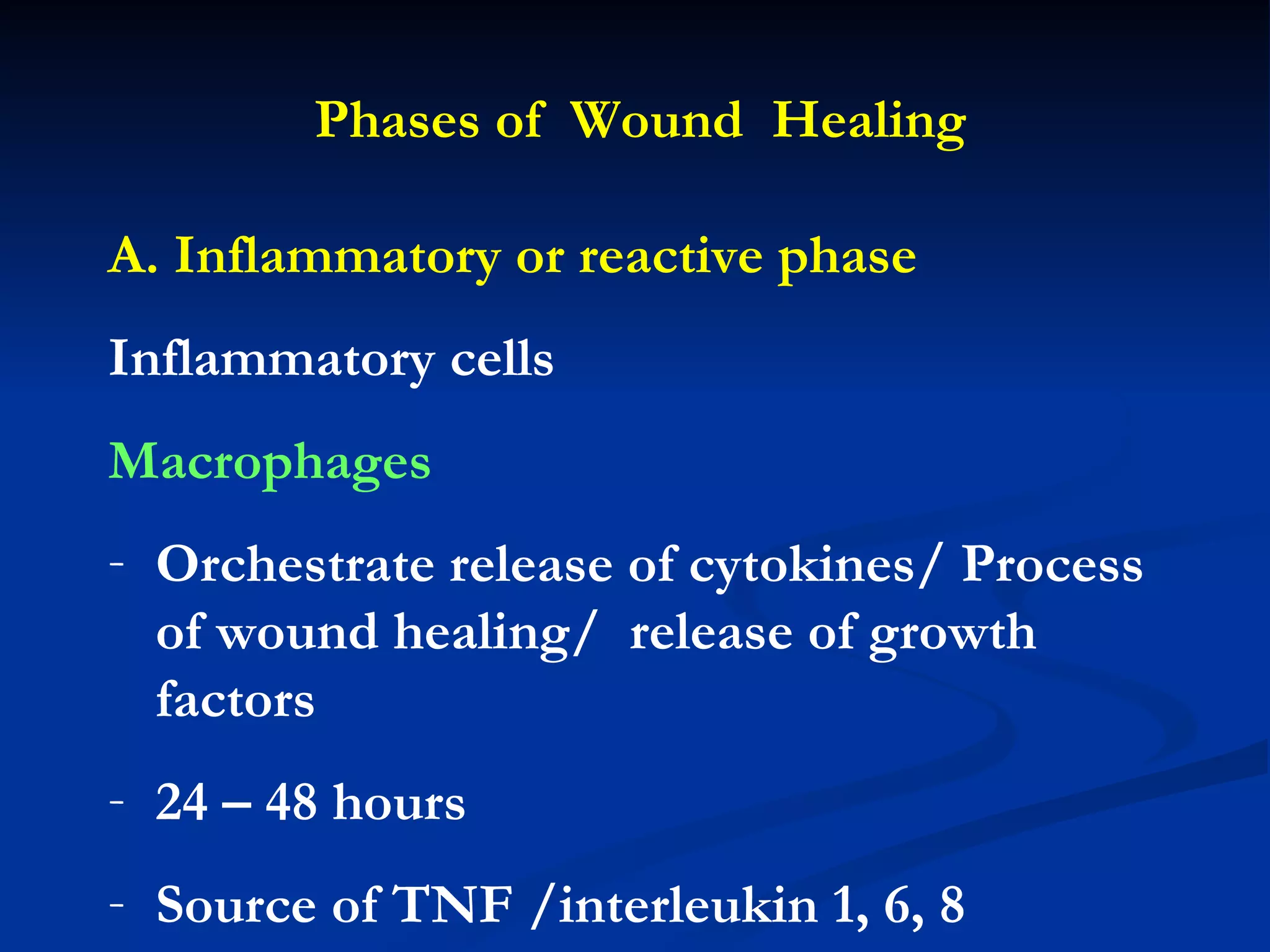
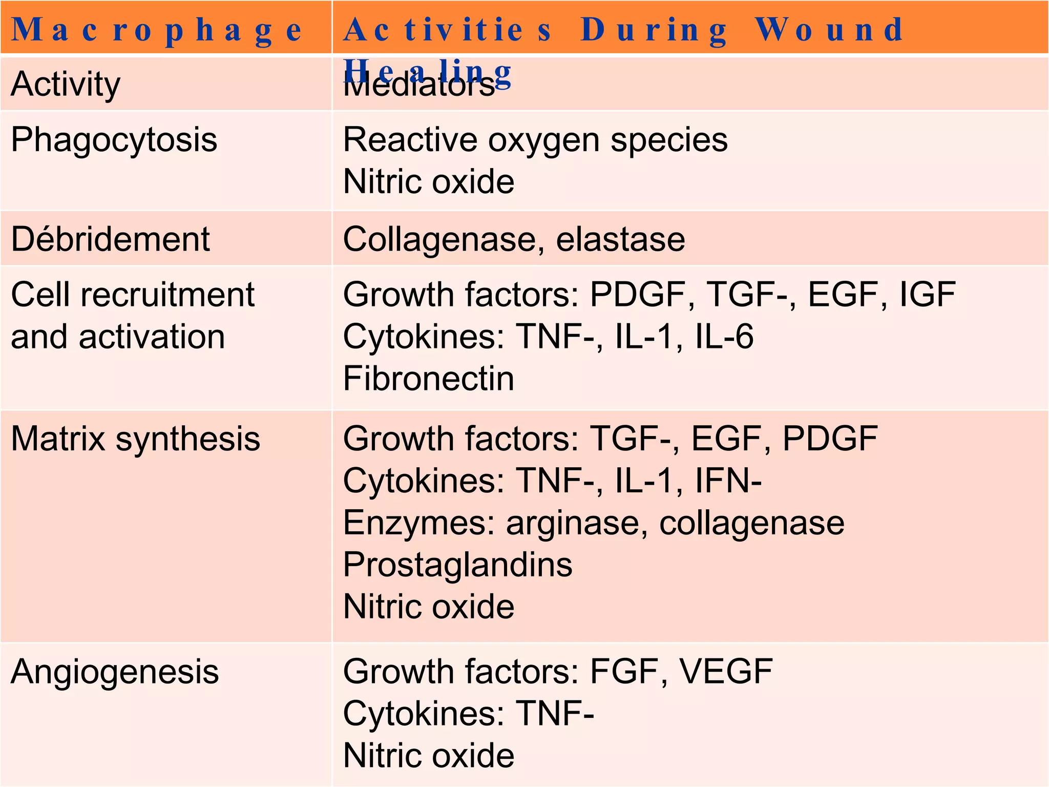
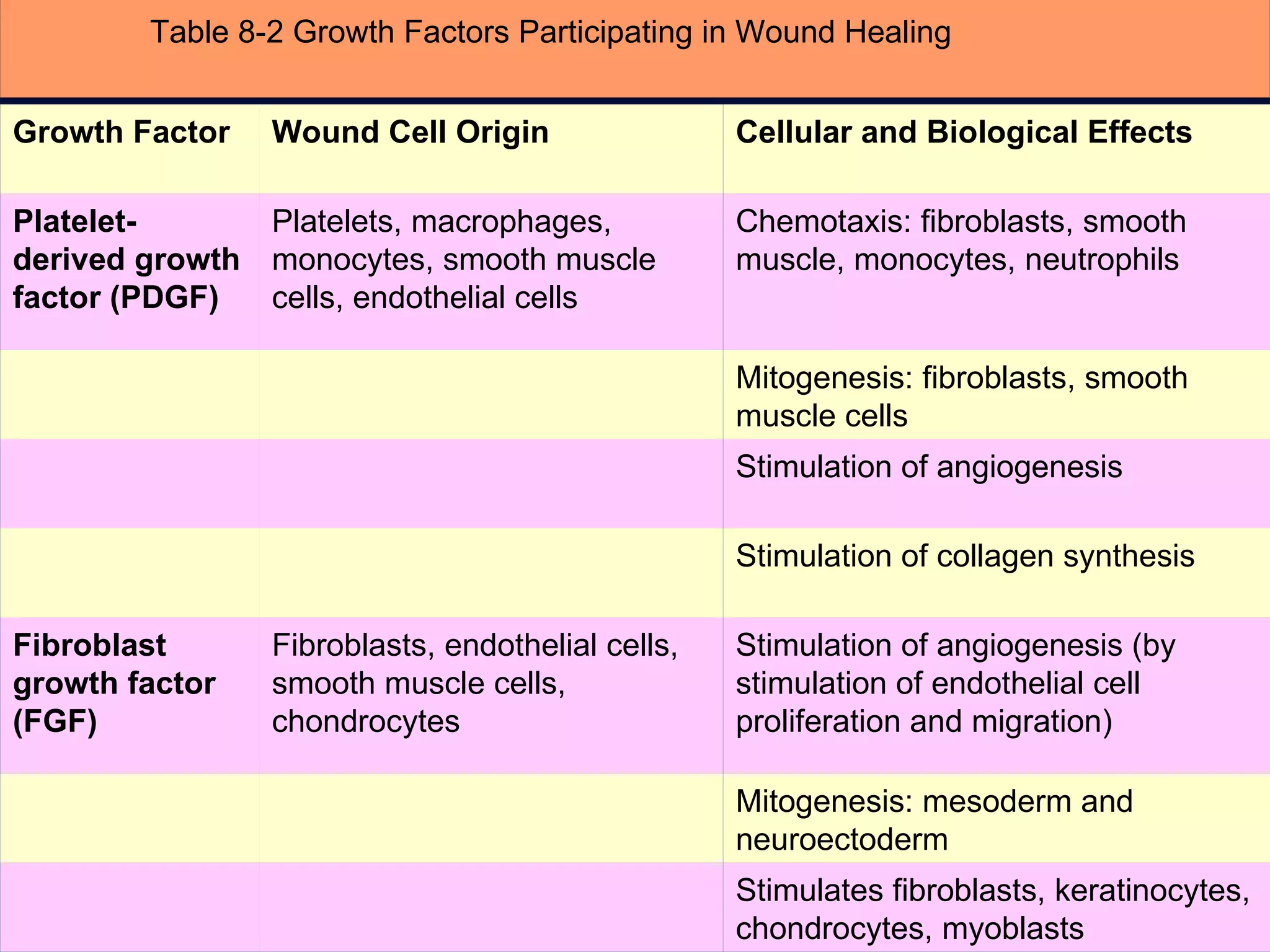
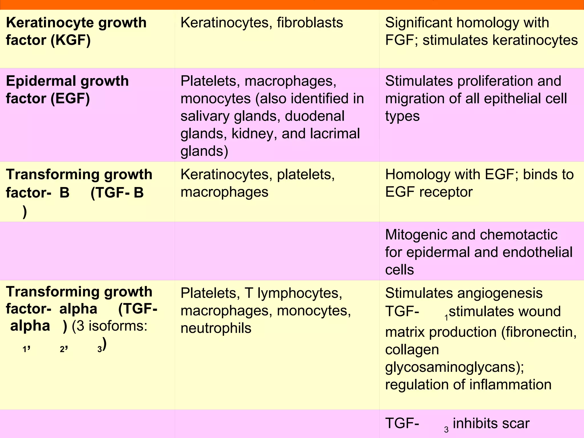
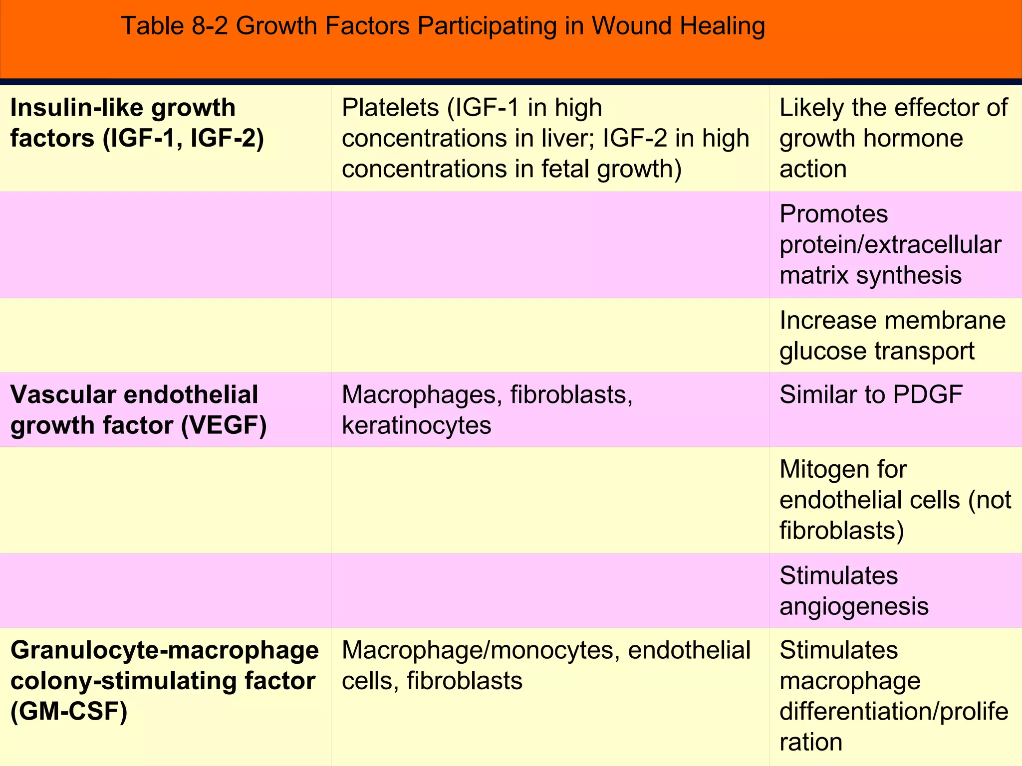
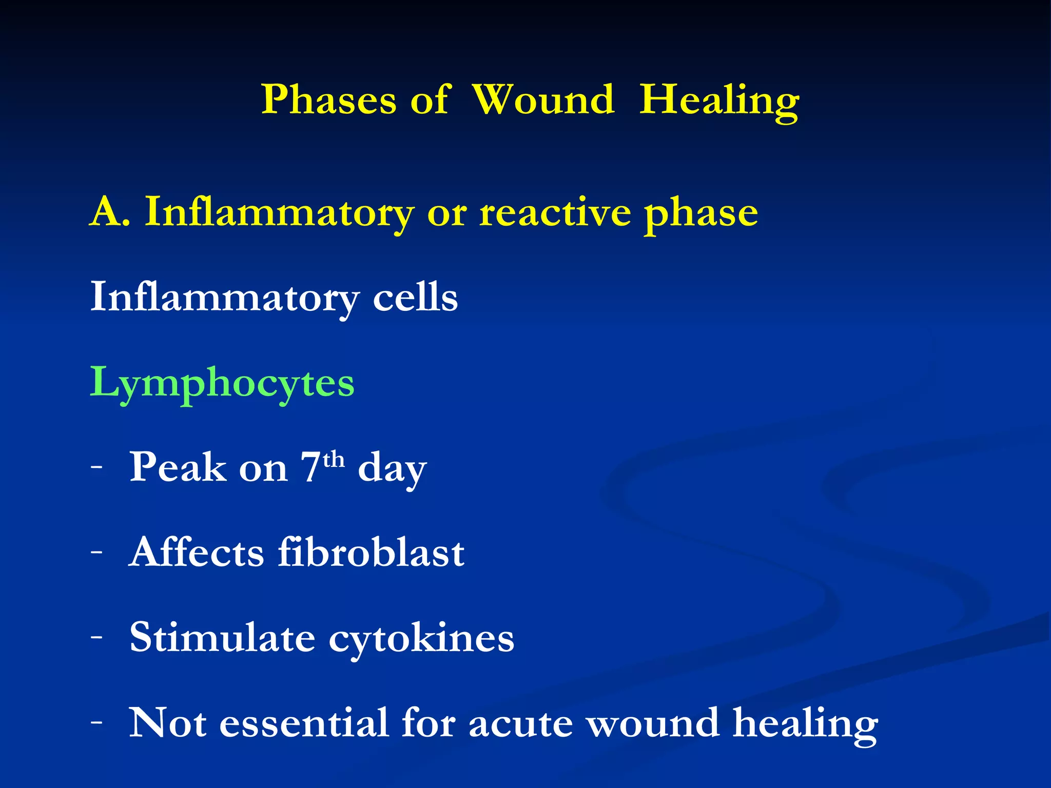
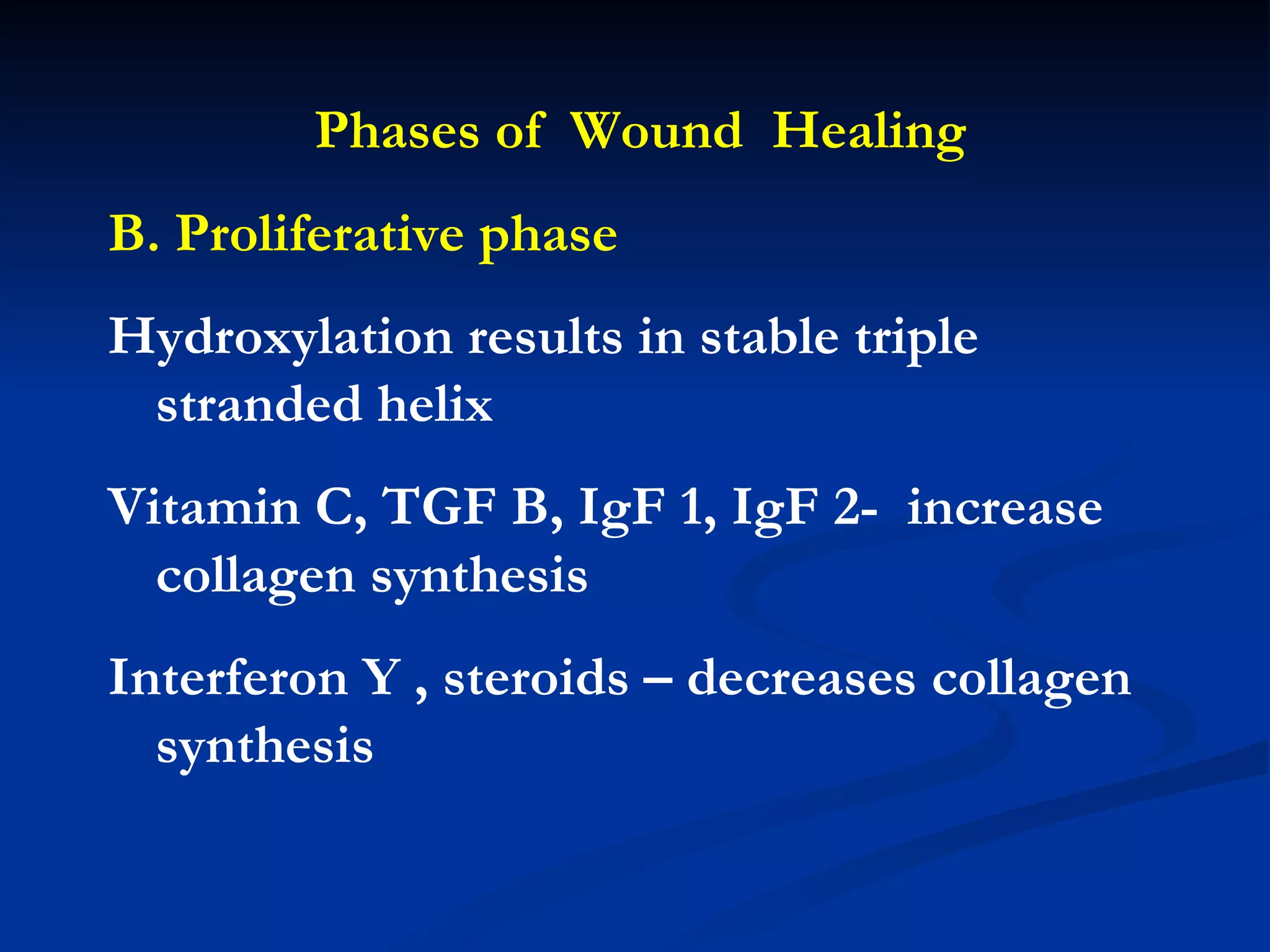
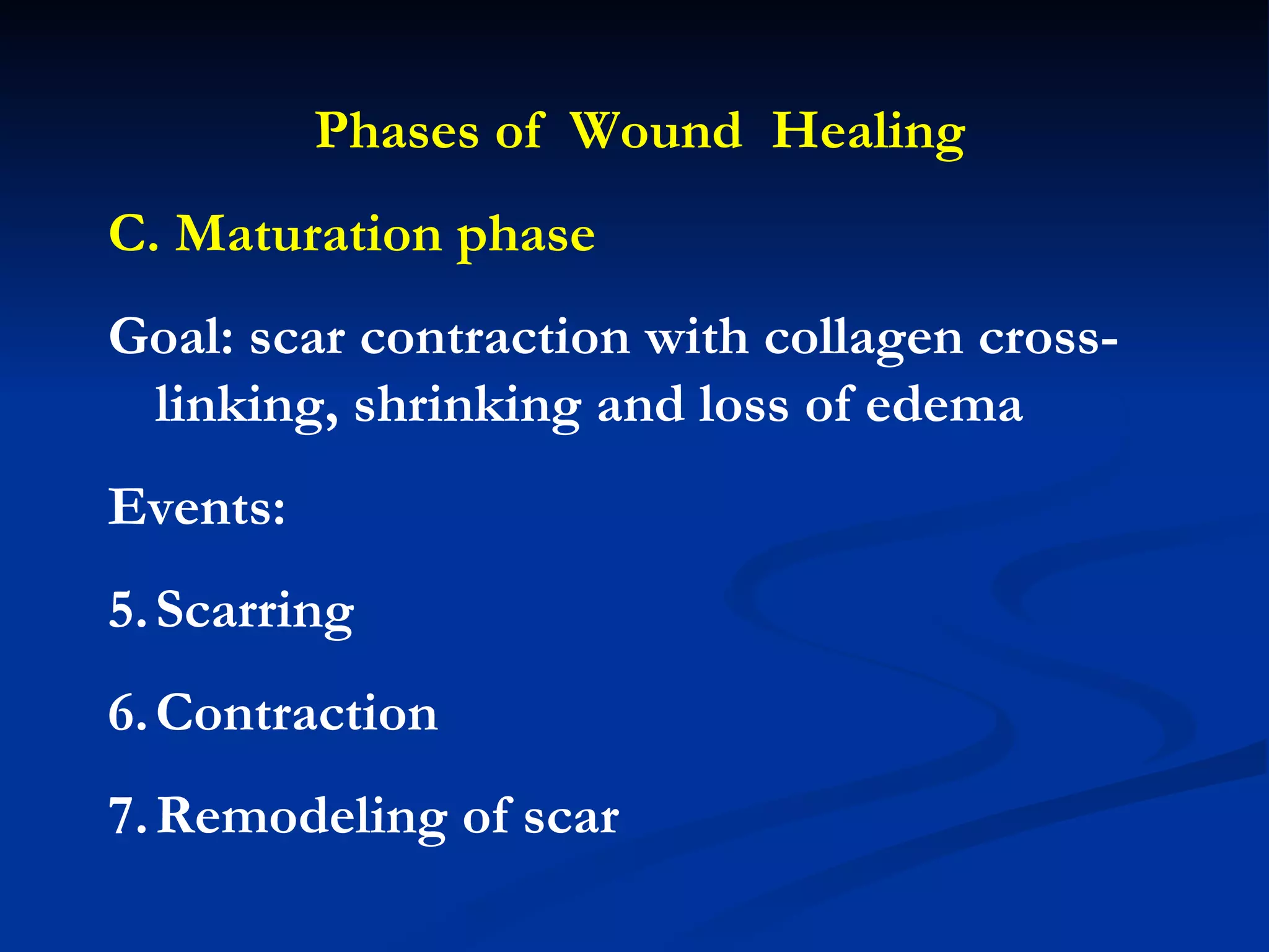
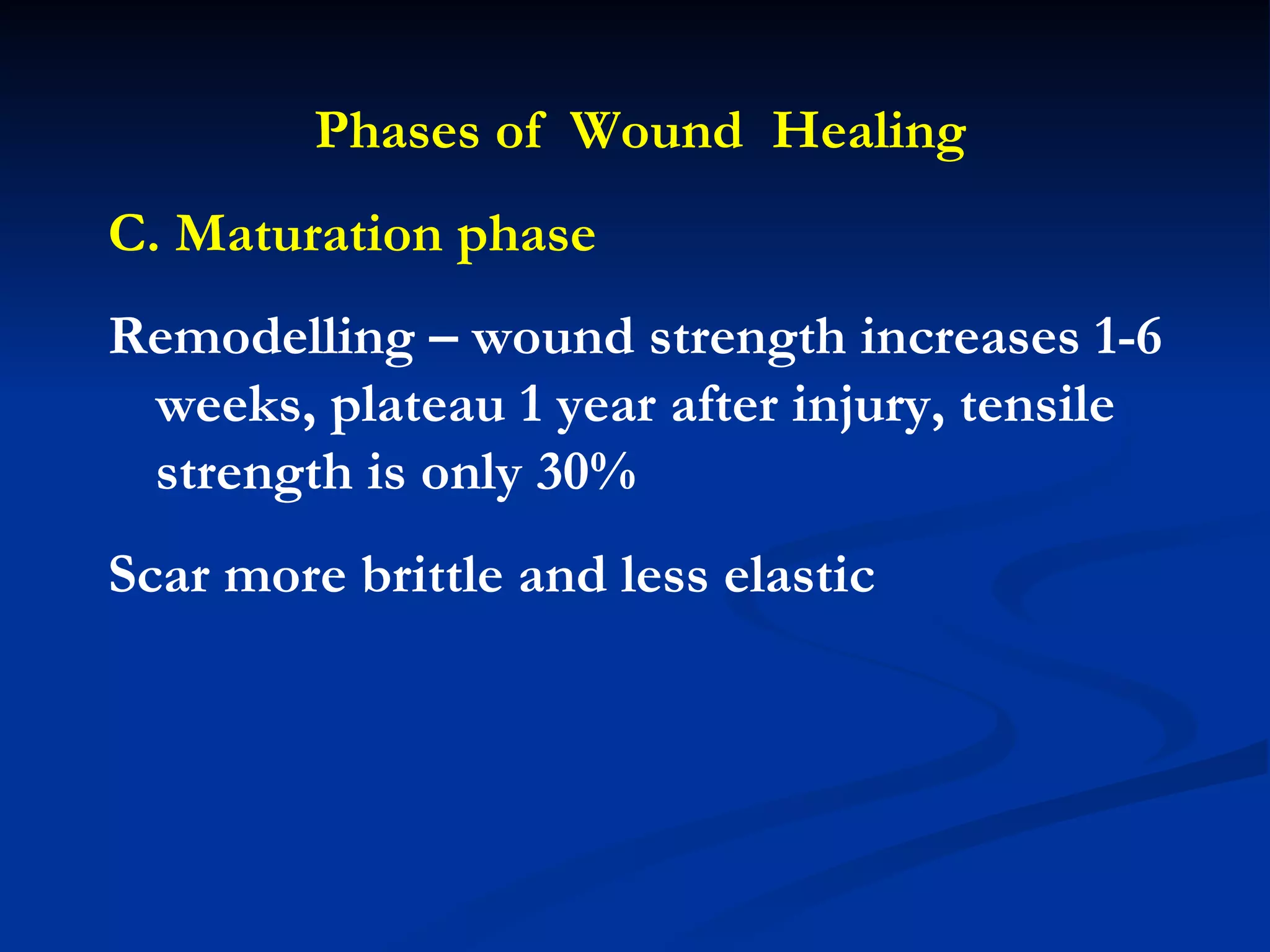
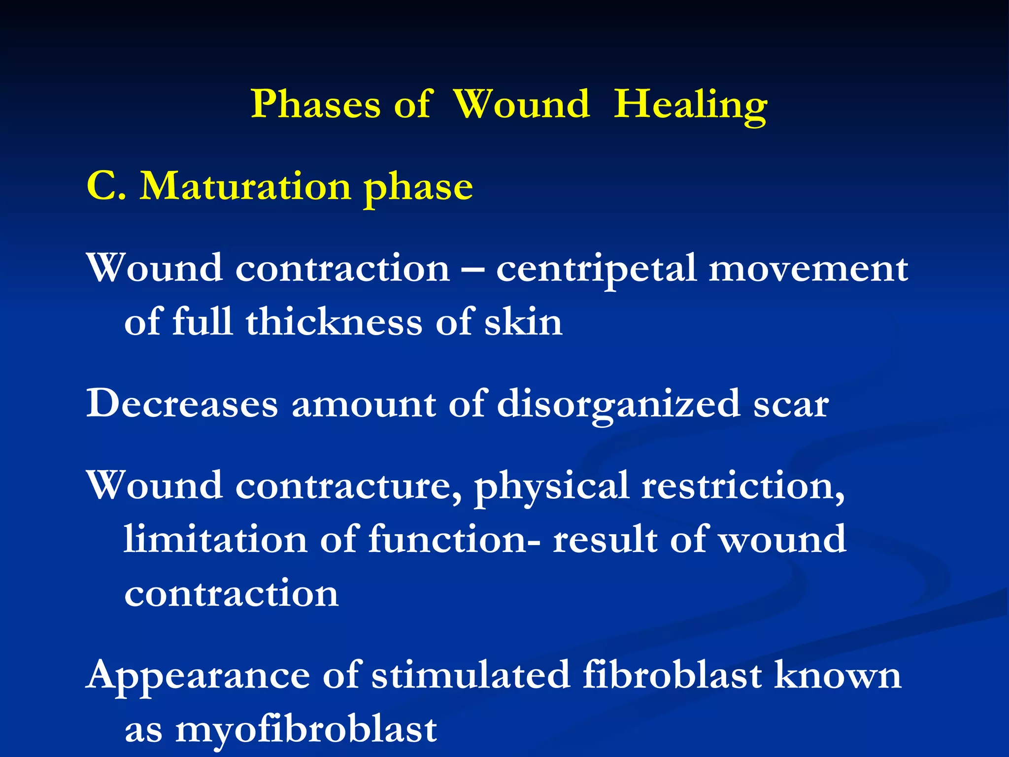
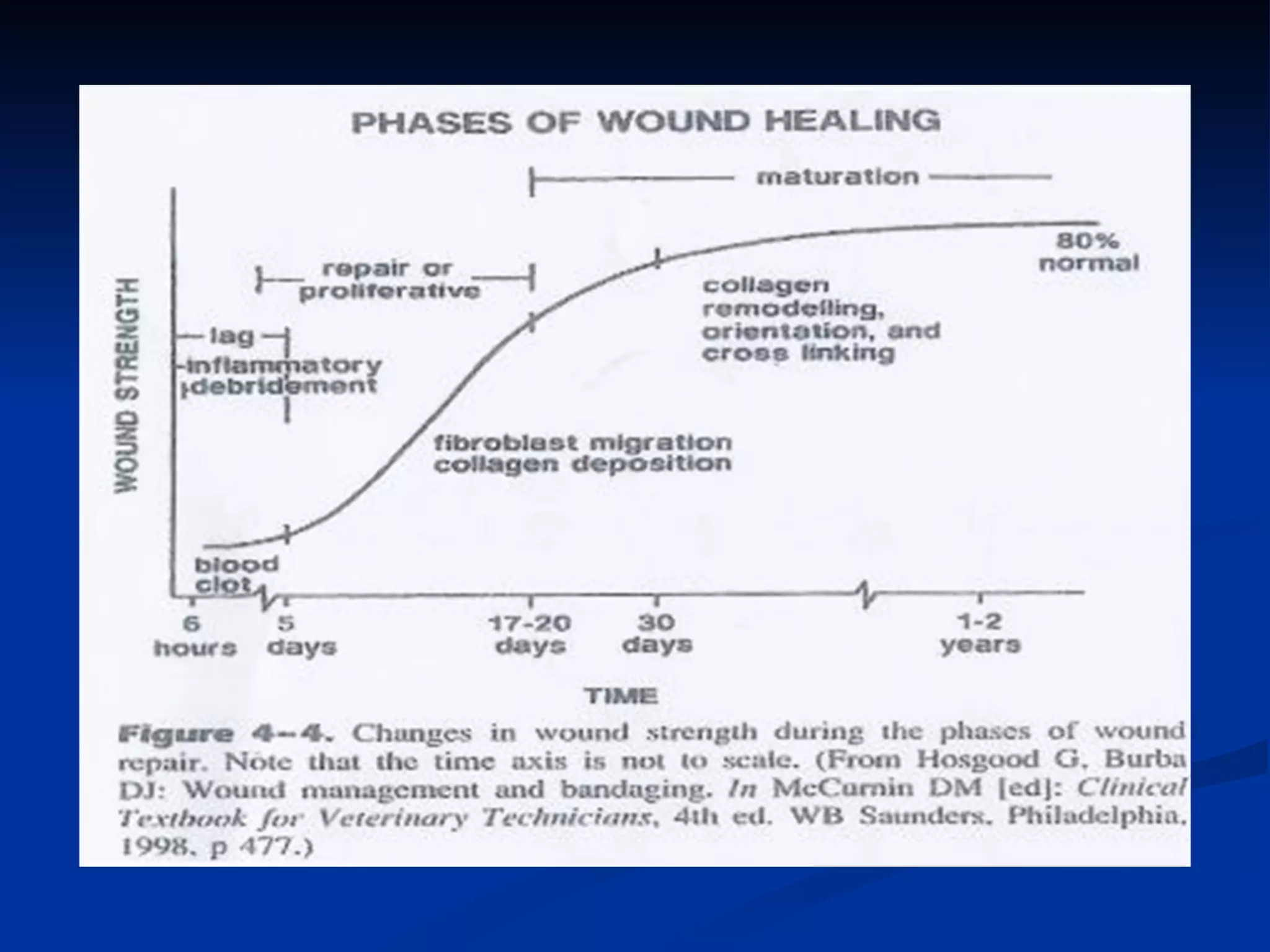
Wound Healing Wallace Medina, MD, FPCS,FPSGS,FPALES
Terminology W ound repair-  ability to restore normal function and structure R egeneration  – perfect restoration, no scar formation * All tissues proceed through the same series of events
Terminology A cute wounds -  orderly and timely reparative process C hronic wounds  – no restoration of functional integrity, stops at inflammatory phase
Types of  Wound  Closure Primary or first intention Secondary or spontaneous Tertiary or delayed primary
Phases of  Wound  Healing A. Inflammatory or reactive phase - immediate response to injury - goals: hemostasis, debridement , sealing  of the wound
Phases of  Wound  Healing A. Inflammatory or reactive phase Events Increase vascular permeability Chemotaxis Secretion of cytokines Growth factor
Phases of  Wound  Healing A. Inflammatory or reactive phase Inflammatory cells PMN  Migration of PMN stops when wound contamination has been controlled Don’t survive more than 24 hours Increase contamination stimulates PMN resulting to delayed wound healing and destruction of tissues. Not essential for wound healing
Phases of  Wound  Healing A. Inflammatory or reactive phase Inflammatory cells Macrophages Orchestrate release of cytokines/ Process of wound healing/  release of growth factors 24 – 48 hours Source of TNF /interleukin 1, 6, 8
Growth factors: FGF, VEGF Cytokines: TNF-  Nitric oxide Angiogenesis Growth factors: TGF-, EGF, PDGF Cytokines: TNF-, IL-1, IFN- Enzymes: arginase, collagenase Prostaglandins Nitric oxide Matrix synthesis Growth factors: PDGF, TGF-, EGF, IGF Cytokines: TNF-, IL-1, IL-6  Fibronectin Cell recruitment and activation Collagenase, elastase Débridement Reactive oxygen species  Nitric oxide Phagocytosis Mediators Activity Activities During Wound Healing Macrophage
Table 8-2 Growth Factors Participating in Wound Healing Stimulates fibroblasts, keratinocytes, chondrocytes, myoblasts     Mitogenesis: mesoderm and neuroectoderm     Stimulation of angiogenesis (by stimulation of endothelial cell proliferation and migration) Fibroblasts, endothelial cells, smooth muscle cells, chondrocytes Fibroblast growth factor (FGF)   Stimulation of collagen synthesis     Stimulation of angiogenesis     Mitogenesis: fibroblasts, smooth muscle cells     Chemotaxis: fibroblasts, smooth muscle, monocytes, neutrophils Platelets, macrophages, monocytes, smooth muscle cells, endothelial cells Platelet-derived growth factor (PDGF)   Cellular and Biological Effects Wound Cell Origin Growth Factor
Table 8-2 Growth Factors Participating in Wound Healing TGF-        3  inhibits scar formation     Stimulates angiogenesis TGF-        1 stimulates wound matrix production (fibronectin, collagen glycosaminoglycans); regulation of inflammation   Platelets, T lymphocytes, macrophages, monocytes, neutrophils Transforming growth factor-  alpha       (TGF-   alpha    )  (3 isoforms:        1 ,        2 ,        3 )   Mitogenic and chemotactic for epidermal and endothelial cells     Homology with EGF; binds to EGF receptor Keratinocytes, platelets, macrophages Transforming growth factor-  B        (TGF- B       )   Stimulates proliferation and migration of all epithelial cell types Platelets, macrophages, monocytes (also identified in salivary glands, duodenal glands, kidney, and lacrimal glands) Epidermal growth factor (EGF)   Significant homology with FGF; stimulates keratinocytes Keratinocytes, fibroblasts Keratinocyte growth factor (KGF)  
Table 8-2 Growth Factors Participating in Wound Healing Stimulates macrophage differentiation/proliferation Macrophage/monocytes, endothelial cells, fibroblasts Granulocyte-macrophage colony-stimulating factor (GM-CSF)   Stimulates angiogenesis     Mitogen for endothelial cells (not fibroblasts)     Similar to PDGF Macrophages, fibroblasts, keratinocytes Vascular endothelial growth factor (VEGF)   Increase membrane glucose transport     Promotes protein/extracellular matrix synthesis     Likely the effector of growth hormone action Platelets (IGF-1 in high concentrations in liver; IGF-2 in high concentrations in fetal growth) Insulin-like growth factors (IGF-1, IGF-2)  
Phases of  Wound  Healing A. Inflammatory or reactive phase Inflammatory cells Lymphocytes Peak on 7 th  day Affects fibroblast Stimulate cytokines Not essential for acute wound healing
Phases of  Wound  Healing B. Proliferative phase Goal: granulation tissue formation Events:  Angiogenesis Fibroplasia Epithelization
Phases of  Wound  Healing B. Proliferative phase Decrease collagen synthesis at 4 weeks after injury Epithelization begins hours after injury, sealed by clot then covered by epithelial eells, establishment of basement membrane
Phases of  Wound  Healing B. Proliferative phase Extracellular matrix  Scaffold for cellular migration Composed of fibrin, fibrinogen, fibronectin, vitronectin Fibronectin and type 3 collagen = early matrix Type 1 collagen – wound strength later
Phases of  Wound  Healing B. Proliferative phase Collagen – 25% total protein Type 1 found in skin and bone -  most common Adults – 80% type 1, 20% type 3 Neonates – type 3 predominates
Phases of  Wound  Healing B. Proliferative phase Hydroxylation results in stable triple stranded helix Vitamin C, TGF B, IgF 1, IgF 2-  increase collagen synthesis Interferon Y , steroids – decreases collagen synthesis
Phases of  Wound  Healing C. Maturation phase Goal: scar contraction with collagen cross-linking, shrinking and loss of edema Events:  Scarring Contraction Remodeling of scar
Phases of  Wound  Healing C. Maturation phase Remodelling – wound strength increases 1-6 weeks, plateau 1 year after injury, tensile strength is only 30% Scar more brittle and less elastic
Phases of  Wound  Healing C. Maturation phase Wound contraction – centripetal movement of full thickness of skin Decreases amount of disorganized scar Wound contracture, physical restriction, limitation of function- result of wound contraction Appearance of stimulated fibroblast known as myofibroblast
 
Factors affecting wound healing
Proliferative Scar Collagen deposition versus Collagen degradation Keloid and hypertrophic scar-excessive collagen deposition Keloid – beyond borders , darkly pigmented individuals, genetic predisposition, clavicle, trunk, upper extremity, face
Wound Dressing Two concepts Occlusion - increase rate of epithelization – acidic pH, low oxygen tension- good environment for fibroblast and granulation tissue B. Absorption
Types of Wound Dressing Non Adherent Absorptive Occlusive Creams/ointment/solution
Covering a wound with a dressing mimics the barrier role of epithelium  Table 8-7 Desired Characteristics of Wound Dressings Convenience Cost-effectiveness Nontraumatic removal Safety Permeability to gas Nonallergenic and nonirritating Odor control Pain control Conformability Promote wound healing (maintain moist environment)

Wound Healing Lec

  • 1.
    Wound Healing WallaceMedina, MD, FPCS,FPSGS,FPALES
  • 2.
    Terminology W oundrepair- ability to restore normal function and structure R egeneration – perfect restoration, no scar formation * All tissues proceed through the same series of events
  • 3.
    Terminology A cutewounds - orderly and timely reparative process C hronic wounds – no restoration of functional integrity, stops at inflammatory phase
  • 4.
    Types of Wound Closure Primary or first intention Secondary or spontaneous Tertiary or delayed primary
  • 5.
    Phases of Wound Healing A. Inflammatory or reactive phase - immediate response to injury - goals: hemostasis, debridement , sealing of the wound
  • 6.
    Phases of Wound Healing A. Inflammatory or reactive phase Events Increase vascular permeability Chemotaxis Secretion of cytokines Growth factor
  • 7.
    Phases of Wound Healing A. Inflammatory or reactive phase Inflammatory cells PMN Migration of PMN stops when wound contamination has been controlled Don’t survive more than 24 hours Increase contamination stimulates PMN resulting to delayed wound healing and destruction of tissues. Not essential for wound healing
  • 8.
    Phases of Wound Healing A. Inflammatory or reactive phase Inflammatory cells Macrophages Orchestrate release of cytokines/ Process of wound healing/ release of growth factors 24 – 48 hours Source of TNF /interleukin 1, 6, 8
  • 9.
    Growth factors: FGF,VEGF Cytokines: TNF- Nitric oxide Angiogenesis Growth factors: TGF-, EGF, PDGF Cytokines: TNF-, IL-1, IFN- Enzymes: arginase, collagenase Prostaglandins Nitric oxide Matrix synthesis Growth factors: PDGF, TGF-, EGF, IGF Cytokines: TNF-, IL-1, IL-6 Fibronectin Cell recruitment and activation Collagenase, elastase Débridement Reactive oxygen species Nitric oxide Phagocytosis Mediators Activity Activities During Wound Healing Macrophage
  • 10.
    Table 8-2 GrowthFactors Participating in Wound Healing Stimulates fibroblasts, keratinocytes, chondrocytes, myoblasts     Mitogenesis: mesoderm and neuroectoderm     Stimulation of angiogenesis (by stimulation of endothelial cell proliferation and migration) Fibroblasts, endothelial cells, smooth muscle cells, chondrocytes Fibroblast growth factor (FGF)   Stimulation of collagen synthesis     Stimulation of angiogenesis     Mitogenesis: fibroblasts, smooth muscle cells     Chemotaxis: fibroblasts, smooth muscle, monocytes, neutrophils Platelets, macrophages, monocytes, smooth muscle cells, endothelial cells Platelet-derived growth factor (PDGF)   Cellular and Biological Effects Wound Cell Origin Growth Factor
  • 11.
    Table 8-2 GrowthFactors Participating in Wound Healing TGF-       3 inhibits scar formation     Stimulates angiogenesis TGF-       1 stimulates wound matrix production (fibronectin, collagen glycosaminoglycans); regulation of inflammation   Platelets, T lymphocytes, macrophages, monocytes, neutrophils Transforming growth factor- alpha       (TGF-  alpha    ) (3 isoforms:       1 ,       2 ,       3 )   Mitogenic and chemotactic for epidermal and endothelial cells     Homology with EGF; binds to EGF receptor Keratinocytes, platelets, macrophages Transforming growth factor- B       (TGF- B      )   Stimulates proliferation and migration of all epithelial cell types Platelets, macrophages, monocytes (also identified in salivary glands, duodenal glands, kidney, and lacrimal glands) Epidermal growth factor (EGF)   Significant homology with FGF; stimulates keratinocytes Keratinocytes, fibroblasts Keratinocyte growth factor (KGF)  
  • 12.
    Table 8-2 GrowthFactors Participating in Wound Healing Stimulates macrophage differentiation/proliferation Macrophage/monocytes, endothelial cells, fibroblasts Granulocyte-macrophage colony-stimulating factor (GM-CSF)   Stimulates angiogenesis     Mitogen for endothelial cells (not fibroblasts)     Similar to PDGF Macrophages, fibroblasts, keratinocytes Vascular endothelial growth factor (VEGF)   Increase membrane glucose transport     Promotes protein/extracellular matrix synthesis     Likely the effector of growth hormone action Platelets (IGF-1 in high concentrations in liver; IGF-2 in high concentrations in fetal growth) Insulin-like growth factors (IGF-1, IGF-2)  
  • 13.
    Phases of Wound Healing A. Inflammatory or reactive phase Inflammatory cells Lymphocytes Peak on 7 th day Affects fibroblast Stimulate cytokines Not essential for acute wound healing
  • 14.
    Phases of Wound Healing B. Proliferative phase Goal: granulation tissue formation Events: Angiogenesis Fibroplasia Epithelization
  • 15.
    Phases of Wound Healing B. Proliferative phase Decrease collagen synthesis at 4 weeks after injury Epithelization begins hours after injury, sealed by clot then covered by epithelial eells, establishment of basement membrane
  • 16.
    Phases of Wound Healing B. Proliferative phase Extracellular matrix Scaffold for cellular migration Composed of fibrin, fibrinogen, fibronectin, vitronectin Fibronectin and type 3 collagen = early matrix Type 1 collagen – wound strength later
  • 17.
    Phases of Wound Healing B. Proliferative phase Collagen – 25% total protein Type 1 found in skin and bone - most common Adults – 80% type 1, 20% type 3 Neonates – type 3 predominates
  • 18.
    Phases of Wound Healing B. Proliferative phase Hydroxylation results in stable triple stranded helix Vitamin C, TGF B, IgF 1, IgF 2- increase collagen synthesis Interferon Y , steroids – decreases collagen synthesis
  • 19.
    Phases of Wound Healing C. Maturation phase Goal: scar contraction with collagen cross-linking, shrinking and loss of edema Events: Scarring Contraction Remodeling of scar
  • 20.
    Phases of Wound Healing C. Maturation phase Remodelling – wound strength increases 1-6 weeks, plateau 1 year after injury, tensile strength is only 30% Scar more brittle and less elastic
  • 21.
    Phases of Wound Healing C. Maturation phase Wound contraction – centripetal movement of full thickness of skin Decreases amount of disorganized scar Wound contracture, physical restriction, limitation of function- result of wound contraction Appearance of stimulated fibroblast known as myofibroblast
  • 22.
  • 23.
  • 24.
    Proliferative Scar Collagendeposition versus Collagen degradation Keloid and hypertrophic scar-excessive collagen deposition Keloid – beyond borders , darkly pigmented individuals, genetic predisposition, clavicle, trunk, upper extremity, face
  • 25.
    Wound Dressing Twoconcepts Occlusion - increase rate of epithelization – acidic pH, low oxygen tension- good environment for fibroblast and granulation tissue B. Absorption
  • 26.
    Types of WoundDressing Non Adherent Absorptive Occlusive Creams/ointment/solution
  • 27.
    Covering a woundwith a dressing mimics the barrier role of epithelium Table 8-7 Desired Characteristics of Wound Dressings Convenience Cost-effectiveness Nontraumatic removal Safety Permeability to gas Nonallergenic and nonirritating Odor control Pain control Conformability Promote wound healing (maintain moist environment)