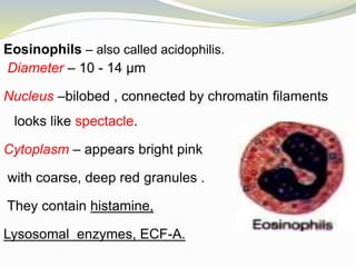
by – Dr .F. B. Irani
Objectives
 Classify leucocytes, with its % in blood
 describe the structure and functions of
leucocytes
 normal count
 variations
WBCs
Granulocyte Agranulocyte
1. Neutrophils 1. Lymphocytes
2. Eosinophils 2. Monocytes
3. Basophils
Normal count of WBCs :-
- At birth- 10,000- 25,000/mm3
-Adults – 4,000 – 11,000/mm3
WBCs DLC(%) Absolute
count
(per mm3)
Neutrophils 50 - 70 2,000 – 7,500
Eosinophils 1 – 4 40 – 440
Basophils 0 – 1 20-80
Lymphocytes 20– 40 1,500 – 4,000
Monocytes 2 - 8 500 - 800
Neutrophils- commonly called polymorph.
Diameter – 10 - 14 μm
Nucleus – multilobed ( 2-6), Lobes are connected
by chromatin filaments.
Cytoplasm – pale bluish with
violet-pink fine granules .
Enzymes are filled in granules.
Function – 1) Phagocytosis
 Engulf foreign particles, bacteria and digest them.
 Constitutes first line of defence.
2) Reaction of inflammation
 release leucotrienes, prostaglandins, thromboxanes.
 Which causes vasodilatation, oedema.
3) Febrile reaction –
 produces fever-producing substance called endogenous
pyrogen in response to bacteria.
 4. Sex determination – in females, drumstick
appendage of chromatin is attached to nuclear lobe.
 5. Role of nourishment of tissue cells -neutrophilis
along with plasma proteins produces trephones or
carrel which are essential for tissue cells.
Eosinophils – also called acidophilis.
Diameter – 10 - 14 μm
Nucleus –bilobed , connected by chromatin filaments
looks like spectacle.
Cytoplasm – appears bright pink
with coarse, deep red granules .
They contain histamine,
Lysosomal enzymes, ECF-A.
Functions – 1) Mild Phagocytosis – as they are less motile.
2) Role in parasitic infestations – produces lethal substances
 Major basic protein(MBP) – larvicidal
 Eosinophil cationic protein (ECP)- destroys
helminthes & is neurotoxin.
 Eosinophil peroxidase – destroys adult helminthes,
bacteria and tumour cells.
 Eosinophil derived neurotoxin- destroys nerve
fibers.
3) Role in allergic reaction
 detoxify mediator of allergy
 destroys antigen-antibody complexes
 prevent spread of local inflammation.
4) Role in immunity –
 provide mucosal immunity in GIT,RT, UT
Basophils -
Diameter – 10 - 14 μm
Nucleus – irregular, may be bilobed or trilobed, boundary
not clearly defined & covered by coarse granules.
Cytoplasm – bluish , filled with plenty
coarse, deep blue coloured granules .
containing heparin, histamine and
chondroitin sulphate
Functions - 1) Mild phagocytic function.
2) Role in allergic reaction-release histamine, bradykinin,
SRS-A, 5HT, that causes allergic reaction
3) Role in preventing spread of allergic inflammation
 releases chemotactic factor that causes eosinophils to
migrate towards the inflamed allergic tissue. Which
then prevent.
4) Release of heparin – which prevents clotting of blood.
Lymphocytes – two types -large & small.
Diameter – 10 - 14 μm of large & 7-10 μm of small
lymphocytes.
Nucleus –large round ,single completely filling the cell.
blue coloured like ink-spot appearance.
Cytoplasm –scanty less than nucleus ,
light blue as crescent around nucleus ,
no granules .
Functions – Imp. role in immunity. Functionally are
classified into two types:
B lymphocytes and T lymphocytes.
Functions of B lymphoctes – responsible for humoral
immunity by producing antibodies(gamma globulins)
Functions of T lymphoctes – responsible for cellular
immunity against virus, bacterial infections, tumour cell.
Monocytes -
Diameter –largest, leucocytes in peripheral blood
12 - 20 μm
Nucleus – large, single , eccentric in position kidney
shaped. With chromatin network.
Cytoplasm –abundant, pale
blue clear no granules, may
contain fine purple dust like
granules.(azur granules)
Functions – 1) Phagocytosis
2) Role in tumour immunity – they kill tumour cell after
sensitization by lymphoctes.
3) Synthesis of biological substances – they
synthesize cytokines, complement factors, growth
factors, interleukins and enzymes for wound
healing.
4) Antigen presenting cell – this first step in activation
of cellular immunity.
Variation in WBCs
Leucocytosis - total WBC count > 11,000/mm3
Physiological causes -
1) Age - at birth WBC count is about 18,000/mm3
which decreases to adult level at 5-10 years .
2) Exercise - count is increased due to disruption of
margination.
3) After food intake, Mental stress, anxiety, Exposure to
low temperature - causes increase in count .
4) Pregnancy - at full term pregnancy, menstruation,
lactation.
Pathological causes
1. Acute bacterial infections - by pyogenic organisms,
2. Acute haemorage ,
3. Burns,
4. Postoperative period ,
5. Tuberculosis ,
6. Glandular fever .
Leucopenia - total WBC count < 4000 /mm3 .
Causes of leucopenia -
1. Infection by non – pyogenic bacteria especially typhoid
fever and Paratyphoid fever .
2. Viral infections such as influenza , smallpox , mumps,
3. Protozoal infections .
4. Starvation and malnutrition .
5. Aplasia of bone marrow.
6. Bone marrow depression due to :
- drugs - chloromycetin & cytotoxic drugs
- Exposure to X – rays
- Chemical poison - arsenic ,dinitrophenol & antimony.
Leukaemia - is a malignant disease of blood with
increase in WBCs. Immature WBCs are present in
the peripheral blood .Total WBC count is usually
above 1,00,000 to 3,00,000 /mm3 .
SAQ:- 1. Functions of WBC,
2 Functions of Neutrophilis,
3. Leukaemia, 4. Leukaemoid reaction,
LAQ:- 1. Describe various functions of WBCs.
2. Describe different types of WBCs.

Leucocytes

  • 1.
    by – Dr.F. B. Irani
  • 2.
    Objectives  Classify leucocytes,with its % in blood  describe the structure and functions of leucocytes  normal count  variations
  • 3.
    WBCs Granulocyte Agranulocyte 1. Neutrophils1. Lymphocytes 2. Eosinophils 2. Monocytes 3. Basophils
  • 4.
    Normal count ofWBCs :- - At birth- 10,000- 25,000/mm3 -Adults – 4,000 – 11,000/mm3
  • 5.
    WBCs DLC(%) Absolute count (permm3) Neutrophils 50 - 70 2,000 – 7,500 Eosinophils 1 – 4 40 – 440 Basophils 0 – 1 20-80 Lymphocytes 20– 40 1,500 – 4,000 Monocytes 2 - 8 500 - 800
  • 6.
    Neutrophils- commonly calledpolymorph. Diameter – 10 - 14 μm Nucleus – multilobed ( 2-6), Lobes are connected by chromatin filaments. Cytoplasm – pale bluish with violet-pink fine granules . Enzymes are filled in granules.
  • 7.
    Function – 1)Phagocytosis  Engulf foreign particles, bacteria and digest them.  Constitutes first line of defence. 2) Reaction of inflammation  release leucotrienes, prostaglandins, thromboxanes.  Which causes vasodilatation, oedema. 3) Febrile reaction –  produces fever-producing substance called endogenous pyrogen in response to bacteria.
  • 8.
     4. Sexdetermination – in females, drumstick appendage of chromatin is attached to nuclear lobe.  5. Role of nourishment of tissue cells -neutrophilis along with plasma proteins produces trephones or carrel which are essential for tissue cells.
  • 9.
    Eosinophils – alsocalled acidophilis. Diameter – 10 - 14 μm Nucleus –bilobed , connected by chromatin filaments looks like spectacle. Cytoplasm – appears bright pink with coarse, deep red granules . They contain histamine, Lysosomal enzymes, ECF-A.
  • 10.
    Functions – 1)Mild Phagocytosis – as they are less motile. 2) Role in parasitic infestations – produces lethal substances  Major basic protein(MBP) – larvicidal  Eosinophil cationic protein (ECP)- destroys helminthes & is neurotoxin.  Eosinophil peroxidase – destroys adult helminthes, bacteria and tumour cells.  Eosinophil derived neurotoxin- destroys nerve fibers.
  • 11.
    3) Role inallergic reaction  detoxify mediator of allergy  destroys antigen-antibody complexes  prevent spread of local inflammation. 4) Role in immunity –  provide mucosal immunity in GIT,RT, UT
  • 12.
    Basophils - Diameter –10 - 14 μm Nucleus – irregular, may be bilobed or trilobed, boundary not clearly defined & covered by coarse granules. Cytoplasm – bluish , filled with plenty coarse, deep blue coloured granules . containing heparin, histamine and chondroitin sulphate
  • 13.
    Functions - 1)Mild phagocytic function. 2) Role in allergic reaction-release histamine, bradykinin, SRS-A, 5HT, that causes allergic reaction 3) Role in preventing spread of allergic inflammation  releases chemotactic factor that causes eosinophils to migrate towards the inflamed allergic tissue. Which then prevent. 4) Release of heparin – which prevents clotting of blood.
  • 14.
    Lymphocytes – twotypes -large & small. Diameter – 10 - 14 μm of large & 7-10 μm of small lymphocytes. Nucleus –large round ,single completely filling the cell. blue coloured like ink-spot appearance. Cytoplasm –scanty less than nucleus , light blue as crescent around nucleus , no granules .
  • 15.
    Functions – Imp.role in immunity. Functionally are classified into two types: B lymphocytes and T lymphocytes. Functions of B lymphoctes – responsible for humoral immunity by producing antibodies(gamma globulins) Functions of T lymphoctes – responsible for cellular immunity against virus, bacterial infections, tumour cell.
  • 16.
    Monocytes - Diameter –largest,leucocytes in peripheral blood 12 - 20 μm Nucleus – large, single , eccentric in position kidney shaped. With chromatin network. Cytoplasm –abundant, pale blue clear no granules, may contain fine purple dust like granules.(azur granules)
  • 17.
    Functions – 1)Phagocytosis 2) Role in tumour immunity – they kill tumour cell after sensitization by lymphoctes. 3) Synthesis of biological substances – they synthesize cytokines, complement factors, growth factors, interleukins and enzymes for wound healing. 4) Antigen presenting cell – this first step in activation of cellular immunity.
  • 18.
    Variation in WBCs Leucocytosis- total WBC count > 11,000/mm3 Physiological causes - 1) Age - at birth WBC count is about 18,000/mm3 which decreases to adult level at 5-10 years .
  • 19.
    2) Exercise -count is increased due to disruption of margination. 3) After food intake, Mental stress, anxiety, Exposure to low temperature - causes increase in count . 4) Pregnancy - at full term pregnancy, menstruation, lactation.
  • 20.
    Pathological causes 1. Acutebacterial infections - by pyogenic organisms, 2. Acute haemorage , 3. Burns, 4. Postoperative period , 5. Tuberculosis , 6. Glandular fever .
  • 21.
    Leucopenia - totalWBC count < 4000 /mm3 . Causes of leucopenia - 1. Infection by non – pyogenic bacteria especially typhoid fever and Paratyphoid fever . 2. Viral infections such as influenza , smallpox , mumps, 3. Protozoal infections . 4. Starvation and malnutrition . 5. Aplasia of bone marrow.
  • 22.
    6. Bone marrowdepression due to : - drugs - chloromycetin & cytotoxic drugs - Exposure to X – rays - Chemical poison - arsenic ,dinitrophenol & antimony. Leukaemia - is a malignant disease of blood with increase in WBCs. Immature WBCs are present in the peripheral blood .Total WBC count is usually above 1,00,000 to 3,00,000 /mm3 .
  • 23.
    SAQ:- 1. Functionsof WBC, 2 Functions of Neutrophilis, 3. Leukaemia, 4. Leukaemoid reaction, LAQ:- 1. Describe various functions of WBCs. 2. Describe different types of WBCs.