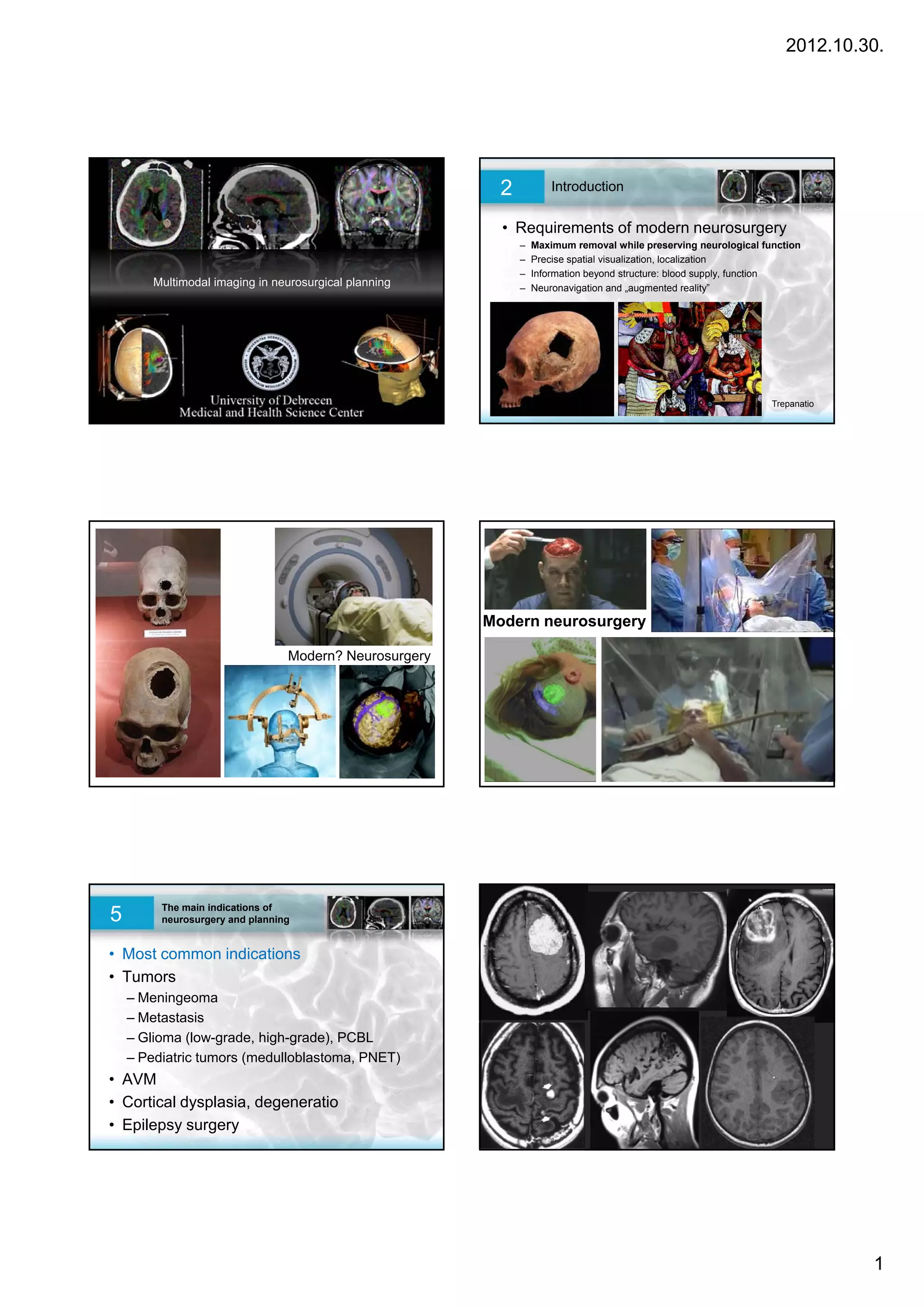
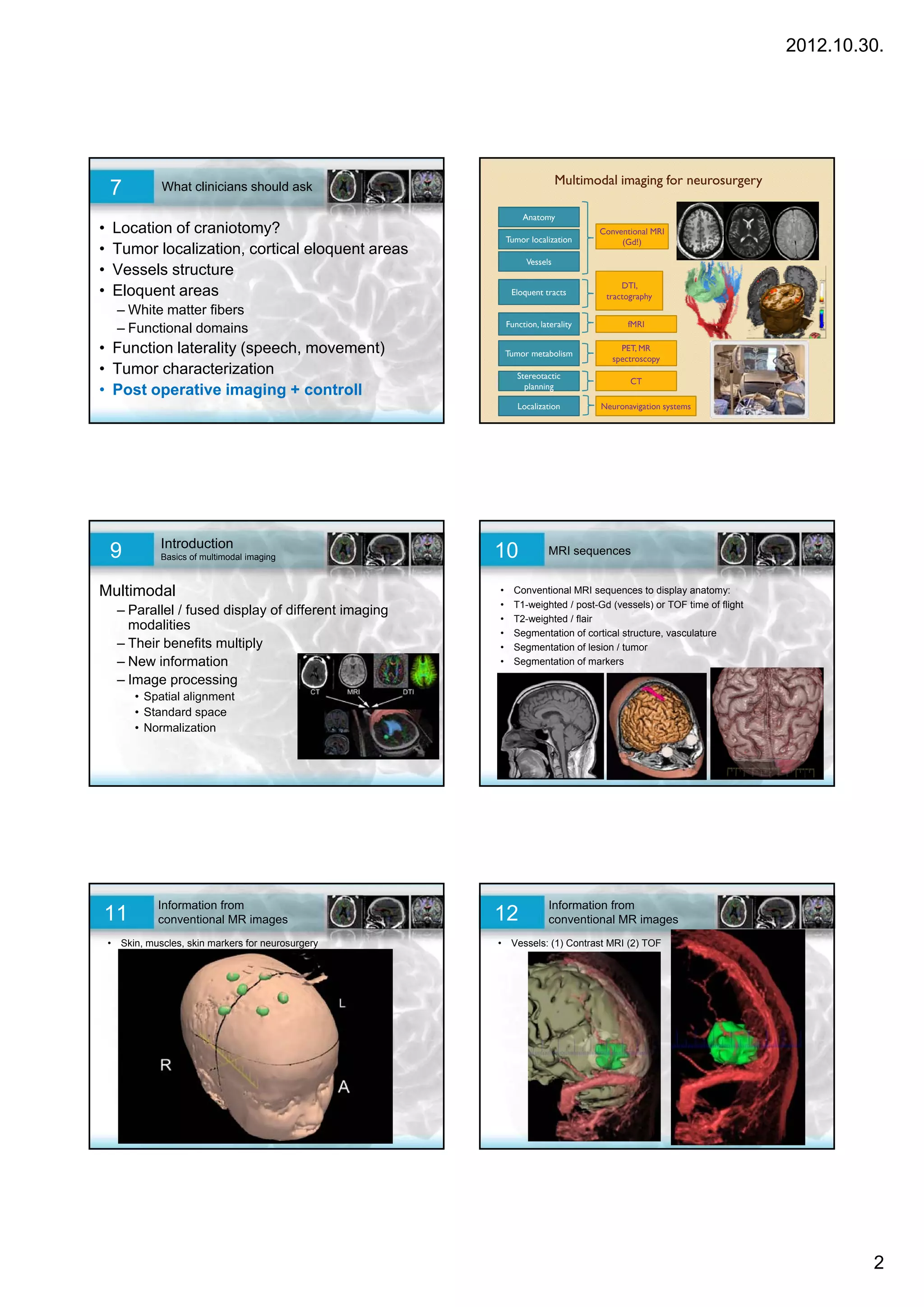
The document discusses the use of multimodal imaging in neurosurgery. It describes how multimodal imaging can provide maximum information beyond just anatomical structures, including blood supply, function, and spatial visualization to help with surgical planning and navigation. It outlines some of the key indications for neurosurgery like tumors, arteriovenous malformations, epilepsy, and discusses how clinicians can utilize different imaging modalities like MRI, DTI, fMRI, and PET to obtain information on anatomy, vessels, eloquent tracts, function and laterality, tumor characterization and metabolism, and localization for stereotactic planning.


![2012.10.30.
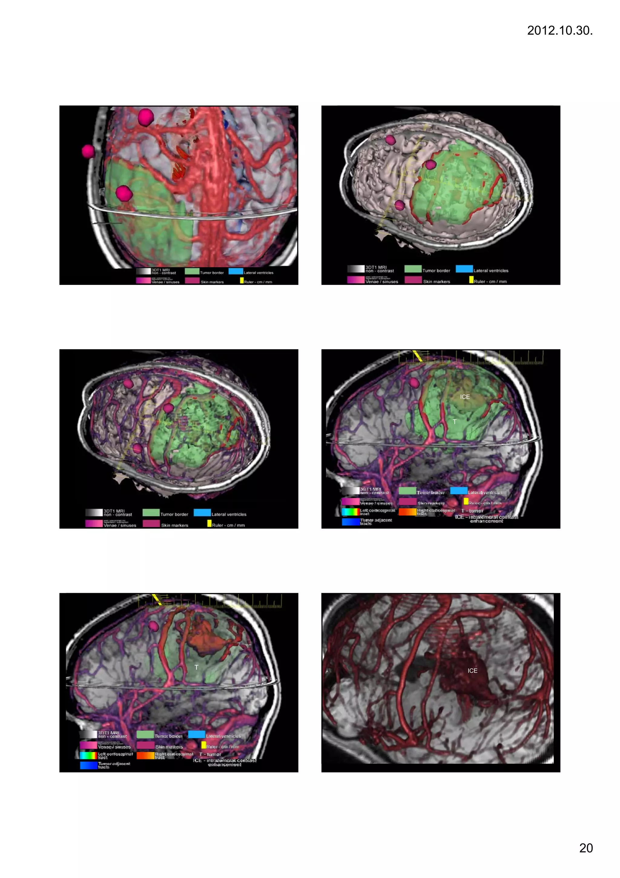
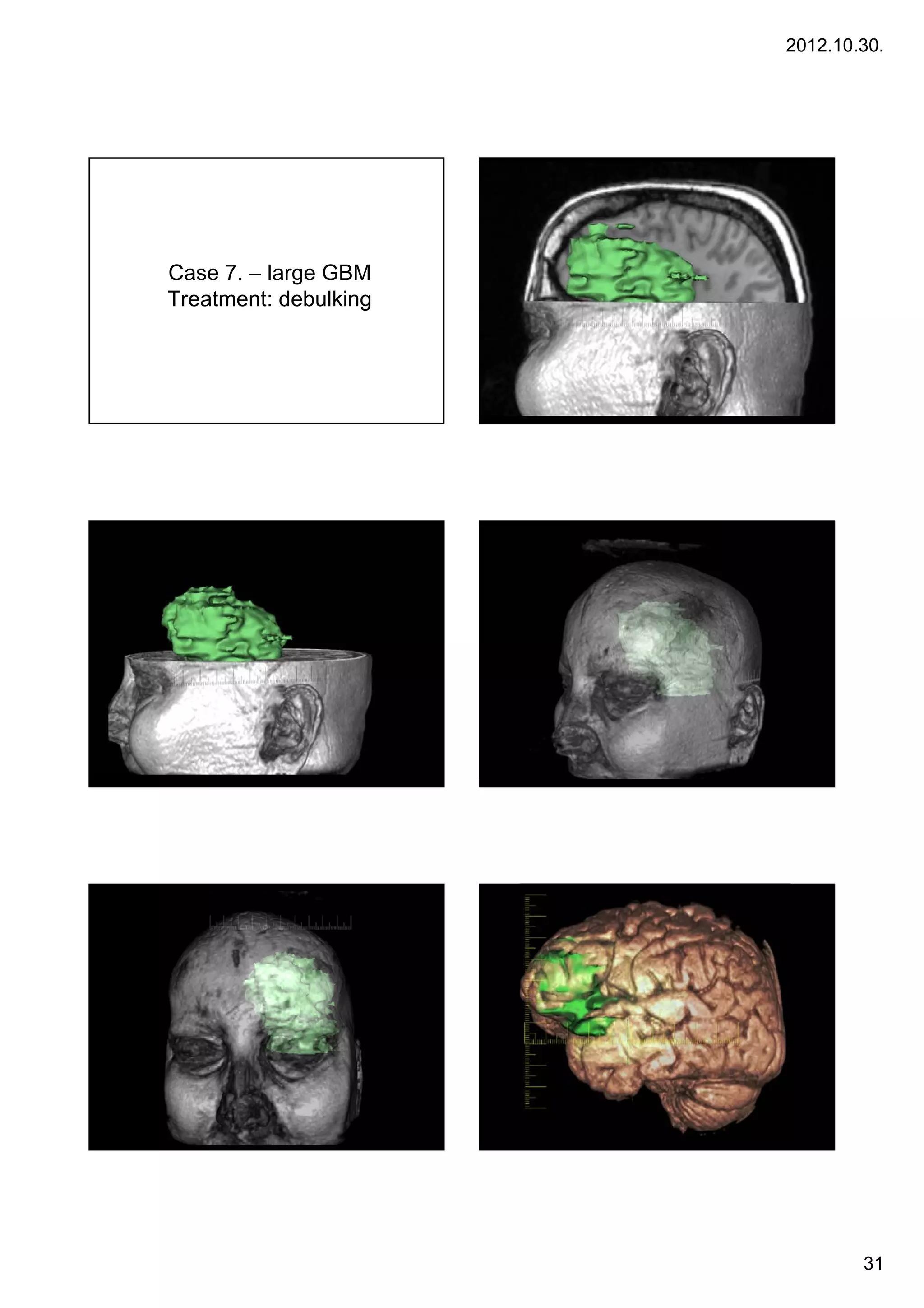
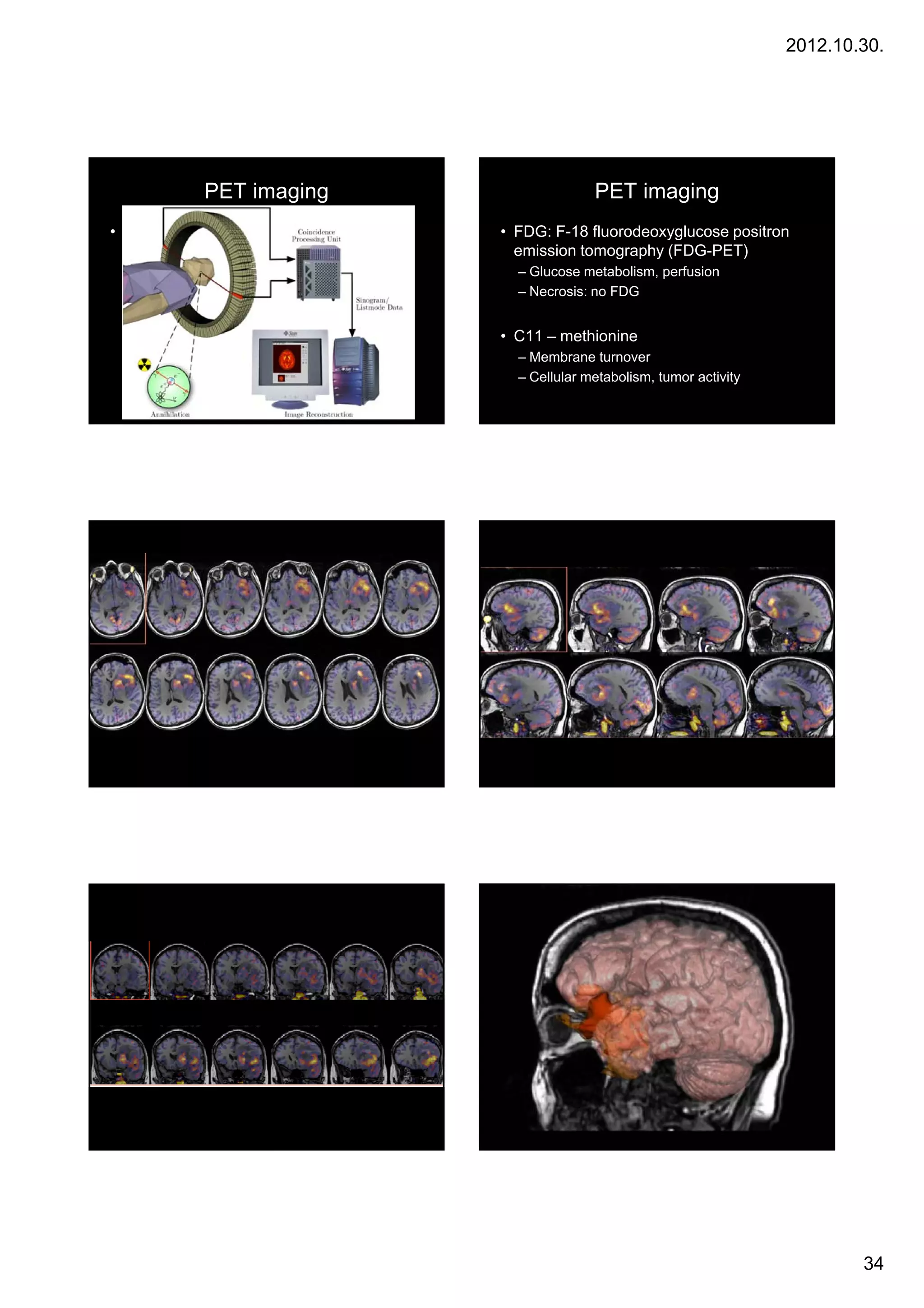
Glioblastoma multiforme
•Glioblastoma multiforme (GBM) is the most common and
most aggressive malignant primary brain tumor in humans,
involving glial cells and accounting for 52% of all functional
tissue brain tumor cases and 20% of all intracranial tumors.
Despite being the most prevalent form of primary brain
Case 3. tumor, GBM incidence is only 2–3 cases per 100,000
, y p ,
people in Europe and North America. According to
the WHO classification of the tumors of the central nervous
system, the standard name for this brain tumor is
"glioblastoma"; it presents two variants: giant cell
glioblastoma and gliosarcoma.
•Treatment can involve chemotherapy, radiation,
radiosurgery, corticosteroids, antiangiogenic therapy,
surgery[1] and experimental approaches such as gene
transfer.[2]
19](https://image.slidesharecdn.com/multimodneurosurg2012engpdf-121030100442-phpapp02/75/Week-3-Neurosurgical-planning-with-multimodal-imaging-19-2048.jpg)
![2012.10.30.
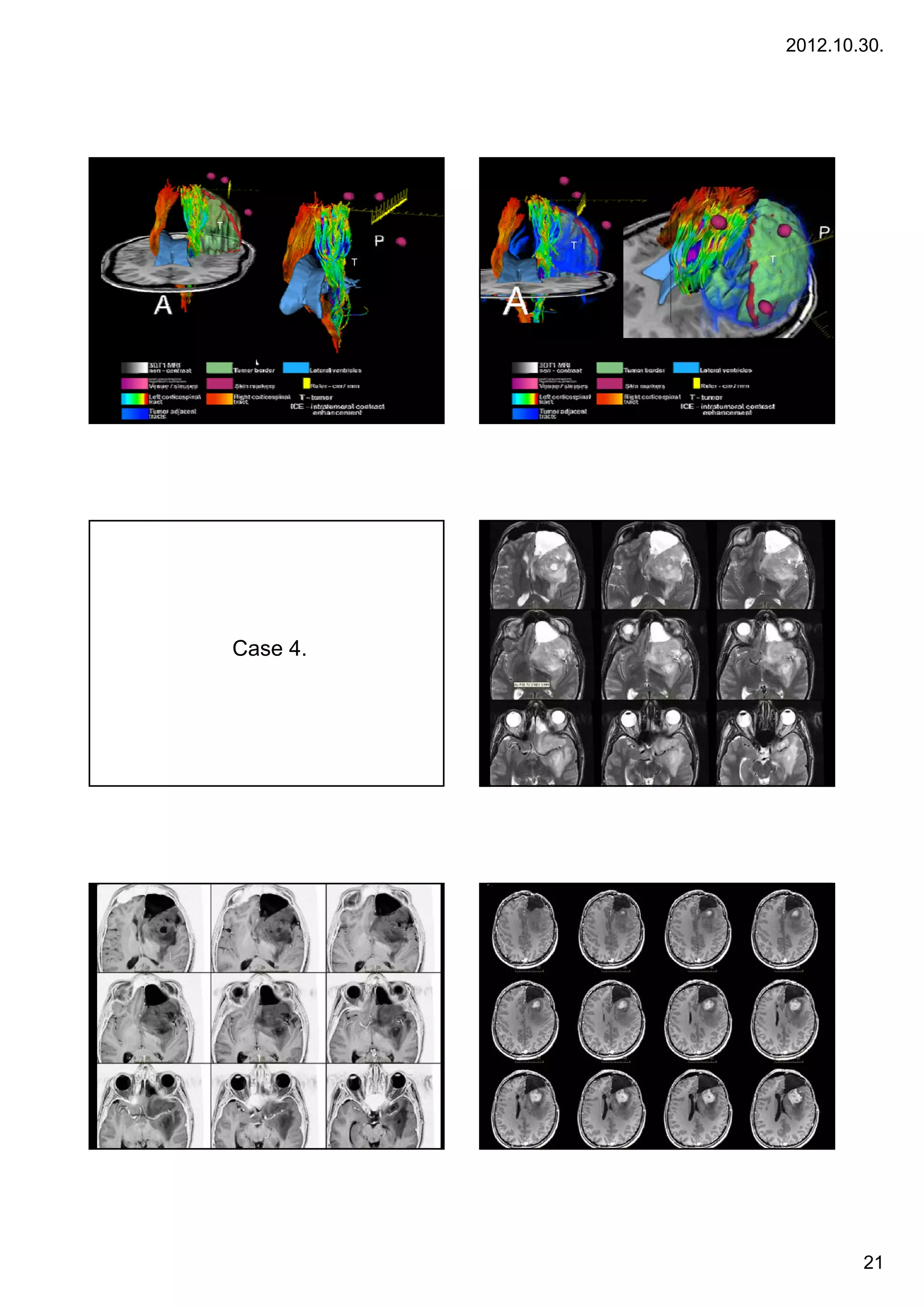
Low grade gliomas
Gliomas are named according to the specific type of cell they share histological
features with, but not necessarily originate from. The main types of gliomas are:
Ependymomas — ependymal cells.
Astrocytomas — astrocytes (glioblastoma multiforme is the most common
astrocytoma).
Oligodendrogliomas — oligodendrocytes.
Case 5
5. Mixed gliomas, such as oligoastrocytomas, contain cells from different types of
glia.
g , g y , yp
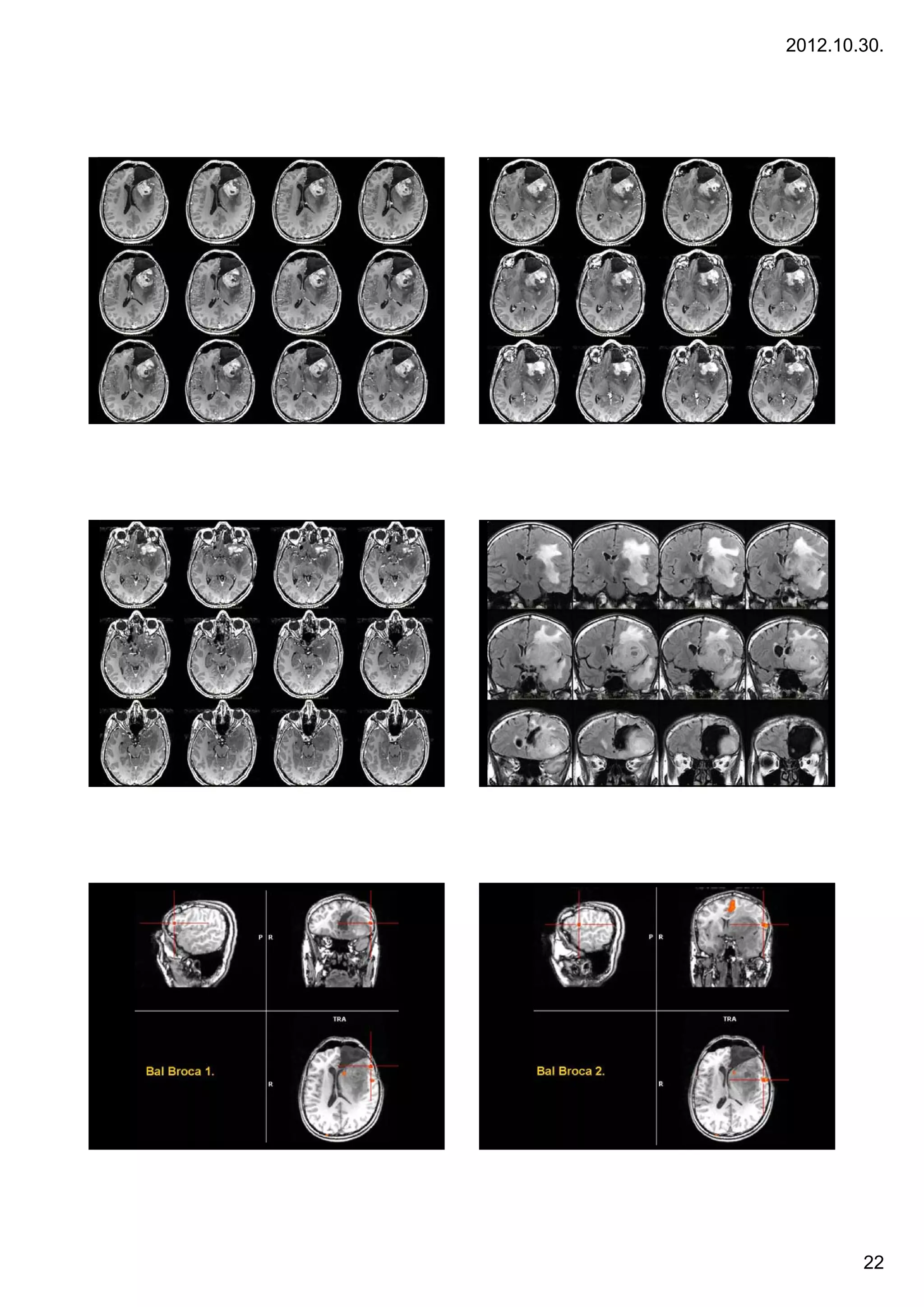
Gliomas are further categorized according to their grade, which is determined
by pathologic evaluation of the tumor.
Low-grade gliomas [WHO grade II] are well-differentiated (not anaplastic);
these are not benign but still portend a better prognosis for the patient.
High-grade [WHO grade III-IV] gliomas are undifferentiated or anaplastic; these
are malignant and carry a worse prognosis.
23](https://image.slidesharecdn.com/multimodneurosurg2012engpdf-121030100442-phpapp02/75/Week-3-Neurosurgical-planning-with-multimodal-imaging-23-2048.jpg)


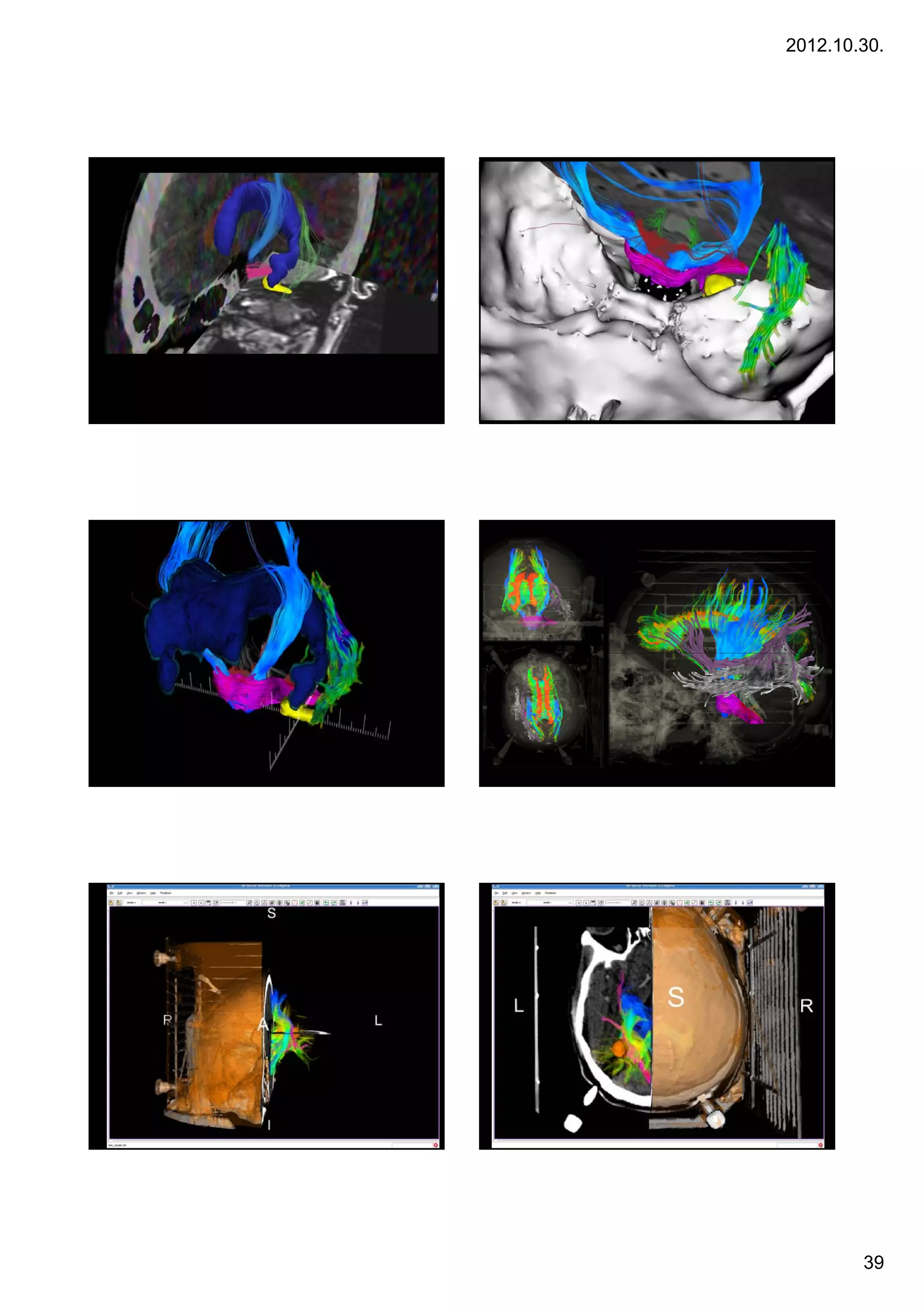
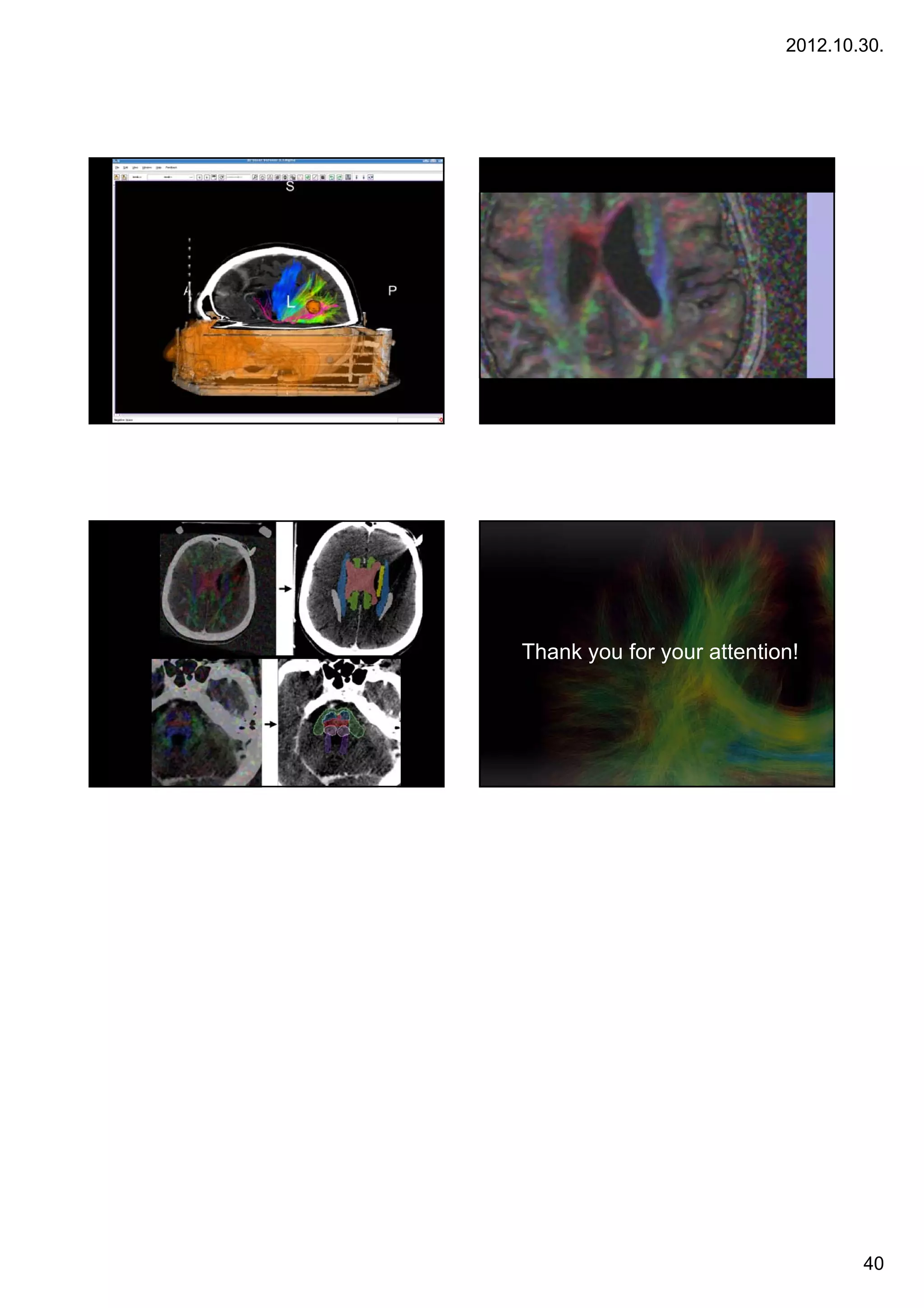
![2012.10.30.
Képregisztrációk (CT + DTI)
Case 8
8.
CT + ADC map CT + colorized FA map
Acoustic neurinomas Acoustic neurinomas
•A vestibular schwannoma, often called an acoustic neuroma,[1] is
a benign primary intracranial tumor of the myelin-forming cells of the vestibulocochlear
nerve(CN VIII).[2] The term "vestibular schwannoma" involves the vestibular portion of
the 8th cranial nerve[3] and arises from Schwann cells, which are responsible for
themyelin sheath in the peripheral nervous system. Approximately 3,000 cases are
diagnosed each year in the United States with a prevalence of about 1 in 100,000
worldwide. It comprises 5-10% of all intracranial neoplasms in adults. Incidence peaks in
the fifth and sixth decades and both sexes are affected equally.
28 éves nő 28 éves nő
Acousticus neurinoma Acousticus neurinoma
Gamma Sugársebészeti Központ Gamma Sugársebészeti Központ
38](https://image.slidesharecdn.com/multimodneurosurg2012engpdf-121030100442-phpapp02/75/Week-3-Neurosurgical-planning-with-multimodal-imaging-38-2048.jpg)