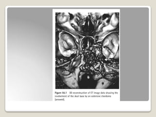
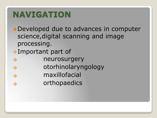
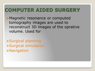
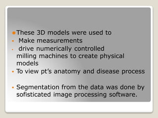
IMAGE GUIDED
SURGERY
Dr.Ramendra singh
NAVIGATION
⚫Developed due to advances in computer
science,digital scanning and image
processing.
Important part of




neurosurgery
otorhinolaryngology
maxillofacial
orthopaedics
COMPUTER AIDED SURGERY
⚫Magnetic resonance or computed
tomography images are used to
reconstruct 3D images of the oprative
volume. Used for
Surgical planning
Surgical simulation
Navigation
⚫These 3D models were used to
 Make measurements
 drive numerically controlled
milling machines to create physical
models
 To view pt’s anatomy and disease process
 Segmentation from the data was done by
sofisticated image processing software.
⚫In OT
, images were presented as 2D slices
within 3 orthogonal plane of spaces.
⚫Surgical tool was developed to enable the
localization of anatomical features to
confirm the precise position of a pointer
within the operative field.
⚫Useful when the disease process or
previous surgery has distorted the normal
anatomy.
⚫The images are vector representations of
previously segmented and processed
data.
⚫3D data set can be superimposed on pt’s
anatomy.
Preoperative imaging
⚫2 purposes
⚫Diagnostic tool
⚫Assess the extent of disease.
⚫Makes surgery safer
⚫Training better
⚫Surgeons plan their approach
maxillofacial surgeons were able to make
better fitting prosthesis.
Navigation in FESS
⚫Improves surgical accuracy-90% accurate
in identifying critical landmarks.
⚫Reduces risk of major
intracranial
o
• intraorbital complications.
• Enhances surgical efficiency
• Accelerates learning curve
• Reduces operative time.
•
IMAGE RECONSTRUCTION
⚫3D data from 2D sections are created
using highly complex algorithms that are
specific to the particular scanner and
scanning protocol.
⚫Slices concept
⚫Pixels and voxels
⚫Volume averaging
Concept of slices
⚫CT scan converts 1D xrays into 2D slices.
⚫Referred to as a single image section
which may be acquired in
axial
coronal
⚫
⚫
⚫ sagital plane
⚫By changing the gantry angle.
⚫Each section has width known as slice
thickness- width of xray beam detector
window used.
⚫Smaller width beams produce higher
resolution 2D images.
⚫Slices are placed along the area to be
scanned with an interslice distancethat
varies depending on
⚫ the scanning protocol to be used
⚫ size of the anatomical str to be imaged.
⚫Spiral CT are better as




faster
less radiation
collect data in helical fashion
reconstructed into 3D data.
Pixels and voxels
⚫Pixel: smallest picture elements
⚫The more pixels in a certain distance,the
better the image.
⚫Highest resolution in CT 512x512 pixels
per image.
⚫Image resolution is stated by no of pixels
in the x and y axis
⚫Hounsfield unit: the value of each pixel
ranging from -1000 to 3096.
⚫CT scans are calibrated as 0=density of
water.
⚫High pixel values are displayed as white.
⚫The lower the value,the lower the density
of the tissue,the darker the pixel would
appear.
⚫VOXELS:3D Blocks or volumnetric picture
elements.
⚫Depending on inter slice distance can be:
⚫
⚫
cube
cuboid
VOLUME AVERAGING
⚫Partial volume averaging:
⚫ if a str falls partially within a pixel,the
true value of the str will be assigned a
value less than the normal value of the
str.
⚫Major problem in delineating where the
edge of a str should lie within a given
image.
⚫Overcome by complex algorithms
VISUALIZATION OF IMAGE
RENDERING:process of generating images which
represent 3D anatomy with some degree of tissue
transperancy.
VOLUME RENDERING
SURFACE RENDERING
 Triplets of data points
are grouped as the
vertices of adjacent
triangles that
interconnect to make
up entire surface
known as facets.
⚫ Projecting each voxel
on to a viewing plane
with a value related to
the physical property
⚫ Operator may choose
to display only the max
contribution for any
voxel along a ray.
⚫ This produces image
max intensity
projection.(MIP)
⚫ Requires preprocessing
⚫ Voxels containing
anatomical surfaces
must be decided
⚫ The derived surfaces
are isosurfaces that
correspond to surfaces
of equal functional
activity.
⚫ Geometric primitives
obtained:
 contour tracing
 Surface extraction
• Segmentation:extractio
n of tissue topology
• These geometric
primitives are displayed
by computer graphics.
⚫ VOLUME RENDERED
images appear diff from
a surface rendered
image in that
anatomical structures
are presented as having
some degree of
transparency.
⚫ Enhances depth
perception
⚫ Increases accuracy
⚫ Placement of surgical
instruments with more
accuracy.
SURGICAL PLANNING
• Enables the surgeon to:
 assess pt’s anatomy objectively
 communicate with other surgeons
 review these films at a later date.
• The data can be segmented and
manipulated to familiarize the surgeons
with specific features pertinent to the pt
and the procedure to be performed.
SURGICAL SIMULATION
⚫Operate using a 2D image on a television
monitor
⚫Develop new hand eye coordination skills
⚫Simulators create virtual surgical
environment.
⚫Esp in skull base surgery.
⚫Able to virtually manipulate and perform
endoscopic surgeries.
Image guidance
⚫ Alerts surgeon about variation in anatomy.
⚫ Use pt’s image data intraoperatively to



determine position
distance from vital organs
hidden anatomical features
• 2 fundamental process
• Registration
• tracking
REGISTRATION
⚫ Once the pt is secured in OT table, registration of
cartesian coordinates of the CT scan to that pt is
done by:
 locating anotomical landmarks visible on the
pt and the image data using a probe that is
visible o the tracking device.
 The position of the tip of the probe is
identified by the tracking device and the
coordinates are fed back into navigation
software.
 other methods use masks and laser scanning
tools.


⚫ Calculations made in real time indicate the point
accuracy.
⚫ Some make use of fiducial markers that are applied
to the pt before scanning on the day of surgery.
⚫ Reference points should be adjacent to surgical field.
⚫ Registration error is only a measure of the accuracy
of correlation beetween selected points in the virtual
data sets and the anatomical markers identified on
the pt.
⚫ Target error:error that could be expected if a probe
was placed on a random point of interest in surgical
field.
influenced by the registration error
lessened if the target is within volume described
by fiducial markers.
⚫
TRACKING
⚫Tracking devices:sensors that provide
dynamic positional information.
⚫Should be:
 very precise
 consistently accurate
 Fast enough to give 25 readings in 1 sec
 Be insensitive to changes in temperature
 unaffected by metal objects
 Able to track 2 objects simultaneously.
1. Earlier we used mechanical arms fitted with
potentiometers
 were fast
 cumbersome
 had restricted range of movements
 hindered the movement of the tracked
object
2.Based on magnetic field distribution


effective and cheap.
affected by metal
3 .Infra red light sensors:
 most commonly used
⚫ Active devices
⚫ Sense infrared light
from LED attached to
the pt or location
probe.
⚫ Passive devices
⚫ Detect infrared light
reflected from
metallic balls
attached to pt or
probe.
⚫ To detect changes in pt’s position ,arches with LED an be
fitted to Mayfield clamp.
⚫ Accuracy of 2-5 mm .
4. Inertial trackers:
⚫ provide one rate of change of rotational measurement
only.
⚫ Not accurate for slow position changes.
5. Based on ultrasound signals:
• achieve greater accuracy.
• susceptible to changes in temperature and air currents.
• long lag times
• Interference from echoes and noises.
CLINICAL APPLICATION
⚫In the areas of skull base and endoscopic
surgeries.
Skull base surgery:
 pre op planning
 Design of bone flaps
 Identification of imp str
 Finding small tumours in obscure parts
Rhinology:
⚫ Difficult revision fess.
⚫ Localize frontal recess in DRAF procedures.
⚫ Trans nasal, trans sphenoidal
hypophysectomy.
Otology :
• Locating facial nerve
• Identifying lesions of petrous apex
• Tumours of IAC.
• meningiomas
• vestibular schwannomas
• Mastoid surgery
• locate dura ,brain,jugular bulb.
•:
THANK YOU

navigation-190721152611 (1).pptx for pg students

  • 1.
  • 3.
    NAVIGATION ⚫Developed due toadvances in computer science,digital scanning and image processing. Important part of     neurosurgery otorhinolaryngology maxillofacial orthopaedics
  • 4.
    COMPUTER AIDED SURGERY ⚫Magneticresonance or computed tomography images are used to reconstruct 3D images of the oprative volume. Used for Surgical planning Surgical simulation Navigation
  • 5.
    ⚫These 3D modelswere used to  Make measurements  drive numerically controlled milling machines to create physical models  To view pt’s anatomy and disease process  Segmentation from the data was done by sofisticated image processing software.
  • 6.
    ⚫In OT , imageswere presented as 2D slices within 3 orthogonal plane of spaces. ⚫Surgical tool was developed to enable the localization of anatomical features to confirm the precise position of a pointer within the operative field.
  • 7.
    ⚫Useful when thedisease process or previous surgery has distorted the normal anatomy. ⚫The images are vector representations of previously segmented and processed data. ⚫3D data set can be superimposed on pt’s anatomy.
  • 9.
    Preoperative imaging ⚫2 purposes ⚫Diagnostictool ⚫Assess the extent of disease. ⚫Makes surgery safer ⚫Training better ⚫Surgeons plan their approach maxillofacial surgeons were able to make better fitting prosthesis.
  • 10.
    Navigation in FESS ⚫Improvessurgical accuracy-90% accurate in identifying critical landmarks. ⚫Reduces risk of major intracranial o • intraorbital complications. • Enhances surgical efficiency • Accelerates learning curve • Reduces operative time. •
  • 11.
    IMAGE RECONSTRUCTION ⚫3D datafrom 2D sections are created using highly complex algorithms that are specific to the particular scanner and scanning protocol. ⚫Slices concept ⚫Pixels and voxels ⚫Volume averaging
  • 12.
    Concept of slices ⚫CTscan converts 1D xrays into 2D slices. ⚫Referred to as a single image section which may be acquired in axial coronal ⚫ ⚫ ⚫ sagital plane ⚫By changing the gantry angle. ⚫Each section has width known as slice thickness- width of xray beam detector window used. ⚫Smaller width beams produce higher resolution 2D images.
  • 13.
    ⚫Slices are placedalong the area to be scanned with an interslice distancethat varies depending on ⚫ the scanning protocol to be used ⚫ size of the anatomical str to be imaged. ⚫Spiral CT are better as     faster less radiation collect data in helical fashion reconstructed into 3D data.
  • 15.
    Pixels and voxels ⚫Pixel:smallest picture elements ⚫The more pixels in a certain distance,the better the image. ⚫Highest resolution in CT 512x512 pixels per image. ⚫Image resolution is stated by no of pixels in the x and y axis ⚫Hounsfield unit: the value of each pixel ranging from -1000 to 3096.
  • 16.
    ⚫CT scans arecalibrated as 0=density of water. ⚫High pixel values are displayed as white. ⚫The lower the value,the lower the density of the tissue,the darker the pixel would appear. ⚫VOXELS:3D Blocks or volumnetric picture elements. ⚫Depending on inter slice distance can be: ⚫ ⚫ cube cuboid
  • 17.
    VOLUME AVERAGING ⚫Partial volumeaveraging: ⚫ if a str falls partially within a pixel,the true value of the str will be assigned a value less than the normal value of the str. ⚫Major problem in delineating where the edge of a str should lie within a given image. ⚫Overcome by complex algorithms
  • 19.
    VISUALIZATION OF IMAGE RENDERING:processof generating images which represent 3D anatomy with some degree of tissue transperancy. VOLUME RENDERING SURFACE RENDERING  Triplets of data points are grouped as the vertices of adjacent triangles that interconnect to make up entire surface known as facets. ⚫ Projecting each voxel on to a viewing plane with a value related to the physical property ⚫ Operator may choose to display only the max contribution for any voxel along a ray. ⚫ This produces image max intensity projection.(MIP)
  • 20.
    ⚫ Requires preprocessing ⚫Voxels containing anatomical surfaces must be decided ⚫ The derived surfaces are isosurfaces that correspond to surfaces of equal functional activity. ⚫ Geometric primitives obtained:  contour tracing  Surface extraction • Segmentation:extractio n of tissue topology • These geometric primitives are displayed by computer graphics. ⚫ VOLUME RENDERED images appear diff from a surface rendered image in that anatomical structures are presented as having some degree of transparency. ⚫ Enhances depth perception ⚫ Increases accuracy ⚫ Placement of surgical instruments with more accuracy.
  • 21.
    SURGICAL PLANNING • Enablesthe surgeon to:  assess pt’s anatomy objectively  communicate with other surgeons  review these films at a later date. • The data can be segmented and manipulated to familiarize the surgeons with specific features pertinent to the pt and the procedure to be performed.
  • 22.
    SURGICAL SIMULATION ⚫Operate usinga 2D image on a television monitor ⚫Develop new hand eye coordination skills ⚫Simulators create virtual surgical environment. ⚫Esp in skull base surgery. ⚫Able to virtually manipulate and perform endoscopic surgeries.
  • 23.
    Image guidance ⚫ Alertssurgeon about variation in anatomy. ⚫ Use pt’s image data intraoperatively to    determine position distance from vital organs hidden anatomical features • 2 fundamental process • Registration • tracking
  • 24.
    REGISTRATION ⚫ Once thept is secured in OT table, registration of cartesian coordinates of the CT scan to that pt is done by:  locating anotomical landmarks visible on the pt and the image data using a probe that is visible o the tracking device.  The position of the tip of the probe is identified by the tracking device and the coordinates are fed back into navigation software.  other methods use masks and laser scanning tools.
  • 26.
      ⚫ Calculations madein real time indicate the point accuracy. ⚫ Some make use of fiducial markers that are applied to the pt before scanning on the day of surgery. ⚫ Reference points should be adjacent to surgical field. ⚫ Registration error is only a measure of the accuracy of correlation beetween selected points in the virtual data sets and the anatomical markers identified on the pt. ⚫ Target error:error that could be expected if a probe was placed on a random point of interest in surgical field. influenced by the registration error lessened if the target is within volume described by fiducial markers. ⚫
  • 27.
    TRACKING ⚫Tracking devices:sensors thatprovide dynamic positional information. ⚫Should be:  very precise  consistently accurate  Fast enough to give 25 readings in 1 sec  Be insensitive to changes in temperature  unaffected by metal objects  Able to track 2 objects simultaneously.
  • 29.
    1. Earlier weused mechanical arms fitted with potentiometers  were fast  cumbersome  had restricted range of movements  hindered the movement of the tracked object 2.Based on magnetic field distribution   effective and cheap. affected by metal 3 .Infra red light sensors:  most commonly used
  • 30.
    ⚫ Active devices ⚫Sense infrared light from LED attached to the pt or location probe. ⚫ Passive devices ⚫ Detect infrared light reflected from metallic balls attached to pt or probe.
  • 32.
    ⚫ To detectchanges in pt’s position ,arches with LED an be fitted to Mayfield clamp. ⚫ Accuracy of 2-5 mm . 4. Inertial trackers: ⚫ provide one rate of change of rotational measurement only. ⚫ Not accurate for slow position changes. 5. Based on ultrasound signals: • achieve greater accuracy. • susceptible to changes in temperature and air currents. • long lag times • Interference from echoes and noises.
  • 33.
    CLINICAL APPLICATION ⚫In theareas of skull base and endoscopic surgeries. Skull base surgery:  pre op planning  Design of bone flaps  Identification of imp str  Finding small tumours in obscure parts
  • 34.
    Rhinology: ⚫ Difficult revisionfess. ⚫ Localize frontal recess in DRAF procedures. ⚫ Trans nasal, trans sphenoidal hypophysectomy. Otology : • Locating facial nerve • Identifying lesions of petrous apex • Tumours of IAC. • meningiomas • vestibular schwannomas • Mastoid surgery • locate dura ,brain,jugular bulb. •:
  • 35.