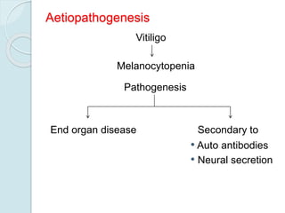
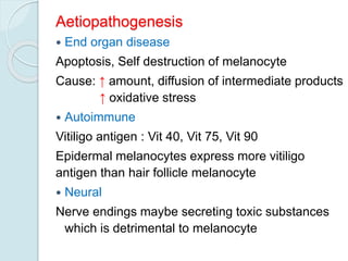
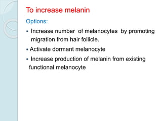
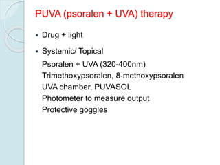
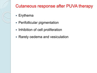
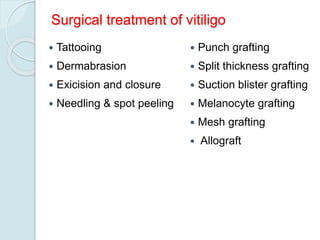
Vitiligo
Definition
Vitiligo is a circumscribed, acquired, idiopathic,
progressive hypomelanosis of skin and hair which
is often familial and is characterized
microscopically by an absence of melanocytes.
Leukoderma is the term applied only to
depigmented patches of known causes
eg: following burns, chemicals, inflammatory
disorder.
Normal Skin Color
 Melanin
 Carotenoids
 Oxyhaemoglobin
 Reduced haemoglobin
Epidermal
Dermal
Normal Melanisation
Neuroectodermal Origin
Migrates to
Cutaneous
Epidermal
Appendageal
Mucous membrane
Follicular Non follicular
Extracutaneous
Eye Brain
Melanocyte
Normal Melanisation
Epidermal melanocyte
 Attached to basement membrane
 Rarely divide
 Require bFGF (Basic fibroblast growth factor)
for growth and multiplication
 Function - Endogenous sunscreen
 Response to injury - Unpredictable
Hair melanocyte
Hair bulb
Hair follicle melanocyte
Mid-follicle + Upper follicle
• Dendritic
• Functional
• DOPA Negative
• Amelanotic & Non dendritic
• Active after trauma
Normal Melanisation
Melanosome
 Membrane bound melanosome inside the
melanocyte
 Site of production and storage of melanin
 Membrane prevents diffusion of intermediate
toxic products of melanin synthesis which are
harmful to melanocyte
Aetiopathogenesis
Vitiligo
Melanocytopenia
Pathogenesis
End organ disease Secondary to
• Auto antibodies
• Neural secretion
Aetiopathogenesis
 End organ disease
Apoptosis, Self destruction of melanocyte
Cause: ↑ amount, diffusion of intermediate products
↑ oxidative stress
 Autoimmune
Vitiligo antigen : Vit 40, Vit 75, Vit 90
Epidermal melanocytes express more vitiligo
antigen than hair follicle melanocyte
 Neural
Nerve endings maybe secreting toxic substances
which is detrimental to melanocyte
Clinical features of Vitiligo
Macule of Vitiligo:
 Round, oval
Milky white
Scalloped margin
 Trichrome or quadrichrome
 Confetti macules
 Inflammatory border in some cases
 Leucotrichia in some cases
Clinical Classification
 Localized
◦ Focal
◦ Segmental
 Generalized
◦ Symmetrical
◦ Acromucosal
◦ Universalis
Cutaneous associations in Vitiligo
 Leucotrichia
 Premature gray hair
 Halo nevi
 Alopecia areata
Systemic associations in Vitiligo
 Thyroid disease
 Diabetes
 Addison's disease
 Pernicious anemia
 Multiple endocrinopathy syndrome
Differential diagnosis
 Piebaldism
 Pityriasis Alba
 Hansens disease
 Pityriasis Versicolor
 Morphoea
 Lichen Sclerosus et Atrophicus
 Post inflammatory leucoderma
Treatment guidelines
Vitiligo is a sign and the cause of melanocyte
destruction may not be the same in every case.
There is no uniform response to treatment.
Aims of treatment
Repigmentation
 Prevention of further depigmentation
To increase melanin
Options:
 Increase number of melanocytes by promoting
migration from hair follicle.
 Activate dormant melanocyte
 Increase production of melanin from existing
functional melanocyte
Medical Treatment
 Psoralen + UVA
 UVB – Narrowband
 Steroids
 Eau de Cologne
 Khellin + UVA
 L-phenylalanine + UVA
 Topical Tacrolimus
 Topical 5 Flurouracil
 Topical Calcipotriol
 Placental extract
 Topical bFGF
 Excimer laser
Treatment options for repigmentation
 Topical steroids - All types of vitiligo
 Topical PUVA - Focal / segmental
 Systemic PUVA - Segmental / Generalized
Prevention of further depigmentation
 Treatment of precipitating cause
 Steroid
◦ Topical (useful for repigmentation also)
◦ Systemic
Oral
 Short course
 Pulse
Injectable
 ACTH
Triamcinolone
PUVA (psoralen + UVA) therapy
 Drug + light
 Systemic/ Topical
Psoralen + UVA (320-400nm)
Trimethoxypsoralen, 8-methoxypsoralen
UVA chamber, PUVASOL
Photometer to measure output
Protective goggles
Cutaneous response after PUVA therapy
 Erythema
 Perifollicular pigmentation
 Inhibition of cell proliferation
 Rarely oedema and vesiculation
Treatment Protocol
TMP 0.6 mg/kg – 25 sittings
No change
TMP 0.9 mg/kg – 25 sittings
No change
8 MOP 0.3 mg/kg – 25 sittings
No change
8 MOP 0.6 mg/kg – 25 sittings
No change
TMP + 8MOP – 25 sittings
No response
Unresponsive case
PUVA Therapy
Follow up
Good response Development of
new patches
Total
pigmentation
Continue
maintenance
PUVA
Increase
dose
Stop
PUVA
PUVA Therapy: Side Effects
Acute
Erythema
Pruritus
Nausea
Headache
Koebner phenomenon
Chronic
Chronic actinic damage
Carcinoma - rare
Immunosuppression
Ophthalmic effect
Premature cataract
Topical steroids
 Isolated macules
 Hydrocortisone
 Mometasone
 Betamethasone
 Clobetasol
 Side effects
 Atrophy
 Striae
Children + Face
Adults + Body
Systemic steroids
 Low dose, long term
◦ Oral
◦ Injectable
 High dose pulse
 ACTH
Permanent depigmentation
 More than 50% area involvement
 Failure of treatment or does not wish to continue
treatment
 20% MBEH (monobenzyl ether of hydroquinone) – 4
to 12 months
 Irreversible
 Eyes, hair spared
 Needs sunscreen afterwards
 Side effect - contact dermatitis
 Rarely accepted by Indian patients
Prognostic factors
 Acrofacial
 Patches on bony prominences
 Lesions on glans penis, palms, soles
 Patches with gray hair
 Patches around nipple
 Long standing cases
 Extensive depigmentation
Cases resistant to medical line of treatment
Failure to respond to medical line of treatment
indicates melanocyte reservoir is no more
available in that area and it is needed to
repopulate that area with melanocytes which can
be achieved by various surgical modalities
Surgical treatment of vitiligo
 Tattooing
 Dermabrasion
 Exicision and closure
 Needling & spot peeling
 Punch grafting
 Split thickness grafting
 Suction blister grafting
 Melanocyte grafting
 Mesh grafting
 Allograft
Thank you

Vitiligo

  • 1.
  • 2.
    Definition Vitiligo is acircumscribed, acquired, idiopathic, progressive hypomelanosis of skin and hair which is often familial and is characterized microscopically by an absence of melanocytes. Leukoderma is the term applied only to depigmented patches of known causes eg: following burns, chemicals, inflammatory disorder.
  • 3.
    Normal Skin Color Melanin  Carotenoids  Oxyhaemoglobin  Reduced haemoglobin Epidermal Dermal
  • 4.
    Normal Melanisation Neuroectodermal Origin Migratesto Cutaneous Epidermal Appendageal Mucous membrane Follicular Non follicular Extracutaneous Eye Brain Melanocyte
  • 5.
    Normal Melanisation Epidermal melanocyte Attached to basement membrane  Rarely divide  Require bFGF (Basic fibroblast growth factor) for growth and multiplication  Function - Endogenous sunscreen  Response to injury - Unpredictable
  • 6.
    Hair melanocyte Hair bulb Hairfollicle melanocyte Mid-follicle + Upper follicle • Dendritic • Functional • DOPA Negative • Amelanotic & Non dendritic • Active after trauma
  • 7.
    Normal Melanisation Melanosome  Membranebound melanosome inside the melanocyte  Site of production and storage of melanin  Membrane prevents diffusion of intermediate toxic products of melanin synthesis which are harmful to melanocyte
  • 8.
    Aetiopathogenesis Vitiligo Melanocytopenia Pathogenesis End organ diseaseSecondary to • Auto antibodies • Neural secretion
  • 9.
    Aetiopathogenesis  End organdisease Apoptosis, Self destruction of melanocyte Cause: ↑ amount, diffusion of intermediate products ↑ oxidative stress  Autoimmune Vitiligo antigen : Vit 40, Vit 75, Vit 90 Epidermal melanocytes express more vitiligo antigen than hair follicle melanocyte  Neural Nerve endings maybe secreting toxic substances which is detrimental to melanocyte
  • 10.
    Clinical features ofVitiligo Macule of Vitiligo:  Round, oval Milky white Scalloped margin  Trichrome or quadrichrome  Confetti macules  Inflammatory border in some cases  Leucotrichia in some cases
  • 11.
    Clinical Classification  Localized ◦Focal ◦ Segmental  Generalized ◦ Symmetrical ◦ Acromucosal ◦ Universalis
  • 12.
    Cutaneous associations inVitiligo  Leucotrichia  Premature gray hair  Halo nevi  Alopecia areata
  • 13.
    Systemic associations inVitiligo  Thyroid disease  Diabetes  Addison's disease  Pernicious anemia  Multiple endocrinopathy syndrome
  • 14.
    Differential diagnosis  Piebaldism Pityriasis Alba  Hansens disease  Pityriasis Versicolor  Morphoea  Lichen Sclerosus et Atrophicus  Post inflammatory leucoderma
  • 15.
    Treatment guidelines Vitiligo isa sign and the cause of melanocyte destruction may not be the same in every case. There is no uniform response to treatment.
  • 16.
    Aims of treatment Repigmentation Prevention of further depigmentation
  • 17.
    To increase melanin Options: Increase number of melanocytes by promoting migration from hair follicle.  Activate dormant melanocyte  Increase production of melanin from existing functional melanocyte
  • 18.
    Medical Treatment  Psoralen+ UVA  UVB – Narrowband  Steroids  Eau de Cologne  Khellin + UVA  L-phenylalanine + UVA  Topical Tacrolimus  Topical 5 Flurouracil  Topical Calcipotriol  Placental extract  Topical bFGF  Excimer laser
  • 19.
    Treatment options forrepigmentation  Topical steroids - All types of vitiligo  Topical PUVA - Focal / segmental  Systemic PUVA - Segmental / Generalized
  • 20.
    Prevention of furtherdepigmentation  Treatment of precipitating cause  Steroid ◦ Topical (useful for repigmentation also) ◦ Systemic Oral  Short course  Pulse Injectable  ACTH Triamcinolone
  • 21.
    PUVA (psoralen +UVA) therapy  Drug + light  Systemic/ Topical Psoralen + UVA (320-400nm) Trimethoxypsoralen, 8-methoxypsoralen UVA chamber, PUVASOL Photometer to measure output Protective goggles
  • 22.
    Cutaneous response afterPUVA therapy  Erythema  Perifollicular pigmentation  Inhibition of cell proliferation  Rarely oedema and vesiculation
  • 23.
    Treatment Protocol TMP 0.6mg/kg – 25 sittings No change TMP 0.9 mg/kg – 25 sittings No change 8 MOP 0.3 mg/kg – 25 sittings No change 8 MOP 0.6 mg/kg – 25 sittings No change TMP + 8MOP – 25 sittings No response Unresponsive case
  • 24.
    PUVA Therapy Follow up Goodresponse Development of new patches Total pigmentation Continue maintenance PUVA Increase dose Stop PUVA
  • 25.
    PUVA Therapy: SideEffects Acute Erythema Pruritus Nausea Headache Koebner phenomenon Chronic Chronic actinic damage Carcinoma - rare Immunosuppression Ophthalmic effect Premature cataract
  • 26.
    Topical steroids  Isolatedmacules  Hydrocortisone  Mometasone  Betamethasone  Clobetasol  Side effects  Atrophy  Striae Children + Face Adults + Body
  • 27.
    Systemic steroids  Lowdose, long term ◦ Oral ◦ Injectable  High dose pulse  ACTH
  • 28.
    Permanent depigmentation  Morethan 50% area involvement  Failure of treatment or does not wish to continue treatment  20% MBEH (monobenzyl ether of hydroquinone) – 4 to 12 months  Irreversible  Eyes, hair spared  Needs sunscreen afterwards  Side effect - contact dermatitis  Rarely accepted by Indian patients
  • 29.
    Prognostic factors  Acrofacial Patches on bony prominences  Lesions on glans penis, palms, soles  Patches with gray hair  Patches around nipple  Long standing cases  Extensive depigmentation Cases resistant to medical line of treatment
  • 30.
    Failure to respondto medical line of treatment indicates melanocyte reservoir is no more available in that area and it is needed to repopulate that area with melanocytes which can be achieved by various surgical modalities
  • 31.
    Surgical treatment ofvitiligo  Tattooing  Dermabrasion  Exicision and closure  Needling & spot peeling  Punch grafting  Split thickness grafting  Suction blister grafting  Melanocyte grafting  Mesh grafting  Allograft
  • 32.