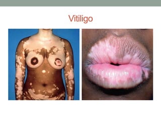
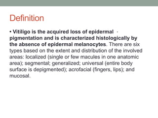
VITILIGO
Prepared By :- Dr Monther Fadel Nagi
Dermatology Resident
Vitiligo
Definition
•
• Vitiligo is the acquired loss of epidermal
pigmentation and is characterized histologically by
the absence of epidermal melanocytes. There are six
types based on the extent and distribution of the involved
areas: localized (single or few macules in one anatomic
area); segmental; generalized; universal (entire body
surface is depigmented); acrofacial (fingers, lips); and
mucosal.
Etiology
•
• Three pathophysiologic theories suggest possible
etiologies:
•
• Autoimmune theory (autoantibodies against melanocytes)
•
• Neural theory (neurochemical mediator selectively destroys
melanocytes)
•
• Self-destructive process whereby melanocytes fail to protect
themselves
•
against cytotoxic melanin precursors
•
• Although vitiligo is considered to be an acquired disease, 25%
to 30% is familial; the mode of transmission is unknown
(polygenic or autosomal dominant with incomplete
•
penetrance and variable expression).
Clinical Manifestation(s)
•
• Initially the disease is limited, but the lesions tend to
become more extensive over the years.
•
Physical Examination
•
• Hypopigmented and depigmented lesions favor sun-
exposed regions, intertriginous areas, genitalia, and sites
over bony prominences (type A vitiligo) .
•
• Areas around body orifices are also commonly
involved.
Clinical Manifestation(s)
•
• The lesions tend to be symmetric.
•
• Occasionally, the lesions are linear o pseudodermatomal
(type B vitiligo).
•
• Vitiligo lesions may occur at trauma sites (Koebner’s
phenomenon).
•
• The hair in affected areas may be white.
•
• The margins of the lesions are usually well demarcated,
and when a ring of hyperpigmentation is seen, the term
trichrome vitiligo is used.
•
• The term marginal inflammatory vitiligo is used to
describe lesions with raised borders.
Diagnostic Tests
•
• Physical examination
•
• Wood’s light examination may enhance lesions in
light-skinned individuals.
DIFFERENTIAL DIAGNOSIS
•
Acquired
•
• Chemical induced
•
• Halo nevus
•
• Idiopathic guttate hypomelanosis
•
• Leprosy
•
• Leukoderma associated with melanoma
•
• Pityriasis alba
•
• Postinflammatory hypopigmentation
•
• Tinea versicolor
•
• Vogt-Koyanagi syndrome (vitiligo, uveitis, and deafness)
DIFFERENTIAL DIAGNOSIS
•
Congenital
•
• Albinism, partial (piebaldism)
•
• Albinism, total
•
• Nevus anemicus
•
• Nevus depigmentosus
•
• Tuberous sclerosis
TREATMENT
•
First Line
•
• Treatment is indicated primarily for cosmetic purposes
when depigmentation causes emotional or social distress.
Depigmentation is more noticeable in darker complexions.
•
• Cosmetic masking agents (Dermablend, Covermark) or
stains (Dy-O-Derm, Vita-Dye) or sunless tanning lotions
(dihydroxyacetone)
•
• Topical midpotency steroids (e.g., triamcinolone 0.1% or
desonide 0.05% cream QD for 3–4 months)
TREATMENT
•
Second Line
•
• PUVA (psoralen phototherapy): oral or topical psoralen
administration followed by phototherapy with UVA (150–200
treatments required over 1–2 years) or narrowband
•
UVB phototherapy
•
• Excimer laser (best for small or localized areas of vitiligo)
•
• Total depigmentation (in cases of extensive vitiligo) with 20%
monobenzyl ether or hydroquinone. This is a permanent
procedure, and patients will require lifelong protection from sun
exposure.
•
• Intralesional steroid injection
•
• Systemic prednisone (5 mg QD on 2 consecutive days per
week for 2–4 months)
TREATMENT
•
Third Line
•
• Topical immunomodulators (tacrolimus, pimecrolimus)
can also induce repigmentation of vitiliginous skin lesions.
Their potential for systemic immunosuppression or
increased risk of skin or other malignancies remains to be
defined.
•
• Calcipotriol, a synthetic analogue of vitamin D3, has also
been used in combination with UV light or clobetasol, with
limited results
Vitiligo

Vitiligo

  • 1.
    VITILIGO Prepared By :-Dr Monther Fadel Nagi Dermatology Resident
  • 2.
  • 3.
    Definition • • Vitiligo isthe acquired loss of epidermal pigmentation and is characterized histologically by the absence of epidermal melanocytes. There are six types based on the extent and distribution of the involved areas: localized (single or few macules in one anatomic area); segmental; generalized; universal (entire body surface is depigmented); acrofacial (fingers, lips); and mucosal.
  • 4.
    Etiology • • Three pathophysiologictheories suggest possible etiologies: • • Autoimmune theory (autoantibodies against melanocytes) • • Neural theory (neurochemical mediator selectively destroys melanocytes) • • Self-destructive process whereby melanocytes fail to protect themselves • against cytotoxic melanin precursors • • Although vitiligo is considered to be an acquired disease, 25% to 30% is familial; the mode of transmission is unknown (polygenic or autosomal dominant with incomplete • penetrance and variable expression).
  • 5.
    Clinical Manifestation(s) • • Initiallythe disease is limited, but the lesions tend to become more extensive over the years. • Physical Examination • • Hypopigmented and depigmented lesions favor sun- exposed regions, intertriginous areas, genitalia, and sites over bony prominences (type A vitiligo) . • • Areas around body orifices are also commonly involved.
  • 6.
    Clinical Manifestation(s) • • Thelesions tend to be symmetric. • • Occasionally, the lesions are linear o pseudodermatomal (type B vitiligo). • • Vitiligo lesions may occur at trauma sites (Koebner’s phenomenon). • • The hair in affected areas may be white. • • The margins of the lesions are usually well demarcated, and when a ring of hyperpigmentation is seen, the term trichrome vitiligo is used. • • The term marginal inflammatory vitiligo is used to describe lesions with raised borders.
  • 7.
    Diagnostic Tests • • Physicalexamination • • Wood’s light examination may enhance lesions in light-skinned individuals.
  • 8.
    DIFFERENTIAL DIAGNOSIS • Acquired • • Chemicalinduced • • Halo nevus • • Idiopathic guttate hypomelanosis • • Leprosy • • Leukoderma associated with melanoma • • Pityriasis alba • • Postinflammatory hypopigmentation • • Tinea versicolor • • Vogt-Koyanagi syndrome (vitiligo, uveitis, and deafness)
  • 9.
    DIFFERENTIAL DIAGNOSIS • Congenital • • Albinism,partial (piebaldism) • • Albinism, total • • Nevus anemicus • • Nevus depigmentosus • • Tuberous sclerosis
  • 10.
    TREATMENT • First Line • • Treatmentis indicated primarily for cosmetic purposes when depigmentation causes emotional or social distress. Depigmentation is more noticeable in darker complexions. • • Cosmetic masking agents (Dermablend, Covermark) or stains (Dy-O-Derm, Vita-Dye) or sunless tanning lotions (dihydroxyacetone) • • Topical midpotency steroids (e.g., triamcinolone 0.1% or desonide 0.05% cream QD for 3–4 months)
  • 11.
    TREATMENT • Second Line • • PUVA(psoralen phototherapy): oral or topical psoralen administration followed by phototherapy with UVA (150–200 treatments required over 1–2 years) or narrowband • UVB phototherapy • • Excimer laser (best for small or localized areas of vitiligo) • • Total depigmentation (in cases of extensive vitiligo) with 20% monobenzyl ether or hydroquinone. This is a permanent procedure, and patients will require lifelong protection from sun exposure. • • Intralesional steroid injection • • Systemic prednisone (5 mg QD on 2 consecutive days per week for 2–4 months)
  • 12.
    TREATMENT • Third Line • • Topicalimmunomodulators (tacrolimus, pimecrolimus) can also induce repigmentation of vitiliginous skin lesions. Their potential for systemic immunosuppression or increased risk of skin or other malignancies remains to be defined. • • Calcipotriol, a synthetic analogue of vitamin D3, has also been used in combination with UV light or clobetasol, with limited results