The document summarizes key anatomical and physiological details of the extraocular muscles and eye movements:
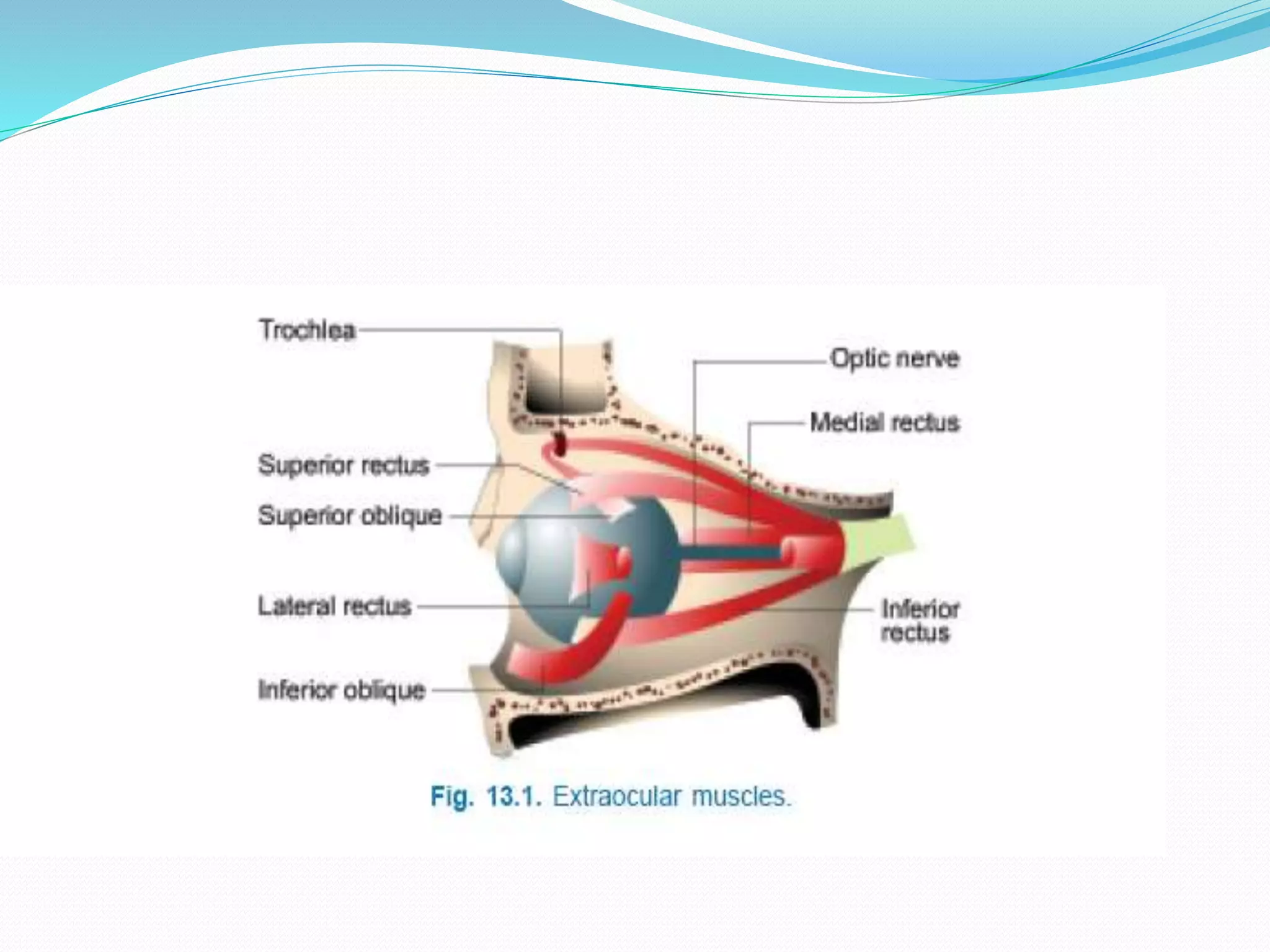
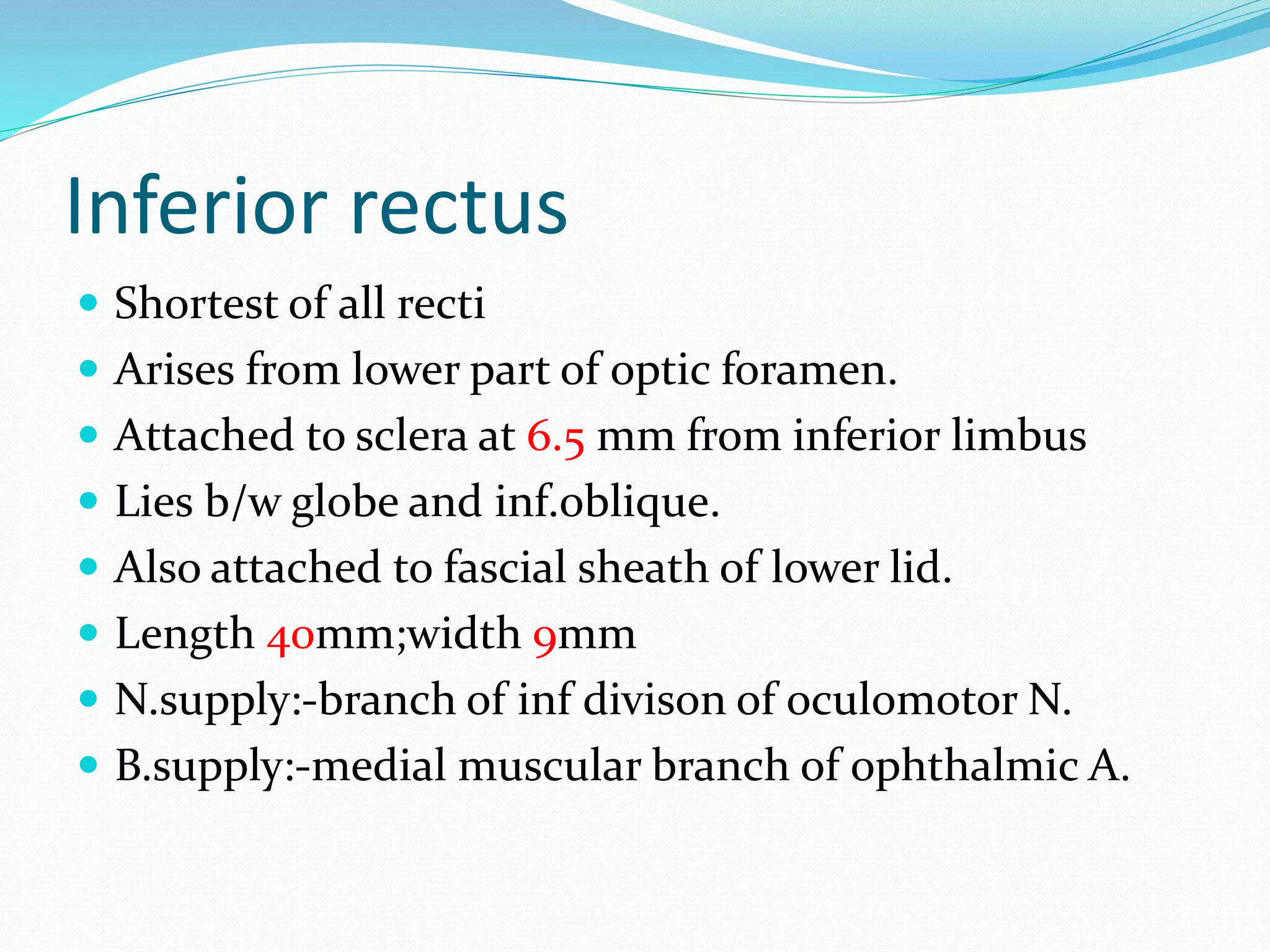
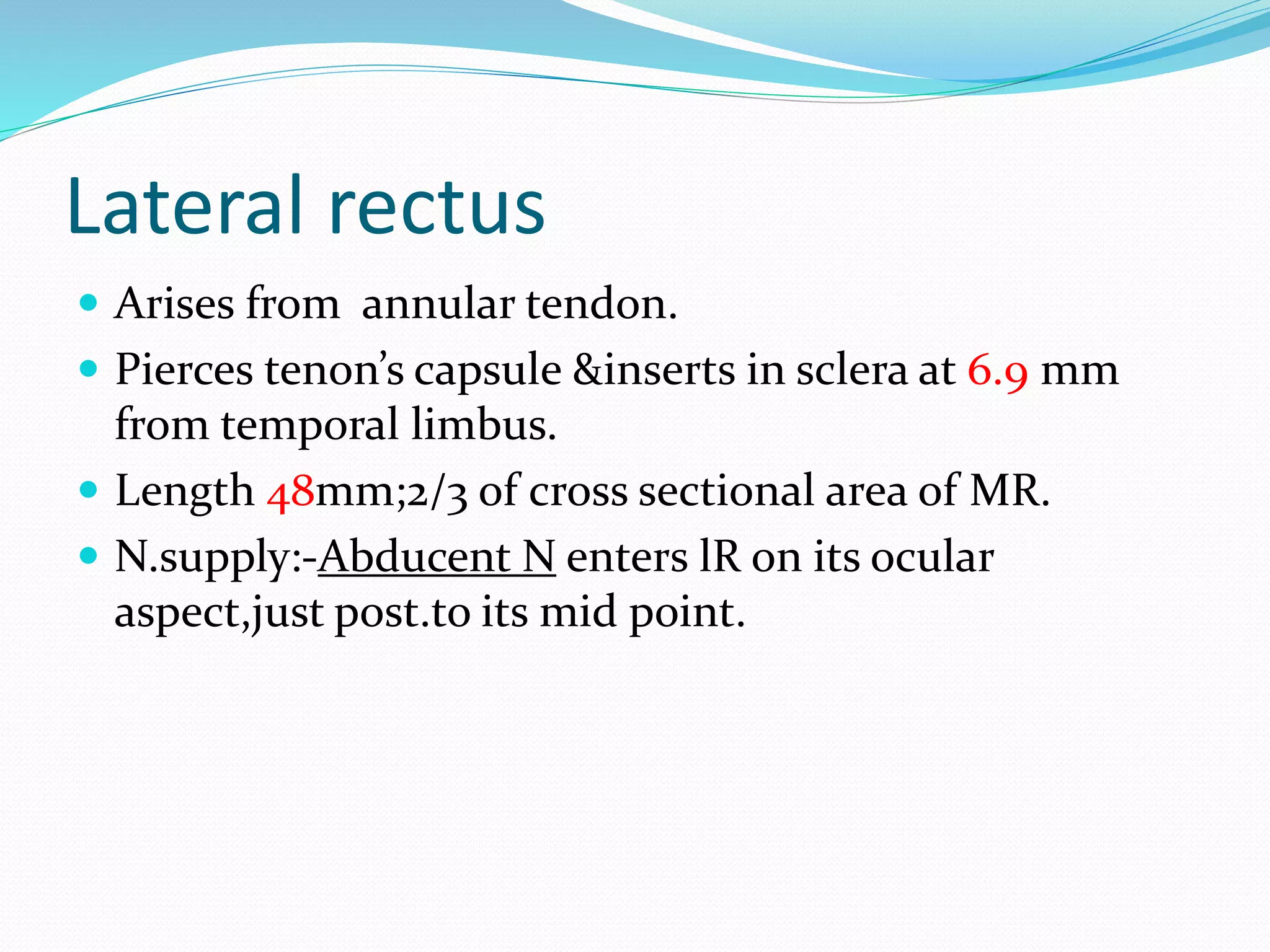
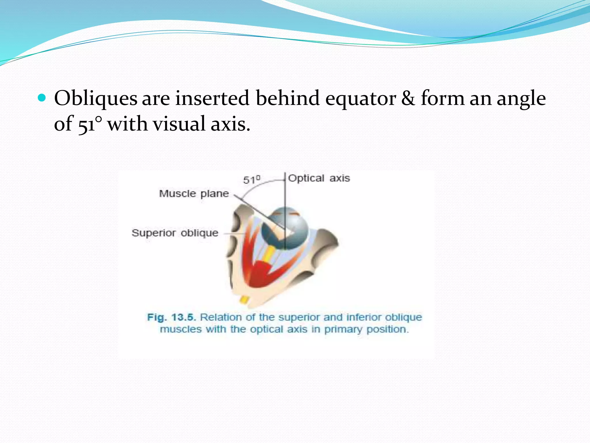
1. It describes the bony orbit anatomy, six extraocular muscles and their actions, innervation and blood supply. The four rectus muscles control horizontal and vertical eye movements, while the two oblique muscles enable torsional movements.
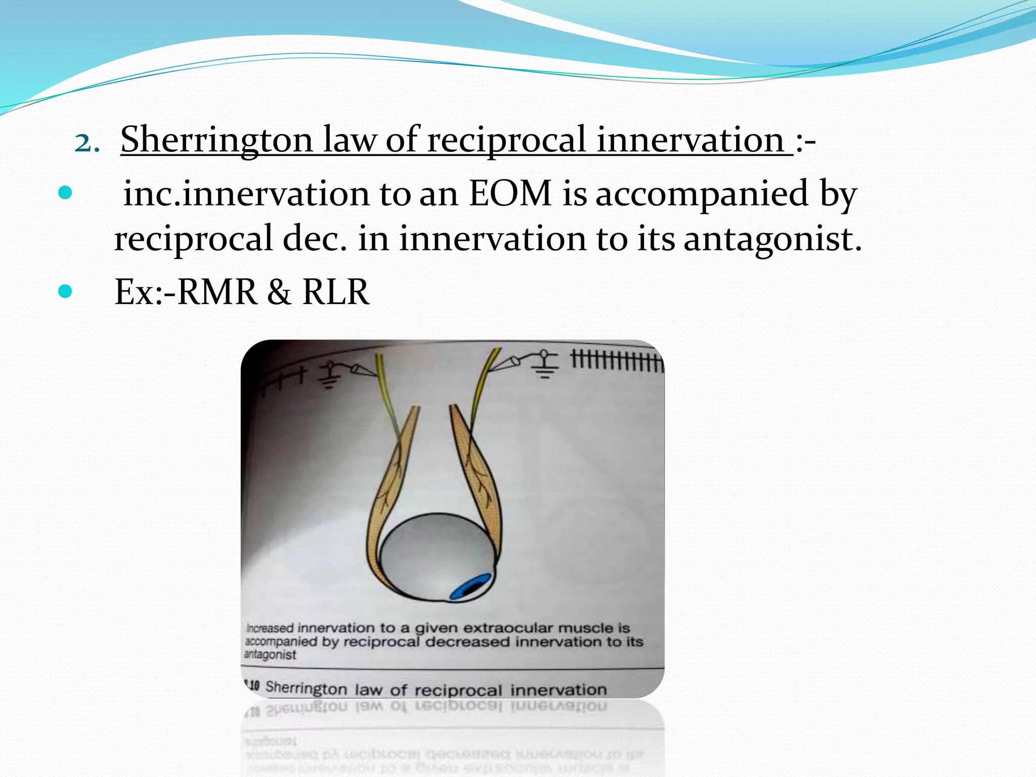
2. The document outlines uniocular and binocular eye movements including versions, vergences, and diagnostic positions of gaze. Hering's and Sherrington's laws govern coordinated eye movements between the eyes.
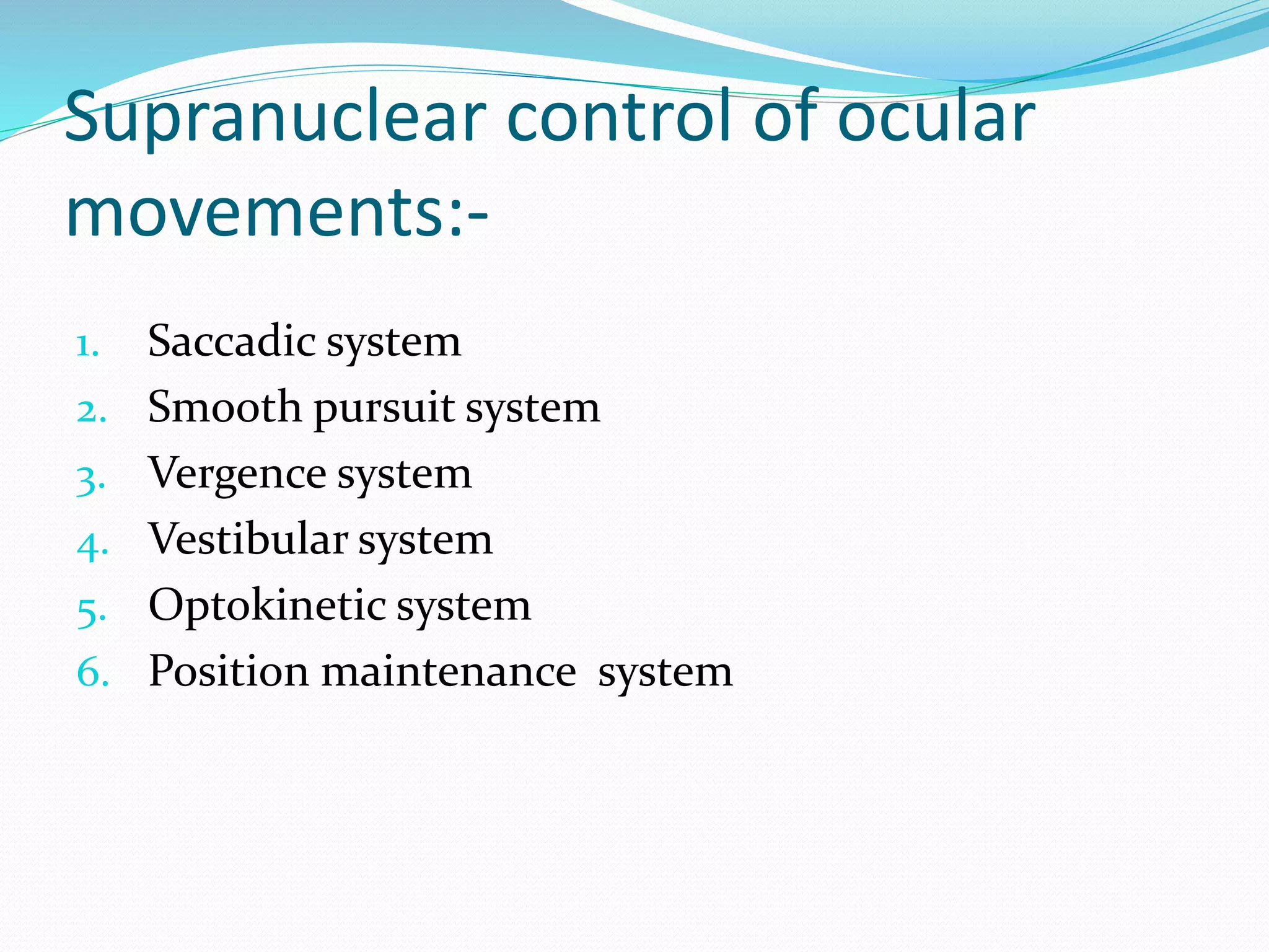
3. Supranuclear control systems like saccadic, smooth pursuit, vergence and vestibulo-ocular pathways mediate voluntary and reflexive eye movements
Each eyelid contains a fibrous plate, called a tarsus, that gives it structure and shape; muscles, which move the eyelids; and meibomian (or tarsal) glands, which secrete lubricating fluids. The lids are covered with skin, lined with mucous membrane, and bordered with a fringe of hairs, the eyelashes.
1. Introduction Gross anatomy Layers Blood supply, drainage and nerve supply
2. INTRODUCTION • Sclera forms posterior 5/6th of external tunic , connective tissue coat of eyeball. • it continues with duramater and cornea • Its whole surface covered by tenon’s capsule • Anteriorly covered by- bulbar conjunctiva • Inner surface lies in contact with choroid • With a potential suprachoroidal space in between
3. Equa THICKNESS OF SCLERA
4. • Thickness varies with individual, with age • Thinner- children, elder, F> M • Thickest posteriorly • Gradually becomes thinner when traced anteriorly • Thin at insertion of extraocular muscle
Extraocular musles(EOM) They are six in number Four recti: Superior rectus Inferior rectus Medial rectus Lateral rectus Two oblique muscles: Superior oblique Inferior oblique
3. SUPERIOR RECTUS MUSCLE . Origin Superior part of common annular tendon of Zinn Course Passes anterolaterally beneath the levator At 23 degrees with the globe ‘s AP axis Pierces Tenon s capsule Insertion into sclera by flat tendinous 10 mm broad insertion 7.7 mm behind sclero-corneal junction. 42 mm long 9 mm wide
4. Nerve supply Sup division of 3rd N Blood Supply Lateral Muscular br. of Ophthalmic A APPLIED: SR loosely bound to LPS muscle. • During SR resection- eyelid may be pulled forward narr owing palpebral fissure • In hypotropia pseudoptosis may be present Origin of SR and MR are closely attached to the dural sheat h of the optic nerve pain during upward & inward movements of the globe in RETROBULBAR NEURITIS
ANATOMY AND PHYSIOLOGY OF EXTRAOCULAR MUSCLES.pptANUJA DHAKAL
The presentation begins with an overview of the extraocular muscles, highlighting their crucial role in controlling eye movements and maintaining proper vision. Emphasized the significance of these muscles in daily activities and visual perception.
you can get information about the extraocular muscles which are responsible for the movement of the eyes in different direction, near and distance.
you will know how many extraocular muscles and how they work....
you will get information about the different position of gazes....
The extraocular muscles are the six muscles that control movement of the eye and one muscle that controls eyelid elevation. The actions of the six muscles responsible for eye movement depend on the position of the eye at the time of muscle contraction.
Human eye is a sense organ that responses to light and allows vision. Eyeball is placed in bony orbit in the skull and protected by eyelids. Eyeball is made up of three layers; Fibrous tunic (cornea and sclera), Vascular tunic (choroid, ciliary body and iris) and Retina. There are six extra ocular muscles to control movement of each eye. Optic nerve for its co-ordination with the brain. Blood is supplied to eye by the branches of internal carotid artery.
MANAGEMENT OF ATRIOVENTRICULAR CONDUCTION BLOCK.pdfJim Jacob Roy
Cardiac conduction defects can occur due to various causes.
Atrioventricular conduction blocks ( AV blocks ) are classified into 3 types.
This document describes the acute management of AV block.
micro teaching on communication m.sc nursing.pdfAnurag Sharma
Microteaching is a unique model of practice teaching. It is a viable instrument for the. desired change in the teaching behavior or the behavior potential which, in specified types of real. classroom situations, tends to facilitate the achievement of specified types of objectives.
TEST BANK for Operations Management, 14th Edition by William J. Stevenson, Ve...kevinkariuki227
TEST BANK for Operations Management, 14th Edition by William J. Stevenson, Verified Chapters 1 - 19, Complete Newest Version.pdf
These simplified slides by Dr. Sidra Arshad present an overview of the non-respiratory functions of the respiratory tract.
Learning objectives:
1. Enlist the non-respiratory functions of the respiratory tract
2. Briefly explain how these functions are carried out
3. Discuss the significance of dead space
4. Differentiate between minute ventilation and alveolar ventilation
5. Describe the cough and sneeze reflexes
Study Resources:
1. Chapter 39, Guyton and Hall Textbook of Medical Physiology, 14th edition
2. Chapter 34, Ganong’s Review of Medical Physiology, 26th edition
3. Chapter 17, Human Physiology by Lauralee Sherwood, 9th edition
4. Non-respiratory functions of the lungs https://academic.oup.com/bjaed/article/13/3/98/278874
HOT NEW PRODUCT! BIG SALES FAST SHIPPING NOW FROM CHINA!! EU KU DB BK substit...GL Anaacs
Contact us if you are interested:
Email / Skype : kefaya1771@gmail.com
Threema: PXHY5PDH
New BATCH Ku !!! MUCH IN DEMAND FAST SALE EVERY BATCH HAPPY GOOD EFFECT BIG BATCH !
Contact me on Threema or skype to start big business!!
Hot-sale products:
NEW HOT EUTYLONE WHITE CRYSTAL!!
5cl-adba precursor (semi finished )
5cl-adba raw materials
ADBB precursor (semi finished )
ADBB raw materials
APVP powder
5fadb/4f-adb
Jwh018 / Jwh210
Eutylone crystal
Protonitazene (hydrochloride) CAS: 119276-01-6
Flubrotizolam CAS: 57801-95-3
Metonitazene CAS: 14680-51-4
Payment terms: Western Union,MoneyGram,Bitcoin or USDT.
Deliver Time: Usually 7-15days
Shipping method: FedEx, TNT, DHL,UPS etc.Our deliveries are 100% safe, fast, reliable and discreet.
Samples will be sent for your evaluation!If you are interested in, please contact me, let's talk details.
We specializes in exporting high quality Research chemical, medical intermediate, Pharmaceutical chemicals and so on. Products are exported to USA, Canada, France, Korea, Japan,Russia, Southeast Asia and other countries.
- Video recording of this lecture in English language: https://youtu.be/lK81BzxMqdo
- Video recording of this lecture in Arabic language: https://youtu.be/Ve4P0COk9OI
- Link to download the book free: https://nephrotube.blogspot.com/p/nephrotube-nephrology-books.html
- Link to NephroTube website: www.NephroTube.com
- Link to NephroTube social media accounts: https://nephrotube.blogspot.com/p/join-nephrotube-on-social-media.html
Pulmonary Thromboembolism - etilogy, types, medical- Surgical and nursing man...VarunMahajani
Disruption of blood supply to lung alveoli due to blockage of one or more pulmonary blood vessels is called as Pulmonary thromboembolism. In this presentation we will discuss its causes, types and its management in depth.
Title: Sense of Taste
Presenter: Dr. Faiza, Assistant Professor of Physiology
Qualifications:
MBBS (Best Graduate, AIMC Lahore)
FCPS Physiology
ICMT, CHPE, DHPE (STMU)
MPH (GC University, Faisalabad)
MBA (Virtual University of Pakistan)
Learning Objectives:
Describe the structure and function of taste buds.
Describe the relationship between the taste threshold and taste index of common substances.
Explain the chemical basis and signal transduction of taste perception for each type of primary taste sensation.
Recognize different abnormalities of taste perception and their causes.
Key Topics:
Significance of Taste Sensation:
Differentiation between pleasant and harmful food
Influence on behavior
Selection of food based on metabolic needs
Receptors of Taste:
Taste buds on the tongue
Influence of sense of smell, texture of food, and pain stimulation (e.g., by pepper)
Primary and Secondary Taste Sensations:
Primary taste sensations: Sweet, Sour, Salty, Bitter, Umami
Chemical basis and signal transduction mechanisms for each taste
Taste Threshold and Index:
Taste threshold values for Sweet (sucrose), Salty (NaCl), Sour (HCl), and Bitter (Quinine)
Taste index relationship: Inversely proportional to taste threshold
Taste Blindness:
Inability to taste certain substances, particularly thiourea compounds
Example: Phenylthiocarbamide
Structure and Function of Taste Buds:
Composition: Epithelial cells, Sustentacular/Supporting cells, Taste cells, Basal cells
Features: Taste pores, Taste hairs/microvilli, and Taste nerve fibers
Location of Taste Buds:
Found in papillae of the tongue (Fungiform, Circumvallate, Foliate)
Also present on the palate, tonsillar pillars, epiglottis, and proximal esophagus
Mechanism of Taste Stimulation:
Interaction of taste substances with receptors on microvilli
Signal transduction pathways for Umami, Sweet, Bitter, Sour, and Salty tastes
Taste Sensitivity and Adaptation:
Decrease in sensitivity with age
Rapid adaptation of taste sensation
Role of Saliva in Taste:
Dissolution of tastants to reach receptors
Washing away the stimulus
Taste Preferences and Aversions:
Mechanisms behind taste preference and aversion
Influence of receptors and neural pathways
Impact of Sensory Nerve Damage:
Degeneration of taste buds if the sensory nerve fiber is cut
Abnormalities of Taste Detection:
Conditions: Ageusia, Hypogeusia, Dysgeusia (parageusia)
Causes: Nerve damage, neurological disorders, infections, poor oral hygiene, adverse drug effects, deficiencies, aging, tobacco use, altered neurotransmitter levels
Neurotransmitters and Taste Threshold:
Effects of serotonin (5-HT) and norepinephrine (NE) on taste sensitivity
Supertasters:
25% of the population with heightened sensitivity to taste, especially bitterness
Increased number of fungiform papillae
Lung Cancer: Artificial Intelligence, Synergetics, Complex System Analysis, S...Oleg Kshivets
RESULTS: Overall life span (LS) was 2252.1±1742.5 days and cumulative 5-year survival (5YS) reached 73.2%, 10 years – 64.8%, 20 years – 42.5%. 513 LCP lived more than 5 years (LS=3124.6±1525.6 days), 148 LCP – more than 10 years (LS=5054.4±1504.1 days).199 LCP died because of LC (LS=562.7±374.5 days). 5YS of LCP after bi/lobectomies was significantly superior in comparison with LCP after pneumonectomies (78.1% vs.63.7%, P=0.00001 by log-rank test). AT significantly improved 5YS (66.3% vs. 34.8%) (P=0.00000 by log-rank test) only for LCP with N1-2. Cox modeling displayed that 5YS of LCP significantly depended on: phase transition (PT) early-invasive LC in terms of synergetics, PT N0—N12, cell ratio factors (ratio between cancer cells- CC and blood cells subpopulations), G1-3, histology, glucose, AT, blood cell circuit, prothrombin index, heparin tolerance, recalcification time (P=0.000-0.038). Neural networks, genetic algorithm selection and bootstrap simulation revealed relationships between 5YS and PT early-invasive LC (rank=1), PT N0—N12 (rank=2), thrombocytes/CC (3), erythrocytes/CC (4), eosinophils/CC (5), healthy cells/CC (6), lymphocytes/CC (7), segmented neutrophils/CC (8), stick neutrophils/CC (9), monocytes/CC (10); leucocytes/CC (11). Correct prediction of 5YS was 100% by neural networks computing (area under ROC curve=1.0; error=0.0).
CONCLUSIONS: 5YS of LCP after radical procedures significantly depended on: 1) PT early-invasive cancer; 2) PT N0--N12; 3) cell ratio factors; 4) blood cell circuit; 5) biochemical factors; 6) hemostasis system; 7) AT; 8) LC characteristics; 9) LC cell dynamics; 10) surgery type: lobectomy/pneumonectomy; 11) anthropometric data. Optimal diagnosis and treatment strategies for LC are: 1) screening and early detection of LC; 2) availability of experienced thoracic surgeons because of complexity of radical procedures; 3) aggressive en block surgery and adequate lymph node dissection for completeness; 4) precise prediction; 5) adjuvant chemoimmunoradiotherapy for LCP with unfavorable prognosis.
2. Bony orbit
Angle of the medial and lateral walls of the orbit is 45°
so the optical axis forms approimately 23 °with both
medial &lateral walls.
The medial walls of the 2 orbits are parallel to each
other.
5. Two oblique muscles:-
Superior oblique
Inferior oblique
In addition levator palpebrae superioris also present
&it inserts into upper eye lid for elevating palpebral
fissure.
The 4 rectii arises from fibrous ring (annulus of
zinn)around optic foramen.
6. Vertical recti(sup.&inf. Rectus ) run in line with orbital
axis & are inserted infront of equator.
They form an angle of 23° with visual axis.
7. Superior rectus
Arises from upper part of annulus o zinn.
Below the attachment of levator M.
Continuous with attachment of med.,&lat. Recti
Pierces tenon’s capsule &it is inserted into sclera 7.7
mm from superior limbus.
Length 48 mm;width 9mm.
N.supply:-sup.divison of oculomotor N.
B.supply:-lat. Muscular branch of ophthalmic A.
8. Inferior rectus
Shortest of all recti
Arises from lower part of optic foramen.
Attached to sclera at 6.5 mm from inferior limbus
Lies b/w globe and inf.oblique.
Also attached to fascial sheath of lower lid.
Length 40mm;width 9mm
N.supply:-branch of inf divison of oculomotor N.
B.supply:-medial muscular branch of ophthalmic A.
9. Medial rectus
Largest ocular M& stronger than lateral rectus.
Arise from medial & inferior sides of optic foramen
Passing along medial wall of orbit ;inserts 5.5mm from
nasal limbus.
Length 40mm;thicker than other EOM.
N.supply:-inf.divison of oculomotor N.
B.supply:-medial muscular branch of ophthalmic A.
10. Lateral rectus
Arises from annular tendon.
Pierces tenon’s capsule &inserts in sclera at 6.9 mm
from temporal limbus.
Length 48mm;2/3 of cross sectional area of MR.
N.supply:-Abducent N enters lR on its ocular
aspect,just post.to its mid point.
11. Spiral of tillaux
Imaginary line joining the insertions of the 4 recti and
is an important anatomical landmark when
performing surgery.
The insertions are located progressively further away
from the limbus in a spiral pattern.
the medial rectus insertion is closest .
Superior rectus is farthest.
12.
13. Obliques are inserted behind equator & form an angle
of 51° with visual axis.
14. Superior oblique
Longest& thinnest EOM.
Arises from common origin at the apex of orbit;
superomedial to optic foramen.
Runs forward to trochlea(cartilaginous ring at
upper&inner angles of orbit)
After threading through this it becomes tendinous
It changes its direction completely and runs over the
globe under SR to attach above & lat, to posterior pole.
15. Ant.fibres of S.O tendon-intorsion
Post.fibres of S.O tendon-extorsion
N.supply:-Trochlear N(4) after dividing into 2-3
branches enters muscle superiorly.
B.supply:-superior muscular branch of ophthalmic A.
16. Inferior oblique
Only EOM not arising from apex of orbit
It arises anteriorly from lower & inner orbital walls
near lacrimal fossa.
Running below inf.rectus& attaches below&lat. to
post.pole of globe.
N.supply:-Inf.divison of oculomotor N.
B.supply:-Infraorbital &medial muscular branches of
ophtalmic A.
17. Action of extraocular muscles
Rotation around centre of rotation
Centre of rotation lies 12/13 mm behind cornea.
3 types of rotation:
1. Rotation around fick vertical axis Z—side to side
2. “ “ fick horizontal axis X– up&down
3. “ “ fick antero posterior axis– torsion
18.
19. Uniocular movements
Ductions – only one eye is open,the other covered/closed
tested by asking the pt. to follow a target in each direction
of gaze.
Types of ductions:-
1. Adduction
2. Abduction
3. Supraduction
4. Infraduction
5. Incycloduction
6. excycloduction
20.
21. Binocular movements
Versions:-both eyes open,attempting to fixate a target
&moving in same direction.
Binocular ,simultaneous,conjugate movements in
same direction.
Abduction of one eye accompanied by adduction of
other eye is called conjugate movements.
22. Types of versions:-
Dextroversion&laevo version
Elevation&depression
Dextro elevation&dextro depression
Laevo elevation& laevo depression
23. Torsional movements/righting reflexes:-
When you tilt head to maintain upright image.
Vergences:-
binocular,simultaneous,disjugate/disjunctive
movements (opp.direction)
Convergence– simultaneous adduction
Divergence– outward movement from convergent
position
25. Actions of EOM
ACTION PRIMARY SECONDARY TERTIARY
MR ADDUCTION ------ ---------
LR ABDUCTION ------ ---------
SR ELEVATION INTORSION ADDUCTION
IR DEPRESSION EXTORSION ADDUCTION
SO INTORSION DEPRESSION ABDUCTION
IO EXTORSION ELEVATION ABDUCTION
26. Both obliques have same tertiary action because
inserted behind the center of rotation,
pull post. pole of globe medially
when they contract ant.portion of eye so it causes
abduction
27. Both recti have same tertiary action bcz they inserted
anterior to centre of rotation
pull ant.portion of globe medially so it causes
adduction
28. Synergists:-ref.to muscles having same primary action
in same eye.
Ex:-sup.rectus & inf.oblique----elevators
inf.rectus&sup.oblique-----depressors
Antagonists:-having opp.action in same eye
Ex:-sup.&inf. Recti
sup.&inf.oblique
29. Yoke muscle(contralateral synergists):-
Ref. to pair of muscles (one from each eye) which
contract simultaneously during version movements.
Ex :-in dextroversion RLR &LMR
Contralateral antagonist:-pair of muscle (one from
each eye)having an opposite action.
Ex:-in dextroversion RLR & LLR
30. Diagnostic positions of gaze:-9
1 Primary position of gaze:-assumed by eyes when
fixating a distant object with head erect.
6 cardinal positions :- to test 12 EOM in their main
field of action
33. Laws of ocular motility
1. Hering’s law of equal innervation:- during any
conjugate movement equal & simultaneous
innervation flows to yoke muscles
34. 2. Sherrington law of reciprocal innervation :-
inc.innervation to an EOM is accompanied by
reciprocal dec. in innervation to its antagonist.
Ex:-RMR & RLR
35. Supranuclear control of ocular
movements:-
1. Saccadic system
2. Smooth pursuit system
3. Vergence system
4. Vestibular system
5. Optokinetic system
6. Position maintenance system
36. Saccadic system:-
saccades are sudden,jerky,conjugate,movements
as the gaze shifts from one object to other.
voluntary(normal)
invoiuntary(peripheral,auditory,visual
stimuli)
37. Smooth pursuit eye movements:-
Tracking movements of eye as they follow moving
object
Voluntary movements
When the velocity of moving object inc. replaced by
small saccades(“catchup saccades”)
38. Vergence movement:
Allow focussing an object which moves away
from/towards observer.
Very slow disjugate movements
39. Vestibular eye movement:-
Effective in compensating for effects of head
movements in disturbing visual fixation
Through vestibular system
40. Optokinetic system:-
a movement following the moving scene , succeeded
by rapid saccade in opp.direction
Position maintenance system:-
Helps to maintain specific gaze by rapid micro
movements called “flicks” & slow micro movements
called “drifts”.