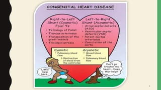
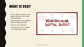
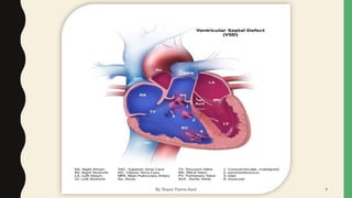
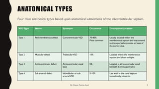
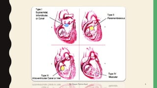
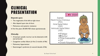
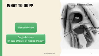
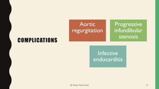
V E N T R I C U L A R
S E P T A L D E F E C T
By: Shayan Fatima Azad 1
By: Shayan Fatima Azad 2
WHAT IS VSD?
• VSD is a defect in interventricular
septum that allows a left to right
shunting of blood.
• Approximately 2-8 out of 1000 live
births are affected withVSD.
• They may occur separately.
• They may also occur as complex
set of cardiac abnormalities.
By: Shayan Fatima Azad 3
By: Shayan Fatima Azad 4
ANATOMICAL TYPES
Four main anatomical types based upon anatomical subsections of the interventricular septum.
VSDType Name Synonym Occurance Description/Location
Type 1 Peri membranous defect ConoventricularVSD 70-80%
Most common
Usually located within the
membranous septum and may extend
to tricuspid valve annulas or base of
the aortic valve.
Type 2 Muscular defect TrabecularVSD 10% Located within the membranous
septum and often multiple.
Type 3 Atrioventricular defect Atrioventricular canal
type
5% Located in atrioventricular canal
beneath the tricuspid valve.
Type 4 Sub arterial defect Infundibular or sub
arterialVSD
5-10% Lies with in the canal septum
immediately subaortic.
By: Shayan Fatima Azad 5
By: Shayan Fatima Azad 6
PATHOPHYSIOLOGY
• A defect in the interventricular septum allows communication between the systemic and pulmonary circulations.
• As a result, flow moves from a region of high pressure to a region of low pressure—that is, from the LV to the RV (a
left-to-right shunt).
• The pathophysiologic effects of aVSD derive from the hemodynamic effects secondary to a left-to-right shunt and
from changes in the pulmonary vasculature.
• A left-to-right shunt at the ventricular level has following hemodynamic consequences:
1. Increased LV volume load
2. Excessive pulmonary blood flow (may lead to progressive edema and congestive cardiac failure).
3. Reduced systemic cardiac output
4. Elevated pulmonary artery pressures (due to persistent elevated pulmonary blood flow and pulmonary vascular
resistance lead to irreversible pulmonary hypertension)
They may also result in reversal of flow across the defect called as Eisenmenger’s syndrome
By: Shayan Fatima Azad 7
PATHOPHYSIOLOGY OF SHUNTING
The pressure generated during
contraction by the left
ventricle is higher than that
generated by the simultaneous
contraction of the right
ventricle. Blood will thus be
pushed through the VSD (also
called "shunted") from the left
ventricle to the right ventricle.
The right ventricle has to do
extra work to handle the
additional blood volume. It
may have trouble keeping up
with the load and enlarge,
affecting its ability to pump
efficiently.
In addition, the lungs receive
too much blood under too
much pressure.
The arterioles (small arteries)
in the lungs thicken in
response to the excess blood
under excess pressure. If this
extra pressure persists,
permanent damage can be
done to the lungs.
It makes a considerable
difference whether the size of
the VSD is small or large
By: Shayan Fatima Azad 8
CLINICAL
PRESENTATION
Depends upon:
• The magnitude of the left-to-right shunt
• Also depend upon size of shunt
• Pulmonary and systemic resistance
In first five years 30-50%VSD closes spontaneously
Clinically:
• A loud pan systolic murmur can be detected at left
sternal border
• Congestive cardiac failure (in first 2 months of life)
• Pulmonary hypertension
• Eisenmenger’s syndrome (in second decade of life)
By: Shayan Fatima Azad 9
SIZES OF VSD
By: Shayan Fatima Azad 10
RISK FACTORS
Factors affecting the
developing fetal heart can
be associated with
development ofVSDs.
Genetic conditions
(chromosomal, single gene
or polygenic), like trisomy
(Edward’s, Patau,Turner’s,
Down’s syndrome)
Environmental influences
Fetal alcohol syndrome
Gestational Diabetes
Mellitus
By: Shayan Fatima Azad 11
DIAGNOSIS
ECG
Chest X-Ray
Echocardiography
(degree of shunting)
Cardiac catheterization
(quantify various pressures
between chambers and to assess
the degree of pulmonary HTN
and Oxygen saturation)
By: Shayan Fatima Azad 12
WHAT TO DO??
Medical therapy
Surgical closure
(in case of failure of medical therapy)
By: Shayan Fatima Azad 13
COMPLICATIONS
Aortic
regurgitation
Progressive
infundibular
stenosis
Infective
endocarditis
By: Shayan Fatima Azad 14
PROGNOSIS
• Long term prognosis is excellent
• If treated within 1-2 years of life
By: Shayan Fatima Azad 15
HOPE YOU HAVE NO QUESTION
By: Shayan Fatima Azad 16
By: Shayan Fatima Azad 17

Ventricular septal defect

  • 1.
    V E NT R I C U L A R S E P T A L D E F E C T By: Shayan Fatima Azad 1
  • 2.
  • 3.
    WHAT IS VSD? •VSD is a defect in interventricular septum that allows a left to right shunting of blood. • Approximately 2-8 out of 1000 live births are affected withVSD. • They may occur separately. • They may also occur as complex set of cardiac abnormalities. By: Shayan Fatima Azad 3
  • 4.
  • 5.
    ANATOMICAL TYPES Four mainanatomical types based upon anatomical subsections of the interventricular septum. VSDType Name Synonym Occurance Description/Location Type 1 Peri membranous defect ConoventricularVSD 70-80% Most common Usually located within the membranous septum and may extend to tricuspid valve annulas or base of the aortic valve. Type 2 Muscular defect TrabecularVSD 10% Located within the membranous septum and often multiple. Type 3 Atrioventricular defect Atrioventricular canal type 5% Located in atrioventricular canal beneath the tricuspid valve. Type 4 Sub arterial defect Infundibular or sub arterialVSD 5-10% Lies with in the canal septum immediately subaortic. By: Shayan Fatima Azad 5
  • 6.
  • 7.
    PATHOPHYSIOLOGY • A defectin the interventricular septum allows communication between the systemic and pulmonary circulations. • As a result, flow moves from a region of high pressure to a region of low pressure—that is, from the LV to the RV (a left-to-right shunt). • The pathophysiologic effects of aVSD derive from the hemodynamic effects secondary to a left-to-right shunt and from changes in the pulmonary vasculature. • A left-to-right shunt at the ventricular level has following hemodynamic consequences: 1. Increased LV volume load 2. Excessive pulmonary blood flow (may lead to progressive edema and congestive cardiac failure). 3. Reduced systemic cardiac output 4. Elevated pulmonary artery pressures (due to persistent elevated pulmonary blood flow and pulmonary vascular resistance lead to irreversible pulmonary hypertension) They may also result in reversal of flow across the defect called as Eisenmenger’s syndrome By: Shayan Fatima Azad 7
  • 8.
    PATHOPHYSIOLOGY OF SHUNTING Thepressure generated during contraction by the left ventricle is higher than that generated by the simultaneous contraction of the right ventricle. Blood will thus be pushed through the VSD (also called "shunted") from the left ventricle to the right ventricle. The right ventricle has to do extra work to handle the additional blood volume. It may have trouble keeping up with the load and enlarge, affecting its ability to pump efficiently. In addition, the lungs receive too much blood under too much pressure. The arterioles (small arteries) in the lungs thicken in response to the excess blood under excess pressure. If this extra pressure persists, permanent damage can be done to the lungs. It makes a considerable difference whether the size of the VSD is small or large By: Shayan Fatima Azad 8
  • 9.
    CLINICAL PRESENTATION Depends upon: • Themagnitude of the left-to-right shunt • Also depend upon size of shunt • Pulmonary and systemic resistance In first five years 30-50%VSD closes spontaneously Clinically: • A loud pan systolic murmur can be detected at left sternal border • Congestive cardiac failure (in first 2 months of life) • Pulmonary hypertension • Eisenmenger’s syndrome (in second decade of life) By: Shayan Fatima Azad 9
  • 10.
    SIZES OF VSD By:Shayan Fatima Azad 10
  • 11.
    RISK FACTORS Factors affectingthe developing fetal heart can be associated with development ofVSDs. Genetic conditions (chromosomal, single gene or polygenic), like trisomy (Edward’s, Patau,Turner’s, Down’s syndrome) Environmental influences Fetal alcohol syndrome Gestational Diabetes Mellitus By: Shayan Fatima Azad 11
  • 12.
    DIAGNOSIS ECG Chest X-Ray Echocardiography (degree ofshunting) Cardiac catheterization (quantify various pressures between chambers and to assess the degree of pulmonary HTN and Oxygen saturation) By: Shayan Fatima Azad 12
  • 13.
    WHAT TO DO?? Medicaltherapy Surgical closure (in case of failure of medical therapy) By: Shayan Fatima Azad 13
  • 14.
  • 15.
    PROGNOSIS • Long termprognosis is excellent • If treated within 1-2 years of life By: Shayan Fatima Azad 15
  • 16.
    HOPE YOU HAVENO QUESTION By: Shayan Fatima Azad 16
  • 17.