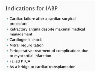
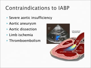
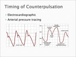
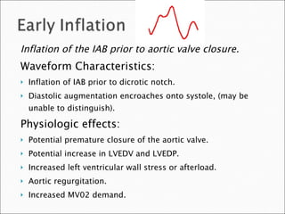
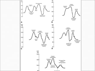
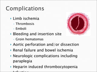
Has a linear relationship to: Systolic wall stress Intraventricular pressure Afterload End diastolic volume Wall thickness
 
Cardiac failure after a cardiac surgical procedure Refractory angina despite maximal medical management Cardiogenic shock Mitral regurgitation Perioperative treatment of complications due to myocardial infarction Failed PTCA As a bridge to cardiac transplantation
Severe aortic insufficiency Aortic aneurysm Aortic dissection Limb ischemia Thromboembolism
The end of the balloon should be just distal to the takeoff of the left subclavian artery Position should be confirmed by fluoroscopy or chest x-ray
Electrocardiographic Arterial pressure tracing
Inflation of the IAB prior to aortic valve closure. Waveform Characteristics: Inflation of IAB prior to dicrotic notch. Diastolic augmentation encroaches onto systole, (may be unable to distinguish). Physiologic effects: Potential premature closure of the aortic valve. Potential increase in LVEDV and LVEDP. Increased left ventricular wall stress or afterload. Aortic regurgitation. Increased MV02 demand.
Inflation of the IAB markedly after closure of the aortic valve. Waveform Characteristics: Inflation of IAB after the dicrotic notch. Absence of sharp V. Physiologic Effects: Sub-optimal coronary artery perfusion.
Premature deflation of the IAB during the diastolic phase. Waveform Characteristics: Deflation of IAB is seen as a sharp drop following diastolic augmentation. Sub-optimal diastolic augmentation. Assisted aortic end diastolic pressure may be <= the unassisted aortic end diastolic pressure. Assisted systolic pressure may rise. Physiologic Effects: •  Sub-optimal coronary perfusion. •  Potential for retrograde coronary and carotid blood flow. Sub-optimal after load reduction & Increased MV02 demand.
Late deflation of the IAB during the diastolic phase. Waveform Characteristics: Assisted aortic end diastolic pressure may be equal to the unassisted aortic end diastolic pressure. Rate of rise of assisted systole is prolonged. Diastolic augmentation may appear widened. Physiologic Effects: Afterload reduction is essentially absent. Increased MV02 consumption due to the left ventricle ejecting against a greater resistance IAB may impede left ventricular ejection and increase the afterload
Arterial pressure waveforms
 
Limb ischemia Thrombosis Emboli Bleeding and insertion site Groin hematomas Aortic perforation and/or dissection Renal failure and bowel ischemia Neurologic complications including paraplegia Heparin induced thrombocytopenia Infection
Timing of weaning Patient should be stable for 24-48 hours Decreasing inotropic support Decreasing pump ratio  From 1:1 to 1:2 or 1:3 Decrease augmentation Monitor patient closely If patient becomes unstable, weaning should be immediately discontinued
Discontinue heparin six hours prior Check platelets and coagulation factors Deflate the balloon Apply manual pressure above and below IABP insertion site Remove and alternate pressure to expel any clots Apply constant pressure to the insertion site for a minimum of 30 minutes Check distal pulses frequently

  • 1.
  • 2.
    Has a linearrelationship to: Systolic wall stress Intraventricular pressure Afterload End diastolic volume Wall thickness
  • 3.
  • 4.
    Cardiac failure aftera cardiac surgical procedure Refractory angina despite maximal medical management Cardiogenic shock Mitral regurgitation Perioperative treatment of complications due to myocardial infarction Failed PTCA As a bridge to cardiac transplantation
  • 5.
    Severe aortic insufficiencyAortic aneurysm Aortic dissection Limb ischemia Thromboembolism
  • 6.
    The end ofthe balloon should be just distal to the takeoff of the left subclavian artery Position should be confirmed by fluoroscopy or chest x-ray
  • 7.
  • 8.
    Inflation of theIAB prior to aortic valve closure. Waveform Characteristics: Inflation of IAB prior to dicrotic notch. Diastolic augmentation encroaches onto systole, (may be unable to distinguish). Physiologic effects: Potential premature closure of the aortic valve. Potential increase in LVEDV and LVEDP. Increased left ventricular wall stress or afterload. Aortic regurgitation. Increased MV02 demand.
  • 9.
    Inflation of theIAB markedly after closure of the aortic valve. Waveform Characteristics: Inflation of IAB after the dicrotic notch. Absence of sharp V. Physiologic Effects: Sub-optimal coronary artery perfusion.
  • 10.
    Premature deflation ofthe IAB during the diastolic phase. Waveform Characteristics: Deflation of IAB is seen as a sharp drop following diastolic augmentation. Sub-optimal diastolic augmentation. Assisted aortic end diastolic pressure may be <= the unassisted aortic end diastolic pressure. Assisted systolic pressure may rise. Physiologic Effects: • Sub-optimal coronary perfusion. • Potential for retrograde coronary and carotid blood flow. Sub-optimal after load reduction & Increased MV02 demand.
  • 11.
    Late deflation ofthe IAB during the diastolic phase. Waveform Characteristics: Assisted aortic end diastolic pressure may be equal to the unassisted aortic end diastolic pressure. Rate of rise of assisted systole is prolonged. Diastolic augmentation may appear widened. Physiologic Effects: Afterload reduction is essentially absent. Increased MV02 consumption due to the left ventricle ejecting against a greater resistance IAB may impede left ventricular ejection and increase the afterload
  • 12.
  • 13.
  • 14.
    Limb ischemia ThrombosisEmboli Bleeding and insertion site Groin hematomas Aortic perforation and/or dissection Renal failure and bowel ischemia Neurologic complications including paraplegia Heparin induced thrombocytopenia Infection
  • 15.
    Timing of weaningPatient should be stable for 24-48 hours Decreasing inotropic support Decreasing pump ratio From 1:1 to 1:2 or 1:3 Decrease augmentation Monitor patient closely If patient becomes unstable, weaning should be immediately discontinued
  • 16.
    Discontinue heparin sixhours prior Check platelets and coagulation factors Deflate the balloon Apply manual pressure above and below IABP insertion site Remove and alternate pressure to expel any clots Apply constant pressure to the insertion site for a minimum of 30 minutes Check distal pulses frequently