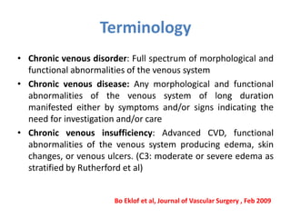
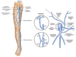
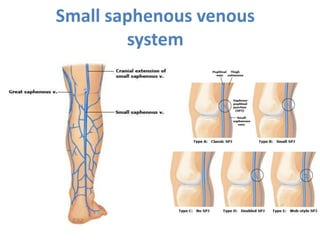
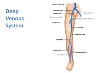
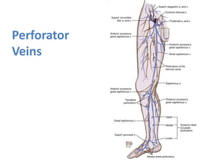
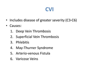
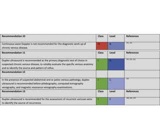
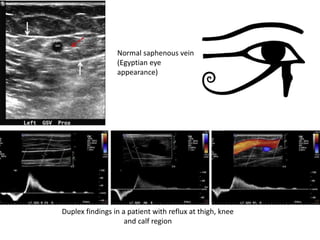
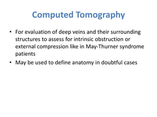
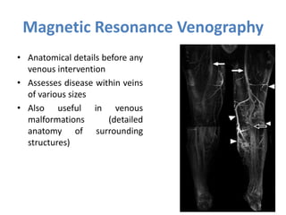
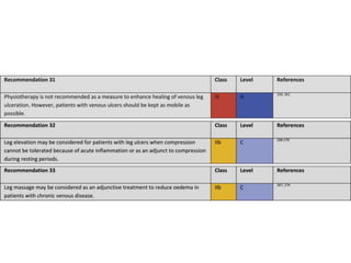
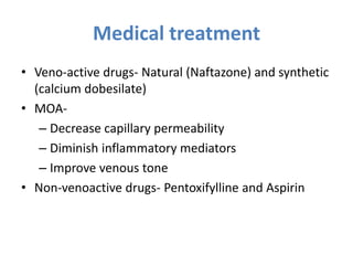
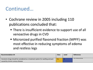
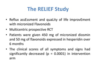
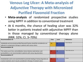
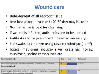
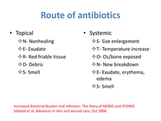
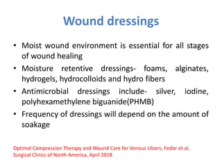
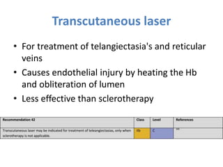
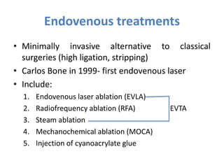
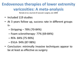
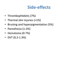
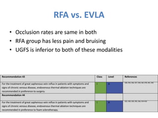
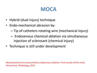
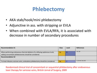
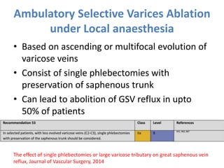
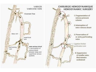
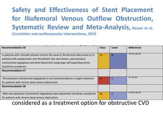
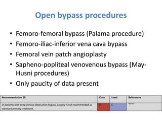
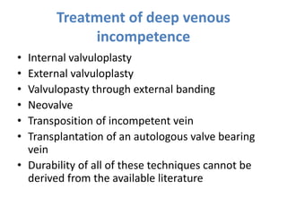
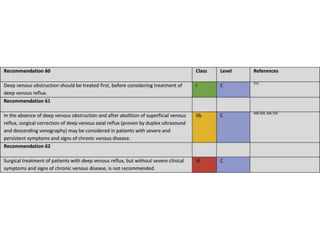
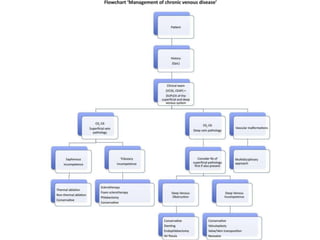
Recent advances in management of
chronic venous insufficiency
Moderator: Dr. Piyush Ranjan
Presenter: Kavindra Singh
Terminology
• Chronic venous disorder: Full spectrum of morphological and
functional abnormalities of the venous system
• Chronic venous disease: Any morphological and functional
abnormalities of the venous system of long duration
manifested either by symptoms and/or signs indicating the
need for investigation and/or care
• Chronic venous insufficiency: Advanced CVD, functional
abnormalities of the venous system producing edema, skin
changes, or venous ulcers. (C3: moderate or severe edema as
stratified by Rutherford et al)
Bo Eklof et al, Journal of Vascular Surgery , Feb 2009
Continued…
• Venous symptoms: tingling, aching, burning, pain, muscle
cramps, swelling, sensations of throbbing or heaviness, itching
skin, restless legs, leg-tiredness and/or fatigue. Suggestive of
CVD, particularly if they are exacerbated by heat or
dependency in the day's course, and relieved with leg rest
and/or elevation.
• Venous signs: include dilated veins (telangiectasia, reticular
veins, varicose veins), leg edema, skin changes, ulcers, post
phlebotomy
Historical perspective
• Hippocrates (460-377 BC)- Upright position inappropriate for
leg with ulceration and also recommended puncture of
varicose veins
• Brodie (17th century) – Symptoms and signs of CVI
• Pravaz (1831)- Basis of sclerotherapy
• Trendelenburg (1891)- test to diff. b/w superficial and deep
reflux and GSV ligature at mid thigh to control distal
varicosities
• Perthes (1896)- Test for patency of deep veins
Anatomy
Small saphenous venous
system
Deep
Venous
System
Perforator
Veins
CVI
• Includes disease of greater severity (C3-C6)
• Causes:
1. Deep Vein Thrombosis
2. Superficial Vein Thrombosis
3. Phlebitis
4. May-Thurner Syndrome
5. Arterio-venous Fistula
6. Varicose Veins
May-Thurner Syndrome
• Ileocaval-venous CS, iliac
vein CS, cocketts
syndrome and venous
spur
• MC variant- compression
of left CIV bw right CIA
and 5th lumbar vertebra
• Intravascular USG is the
imaging modality of
choice
• Needs stenting if
associated with stenosis
and thrombosis
CEAP Classification
• Originally described in 1994
• Revised in 2004
• Descriptive classification
• Not used to measure severity of disease
Clinical Manifestations
• C0- No visible or palpable signs of venous disease
• A- asymptomatic, S- symptomatic
• Pigmentation- because of hemosiderin deposition
• Eczema- manifestation of stasis dermatitis
• Atrophie blanche- atrophic, hypopigmented patches with
focal red punctate dots or telangiectasias, surrounded by
hyperpigmentation. Corresponds to avascular fibrosis
• Lipodermatosclerosis- fibrosing panniculitis of the
subcutaneous tissue
C1-
Telangiectasias
and reticular
veins
C2- Varicose
veins
C3- Pedal
edema
C4a- Pigmentation and eczema C4b-
Lipodermatosclerosis
C5- Healed ulcer C6- Active ulcer
Etiology
• Ec- Congenital venous disorders present at birth or
develop in childhood (e.g. Klippel-Trenaunay
syndrome).
• Ep- Primary venous disorders
• Es- Consequence of another pathology, such as
following venous thrombosis or trauma
• En- origin of venous disease is not identified
Anatomy
• As- Superficial venous system
• Ap- Perforating veins
• Ad- Deep venous system
• An- Anatomy not specified
Pathophysiology
• Pr- Venous reflux
• Po- Venous obstruction
• Pr,o- Venous reflux and obstruction
• Pn- Reflux not specified
Venous Clinical Severity Scale
• Complimentary to CEAP classification
• Uses 10 clinical parameters, each scored from 0-3
1. Pain 6. Induration
2. Varicose vein 7. No. of active ulcers
3. Venous edema 8. Duration of active ulcers
4. Pigmentation 9. Size of active ulcers
5. Inflammation 10. Compliance to
compression therapy
Venous Disability Score
• 0- Asymptomatic
• 1- Asymptomatic, but able to carry out usual
activities without compression therapy
• 2- Symptomatic, able to carry out usual activities
only with compression therapy or limb elevation
• 3- Symptomatic, but unable to carry out usual
activities even with compression therapy or limb
elevation
Villalta scale
• For post thrombotic syndrome(PTS) patients
• Symptoms (5)- pain, cramps, heaviness, paraesthesia,
and pruritus
• Clinical signs (6)- skin edema, skin induration,
hyperpigmentation, redness, venous ectasia, pain on
calf compression
• Each is scored from 0-3
• Diagnosis and severity of PTS is based upon the total
score:
• PTS not present: 0 to 4
• Mild PTS: 5 to 9
• Moderate PTS: 10 to 14
• Severe PTS: >15
Villalta Scale
Pathophysiology
Diagnosis
• Mainly based on history and physical examination
• Tests are mainly done to assess severity and
anatomic distribution to guide further therapy
• Non-invasive tests
• Invasive tests
Venous Duplex Imaging
• Combination of B-mode and color flow imaging
(pulsed doppler assessment for direction of flow)
• Mainstay of non-invasive evaluation
1. Venous thrombosis or obstruction
2. Direction of flow with or without provocative maneuvers
(reflux is defined if reversal of flow >0.5 sec in superficial
and >1 sec in deep venous system)
Duplex findings in a patient with reflux at thigh, knee
and calf region
Normal saphenous vein
(Egyptian eye
appearance)
Computed Tomography
• For evaluation of deep veins and their surrounding
structures to assess for intrinsic obstruction or
external compression like in May-Thurner syndrome
patients
• May be used to define anatomy in doubtful cases
Magnetic Resonance Venography
• Anatomical details before any
venous intervention
• Assesses disease within veins
of various sizes
• Also useful in venous
malformations (detailed
anatomy of surrounding
structures)
Veno/phlebography
• Maybe either ascending or
descending
• Can distinguish primary from
secondary venous disease
• Use is largely replaced by
duplex imaging
• Maybe performed in before
deep venous reconstruction
on in cases of inconclusive
duplex results
Ambulatory Venous Pressure
• Gold-standard for assessment of CVI
• Mean ambulatory venous pressure is 20-30 mmHg
and refill time is 18-20 seconds
• Monitoring severity and clinical outcome
• Provides information on global competence of
venous system
Intravascular Ultrasound
• Catheter based USG probe
• Superior to venography in estimating morphology
and severity of central venous stenosis
• Gold standard for May-Thurner syndrome
Treatment
• Conservative management
• Interventional management
Conservative Management
• Compression therapy
• Physiotherapy, leg elevation and leg massage
• Medical treatment
• Dressing of wounds
Compression therapy
• Cornerstone of conservative management
• Targets the main pathophysiological mechanism of CVD i.e.
venous hypertension
• MOA- is compression of superficial and deep veins and
improvement of muscle pump function
• Elastic bandages, elastic stockings and intermittent pneumatic
compression
• Indicated in all patients with or without ulceration
• In patients with ulceration, unna boot is an alternative
• Recommended in all patients after venous intervention
Types of compression therapy
• Static compression- bandages and custom made
stockings. Most commonly used
• Dynamic compression- Pneumatic compression
devices
• Sustained compression- Pneumatic compression
device with built in pressure sensor for maintaining a
constant pressure
Classes of compression stockings
1. Class 1 (light mild support)
 Compression at ankle 7-14mmhg
 Superficial/early varicose veins
 VV during pregnancy
2. Class 2 (medium or moderate support)
 Pressure 18-24mmhg
 Treatment of, and prevention of recurrent VLU
 Mild edema
3. Class 3 (Strong support)
 Pressure 25-35mmg
 PTS, gross edema
 Treatment of, and prevention of recurrent VLU
Guidelines for compression therapy
• In a patient with a healed VLU, compression therapy is
recommended to decrease the risk of ulcer recurrence
• In patients with a VLU, compression therapy is
recommended over no compression therapy to increase
VLU healing rate
• Use of multicomponent compression bandage over
single-component bandages for the treatment of VLUs
• In a patient with underlying arterial disease, compression
bandages or stockings are to be avoided if the ABPI is </=
0.5 or if absolute ankle pressure is <60 mm Hg
Fedor et al, Surgical Clinics of North America, April 2018
Physiotherapy and leg elevation
• Reduced joint mobility and fixed ankle joint have been
implicated in non-healing of VLU
• Leg elevation reduces leg volume and venous pressure
• Recommended in all patients with leg ulcers
• Compliance is an issue with their implication
• Limited data is present on its efficacy
• But even with the lack of strong evidence, practice and
physiology suggest that leg elevation is helpful
• Leg massage may be considered as adjunctive treatment
to reduce edema
Medical treatment
• Veno-active drugs- Natural (Naftazone) and synthetic
(calcium dobesilate)
• MOA-
– Decrease capillary permeability
– Diminish inflammatory mediators
– Improve venous tone
• Non-venoactive drugs- Pentoxifylline and Aspirin
Continued…
• Cochrane review in 2005 including 110
publications concluded that:
 There is insufficient evidence to support use of all
venoactive drugs in CVD
 Micronized purified flavonoid fraction (MPFF) was
most effective in reducing symptoms of edema
and restless legs
The RELIEF Study
• Reflux assEssment and quaLity of lIfe improvEment
with micronized Flavonoids
• Multicentric prospective RCT
• Patients were given 450 mg of micronized diosmin
and 50 mg of flavonoids expressed in hesperidin over
6 months
• The clinical scores of all symptoms and signs had
significantly decreased (p = 0.0001) in intervention
arm
Venous Leg Ulcer: A Meta-analysis of
Adjunctive Therapy with Micronized
Purified Flavonoid Fraction
• Meta-analysis of randomized prospective studies
using MPFF in addition to conventional treatment
• At 6 months, the chance of healing ulcer was 32%
better in patients treated with adjunctive MPFF than
in those managed by conventional therapy alone
(RRR: 32%; CI, 3–70%)
• Conclusion: venous ulcer healing is accelerated by
MPFF treatment
Wound care
• Debridement of all necrotic tissue
• Low frequency ultrasound (20-60khz) may be used
• Normal saline is best for cleansing
• If wound is infected, antiseptics are to be applied
• Antibiotics to be prescribed if deemed necessary
• Pus swabs to be taken using Levine technique (1cm2)
• Topical medicines include- silver dressings, honey,
mupirocin, iodine compounds etc.
Route of antibiotics
• Topical
N- Nonhealing
E- Exudate
R- Red friable tissue
D- Debris
S- Smell
• Systemic
S- Size enlargement
T- Temperature increase
O- Os/bone exposed
N- New breakdown
E- Exudate, erythema,
edema
S- Smell
Increased Bacterial Burden and Infection: The Story of NERDS and STONES
Sibbald et al, Advances in skin and wound care, Oct 2006
Wound dressings
• Moist wound environment is essential for all stages
of wound healing
• Moisture retentive dressings- foams, alginates,
hydrogels, hydrocolloids and hydro fibers
• Antimicrobial dressings include- silver, iodine,
polyhexamethylene biguanide(PHMB)
• Frequency of dressings will depend on the amount of
soakage
Optimal Compression Therapy and Wound Care for Venous Ulcers, Fedor et al,
Surgical Clinics of North America, April 2018
Interventional Management
• Sclerotherapy
• Transcutaneous laser
• Endovenous treatments
• Surgery of superficial veins
• Treatment of deep venous pathology
Sclerotherapy
• Injection of dilated veins with liquid/foam agents to
damage endothelium and eventual ablation
• Agents:
1. Polidocanol
2. Sodium tetradecyl sulfate
3. Morrhuate sodium
4. Glycerine
5. Hypertonic saline
• Complications- skin necrosis, embolism, DVT,
hyperpigmentation, telangiectatic matting
• Liquid sclerotherapy
a) More effective then
placebo for reticular
veins/VV
b) High chances of
recurrence
• Foam sclerotherapy
a) UGFS is preferred
b) Better then Liquid scl.
c) Adjuvant treatment of
varicose tributaries
after endovenous
ablation or high ligation
d) Higher recanalization
rates if treating veins
are >5-7mm in
diameter
Types of sclerotherapy
Transcutaneous laser
• For treatment of telangiectasia's and reticular
veins
• Causes endothelial injury by heating the Hb
and obliteration of lumen
• Less effective than sclerotherapy
Endovenous treatments
• Minimally invasive alternative to classical
surgeries (high ligation, stripping)
• Carlos Bone in 1999- first endovenous laser
• Include:
1. Endovenous laser ablation (EVLA)
2. Radiofrequency ablation (RFA) EVTA
3. Steam ablation
4. Mechanochemical ablation (MOCA)
5. Injection of cyanoacrylate glue
EVTA
• Requires injection of tumescent liquid around target
vein
1. Acts as heat sink
2. Venous spasm
3. Local compression
4. Anesthesia
• Most commonly used techniques are EVLA and RFA
Technique
• Under USG guidance, a laser fiber or RFA catheter is
inserted
• Tip is positioned 1-2cm distal to SFJ
• Tumescent liquid is injected around vein
• While withdrawing the catheter or fiber, energy is
emitted intraluminally
• Compression post intervention is recommended
Endovenous therapies of lower extremity
varicosities: A meta-analysis
Renate et al, Journal of vascular surgery, Jan 2009
• Included 119 studies
• At 3 years follow up, success rate in different groups
is-
– Stripping – 78% (70-84%)
– Foam sclerotherapy- 77% (69-84%)
– RFA- 84% (75-90%)
– EVLA- 94% (87-98%)
• Conclusion: minimally invasive techniques appear to
be at least as effective as surgery
Side-effects
• Thrombophlebitis (7%)
• Thermal skin injuries (<1%)
• Bruising and hyperpigmentation (5%)
• Paresthesia (1-2%)
• Hematoma (0-7%)
• DVT (0.2-1.3%)
RFA vs. EVLA
• Occlusion rates are same in both
• RFA group has less pain and bruising
• UGFS is inferior to both of these modalities
MOCA
• Hybrid (dual injury) technique
• Endo-mechanical abrasion by-
– Tip of catheters rotating wire (mechanical injury)
– Endovenous chemical ablation via simultaneous
injection of sclerosant (chemical injury)
• Technique is still under development
Mechanochemical tumescentless endovenous ablation: final results of the initial
clinical trial, Phlebology, 2012
Surgery
• High ligation with or without stripping
• Phlebectomy
• Ambulatory Selective Varices Ablation under
Local anesthesia (ASVAL)
• Cure conservatrice et Hémodynamique de
l'Insuffisance Veineuse en Ambulatoire
(CHIVA)
• Powered phlebectomy
High ligation with or without
stripping
• Was gold standard for treatment of varicose veins
• Only high ligation is associated with high rates of
recurrence
• Stripping along with HL leads to lesser recurrences
• High ligation can be combined with endovenous
approaches like EVLA, RFA etc.
• Currently it is indicated where endovenous
techniques are not available for any reason
Randomized clinical trial comparing EVLA, RFA, FS & surgical stripping for GS varicose
veins with 3-year follow-up, Rasmussen et al. JVS, October 2013
A Systematic Review and Meta-analysis of
Randomized Controlled Trials Comparing
Endovenous Ablation and Surgical Intervention in
Patients with Varicose Vein
Siribumrungwong et al. EJVES, August 2012
• Results: Primary failure and recurrences were not
significantly different between endovenous
approaches vs. surgery
• Lower rates of wound infection and hematoma were
seen in endovenous group
• Return to normal activities or work was earlier in
endovenous group
Phlebectomy
• AKA stab/hook/mini phlebectomy
• Adjunctive in ass. with stripping or EVLA
• When combined with EVLA/RFA, it is associated with
decrease in number of secondary procedures
Randomized clinical trial of concomitant or sequential phlebectomy after endovenous
laser therapy for varicose veins, British Jornal of Surgery, 2009
Ambulatory Selective Varices Ablation
under Local anaesthesia
• Based on ascending or multifocal evolution of
varicose veins
• Consist of single phlebectomies with
preservation of saphenous trunk
• Can lead to abolition of GSV reflux in upto
50% of patients
The effect of single phlebectomies or large varicose tributary on great saphenous vein
reflux, Journal of Vascular Surgery, 2014
Cure conservatrice et HĂŠmodynamique de
l'Insuffisance Veineuse en Ambulatoire
(CHIVA)
• AKA ambulatory conservative hemodynamic
treatment of venous incompetence
• Aims to improve the hemodynamics of the
superficial venous network by splitting the column of
hydrostatic pressure and disconnecting venovenous
shunts by interrupting the incompetent trunks at
strategic levels
• Few RCTs have shown that it better then stripping in
terms of recurrence
Varicose vein surgery: stripping versus the CHIVA method: a randomized controlled
trial, Annals of Surgery, 2010
Powered Phlebectomy
• Removal of large varicose vein clusters, with a
decrease in the number of incisions needed
• Combined with ablation procedures
• Principle is same in SEPS also, where instead of
removal, perforators are clipped
• Only limited number of studies are available to
evaluate their efficacy
Transilluminated powered phlebectomy in the treatment of varicose veins,
Vascular, 2007
Treatment of deep venous pathology
• Can be due to DVT, congenital anomalies or
compression syndromes
• Two aspects of treatment include:
1. Treatment of chronic DV obstruction
A. PTA (percutaneous transluminal angioplasty) and
stenting
B. Surgical bypass
2. Treatment of DV incompetence
A. Transposition or transplantation of a vein segment
with valve
B. Neovalve creation from the thickened vein wall
PTA & Stenting
• In patients with ileocaval obstruction post
recanalization
• In veins, PTA is always supplemented with
stenting to avoid collapse of the vein
• Self expandable stents are used
• Usually indicated in patients with clinical score
of >/= 3 post DVT who have not responded to
compression therapy
Ileo-femoral DVT
• For acute event, catheter directed thrombolysis is
recommended
• Correction of venous outflow obstruction after
thrombolysis is recommended by most of the
guidelines
• After stenting, significant improvement is seen in
patients with pain, edema and ulcer
• Stenting was also associated with improvement in
quality of life of these patients
Safety and Effectiveness of Stent Placement
for Iliofemoral Venous Outflow Obstruction,
Systematic Review and Meta-Analysis, Razavi et al.
Circulation and cardiovascular interventions, 2015
A Systematic Review of Endovenous Stenting
in Chronic Venous Disease Secondary to Iliac
Vein Obstruction, Seager et al. European journal of Vascular Surgery,
2016
Conclusion: Endovenous stenting should be
considered as a treatment option for obstructive CVD
Open bypass procedures
• Femoro-femoral bypass (Palama procedure)
• Femoro-iliac-inferior vena cava bypass
• Femoral vein patch angioplasty
• Sapheno-popliteal venovenous bypass (May-
Husni procedures)
• Only paucity of data present
Treatment of deep venous
incompetence
• Internal valvuloplasty
• External valvuloplasty
• Valvulopasty through external banding
• Neovalve
• Transposition of incompetent vein
• Transplantation of an autologous valve bearing
vein
• Durability of all of these techniques cannot be
derived from the available literature
Summary
• CVI is a challenging disease
• Needs multidisciplinary approach
• Minimally invasive methods are preferred as
compared to conventional measures
• There is definitive role of ileofemoral stenting in
patients with symptoms not relieving on
conservative management
varicose veins .pptx

varicose veins .pptx

  • 1.
    Recent advances inmanagement of chronic venous insufficiency Moderator: Dr. Piyush Ranjan Presenter: Kavindra Singh
  • 2.
    Terminology • Chronic venousdisorder: Full spectrum of morphological and functional abnormalities of the venous system • Chronic venous disease: Any morphological and functional abnormalities of the venous system of long duration manifested either by symptoms and/or signs indicating the need for investigation and/or care • Chronic venous insufficiency: Advanced CVD, functional abnormalities of the venous system producing edema, skin changes, or venous ulcers. (C3: moderate or severe edema as stratified by Rutherford et al) Bo Eklof et al, Journal of Vascular Surgery , Feb 2009
  • 3.
    Continued… • Venous symptoms:tingling, aching, burning, pain, muscle cramps, swelling, sensations of throbbing or heaviness, itching skin, restless legs, leg-tiredness and/or fatigue. Suggestive of CVD, particularly if they are exacerbated by heat or dependency in the day's course, and relieved with leg rest and/or elevation. • Venous signs: include dilated veins (telangiectasia, reticular veins, varicose veins), leg edema, skin changes, ulcers, post phlebotomy
  • 4.
    Historical perspective • Hippocrates(460-377 BC)- Upright position inappropriate for leg with ulceration and also recommended puncture of varicose veins • Brodie (17th century) – Symptoms and signs of CVI • Pravaz (1831)- Basis of sclerotherapy • Trendelenburg (1891)- test to diff. b/w superficial and deep reflux and GSV ligature at mid thigh to control distal varicosities • Perthes (1896)- Test for patency of deep veins
  • 5.
  • 6.
  • 7.
  • 8.
  • 9.
    CVI • Includes diseaseof greater severity (C3-C6) • Causes: 1. Deep Vein Thrombosis 2. Superficial Vein Thrombosis 3. Phlebitis 4. May-Thurner Syndrome 5. Arterio-venous Fistula 6. Varicose Veins
  • 10.
    May-Thurner Syndrome • Ileocaval-venousCS, iliac vein CS, cocketts syndrome and venous spur • MC variant- compression of left CIV bw right CIA and 5th lumbar vertebra • Intravascular USG is the imaging modality of choice • Needs stenting if associated with stenosis and thrombosis
  • 11.
    CEAP Classification • Originallydescribed in 1994 • Revised in 2004 • Descriptive classification • Not used to measure severity of disease
  • 12.
    Clinical Manifestations • C0-No visible or palpable signs of venous disease • A- asymptomatic, S- symptomatic • Pigmentation- because of hemosiderin deposition • Eczema- manifestation of stasis dermatitis • Atrophie blanche- atrophic, hypopigmented patches with focal red punctate dots or telangiectasias, surrounded by hyperpigmentation. Corresponds to avascular fibrosis • Lipodermatosclerosis- fibrosing panniculitis of the subcutaneous tissue
  • 13.
  • 14.
    C4a- Pigmentation andeczema C4b- Lipodermatosclerosis
  • 15.
    C5- Healed ulcerC6- Active ulcer
  • 16.
    Etiology • Ec- Congenitalvenous disorders present at birth or develop in childhood (e.g. Klippel-Trenaunay syndrome). • Ep- Primary venous disorders • Es- Consequence of another pathology, such as following venous thrombosis or trauma • En- origin of venous disease is not identified
  • 17.
    Anatomy • As- Superficialvenous system • Ap- Perforating veins • Ad- Deep venous system • An- Anatomy not specified
  • 18.
    Pathophysiology • Pr- Venousreflux • Po- Venous obstruction • Pr,o- Venous reflux and obstruction • Pn- Reflux not specified
  • 19.
    Venous Clinical SeverityScale • Complimentary to CEAP classification • Uses 10 clinical parameters, each scored from 0-3 1. Pain 6. Induration 2. Varicose vein 7. No. of active ulcers 3. Venous edema 8. Duration of active ulcers 4. Pigmentation 9. Size of active ulcers 5. Inflammation 10. Compliance to compression therapy
  • 20.
    Venous Disability Score •0- Asymptomatic • 1- Asymptomatic, but able to carry out usual activities without compression therapy • 2- Symptomatic, able to carry out usual activities only with compression therapy or limb elevation • 3- Symptomatic, but unable to carry out usual activities even with compression therapy or limb elevation
  • 21.
    Villalta scale • Forpost thrombotic syndrome(PTS) patients • Symptoms (5)- pain, cramps, heaviness, paraesthesia, and pruritus • Clinical signs (6)- skin edema, skin induration, hyperpigmentation, redness, venous ectasia, pain on calf compression • Each is scored from 0-3
  • 22.
    • Diagnosis andseverity of PTS is based upon the total score: • PTS not present: 0 to 4 • Mild PTS: 5 to 9 • Moderate PTS: 10 to 14 • Severe PTS: >15 Villalta Scale
  • 23.
  • 25.
    Diagnosis • Mainly basedon history and physical examination • Tests are mainly done to assess severity and anatomic distribution to guide further therapy • Non-invasive tests • Invasive tests
  • 26.
    Venous Duplex Imaging •Combination of B-mode and color flow imaging (pulsed doppler assessment for direction of flow) • Mainstay of non-invasive evaluation 1. Venous thrombosis or obstruction 2. Direction of flow with or without provocative maneuvers (reflux is defined if reversal of flow >0.5 sec in superficial and >1 sec in deep venous system)
  • 28.
    Duplex findings ina patient with reflux at thigh, knee and calf region Normal saphenous vein (Egyptian eye appearance)
  • 29.
    Computed Tomography • Forevaluation of deep veins and their surrounding structures to assess for intrinsic obstruction or external compression like in May-Thurner syndrome patients • May be used to define anatomy in doubtful cases
  • 30.
    Magnetic Resonance Venography •Anatomical details before any venous intervention • Assesses disease within veins of various sizes • Also useful in venous malformations (detailed anatomy of surrounding structures)
  • 32.
    Veno/phlebography • Maybe eitherascending or descending • Can distinguish primary from secondary venous disease • Use is largely replaced by duplex imaging • Maybe performed in before deep venous reconstruction on in cases of inconclusive duplex results
  • 33.
    Ambulatory Venous Pressure •Gold-standard for assessment of CVI • Mean ambulatory venous pressure is 20-30 mmHg and refill time is 18-20 seconds • Monitoring severity and clinical outcome • Provides information on global competence of venous system
  • 34.
    Intravascular Ultrasound • Catheterbased USG probe • Superior to venography in estimating morphology and severity of central venous stenosis • Gold standard for May-Thurner syndrome
  • 35.
  • 36.
    Conservative Management • Compressiontherapy • Physiotherapy, leg elevation and leg massage • Medical treatment • Dressing of wounds
  • 37.
    Compression therapy • Cornerstoneof conservative management • Targets the main pathophysiological mechanism of CVD i.e. venous hypertension • MOA- is compression of superficial and deep veins and improvement of muscle pump function • Elastic bandages, elastic stockings and intermittent pneumatic compression • Indicated in all patients with or without ulceration • In patients with ulceration, unna boot is an alternative • Recommended in all patients after venous intervention
  • 38.
    Types of compressiontherapy • Static compression- bandages and custom made stockings. Most commonly used • Dynamic compression- Pneumatic compression devices • Sustained compression- Pneumatic compression device with built in pressure sensor for maintaining a constant pressure
  • 39.
    Classes of compressionstockings 1. Class 1 (light mild support)  Compression at ankle 7-14mmhg  Superficial/early varicose veins  VV during pregnancy 2. Class 2 (medium or moderate support)  Pressure 18-24mmhg  Treatment of, and prevention of recurrent VLU  Mild edema 3. Class 3 (Strong support)  Pressure 25-35mmg  PTS, gross edema  Treatment of, and prevention of recurrent VLU
  • 40.
    Guidelines for compressiontherapy • In a patient with a healed VLU, compression therapy is recommended to decrease the risk of ulcer recurrence • In patients with a VLU, compression therapy is recommended over no compression therapy to increase VLU healing rate • Use of multicomponent compression bandage over single-component bandages for the treatment of VLUs • In a patient with underlying arterial disease, compression bandages or stockings are to be avoided if the ABPI is </= 0.5 or if absolute ankle pressure is <60 mm Hg Fedor et al, Surgical Clinics of North America, April 2018
  • 42.
    Physiotherapy and legelevation • Reduced joint mobility and fixed ankle joint have been implicated in non-healing of VLU • Leg elevation reduces leg volume and venous pressure • Recommended in all patients with leg ulcers • Compliance is an issue with their implication • Limited data is present on its efficacy • But even with the lack of strong evidence, practice and physiology suggest that leg elevation is helpful • Leg massage may be considered as adjunctive treatment to reduce edema
  • 44.
    Medical treatment • Veno-activedrugs- Natural (Naftazone) and synthetic (calcium dobesilate) • MOA- – Decrease capillary permeability – Diminish inflammatory mediators – Improve venous tone • Non-venoactive drugs- Pentoxifylline and Aspirin
  • 45.
    Continued… • Cochrane reviewin 2005 including 110 publications concluded that:  There is insufficient evidence to support use of all venoactive drugs in CVD  Micronized purified flavonoid fraction (MPFF) was most effective in reducing symptoms of edema and restless legs
  • 46.
    The RELIEF Study •Reflux assEssment and quaLity of lIfe improvEment with micronized Flavonoids • Multicentric prospective RCT • Patients were given 450 mg of micronized diosmin and 50 mg of flavonoids expressed in hesperidin over 6 months • The clinical scores of all symptoms and signs had significantly decreased (p = 0.0001) in intervention arm
  • 47.
    Venous Leg Ulcer:A Meta-analysis of Adjunctive Therapy with Micronized Purified Flavonoid Fraction • Meta-analysis of randomized prospective studies using MPFF in addition to conventional treatment • At 6 months, the chance of healing ulcer was 32% better in patients treated with adjunctive MPFF than in those managed by conventional therapy alone (RRR: 32%; CI, 3–70%) • Conclusion: venous ulcer healing is accelerated by MPFF treatment
  • 48.
    Wound care • Debridementof all necrotic tissue • Low frequency ultrasound (20-60khz) may be used • Normal saline is best for cleansing • If wound is infected, antiseptics are to be applied • Antibiotics to be prescribed if deemed necessary • Pus swabs to be taken using Levine technique (1cm2) • Topical medicines include- silver dressings, honey, mupirocin, iodine compounds etc.
  • 49.
    Route of antibiotics •Topical N- Nonhealing E- Exudate R- Red friable tissue D- Debris S- Smell • Systemic S- Size enlargement T- Temperature increase O- Os/bone exposed N- New breakdown E- Exudate, erythema, edema S- Smell Increased Bacterial Burden and Infection: The Story of NERDS and STONES Sibbald et al, Advances in skin and wound care, Oct 2006
  • 50.
    Wound dressings • Moistwound environment is essential for all stages of wound healing • Moisture retentive dressings- foams, alginates, hydrogels, hydrocolloids and hydro fibers • Antimicrobial dressings include- silver, iodine, polyhexamethylene biguanide(PHMB) • Frequency of dressings will depend on the amount of soakage Optimal Compression Therapy and Wound Care for Venous Ulcers, Fedor et al, Surgical Clinics of North America, April 2018
  • 51.
    Interventional Management • Sclerotherapy •Transcutaneous laser • Endovenous treatments • Surgery of superficial veins • Treatment of deep venous pathology
  • 52.
    Sclerotherapy • Injection ofdilated veins with liquid/foam agents to damage endothelium and eventual ablation • Agents: 1. Polidocanol 2. Sodium tetradecyl sulfate 3. Morrhuate sodium 4. Glycerine 5. Hypertonic saline • Complications- skin necrosis, embolism, DVT, hyperpigmentation, telangiectatic matting
  • 53.
    • Liquid sclerotherapy a)More effective then placebo for reticular veins/VV b) High chances of recurrence • Foam sclerotherapy a) UGFS is preferred b) Better then Liquid scl. c) Adjuvant treatment of varicose tributaries after endovenous ablation or high ligation d) Higher recanalization rates if treating veins are >5-7mm in diameter Types of sclerotherapy
  • 55.
    Transcutaneous laser • Fortreatment of telangiectasia's and reticular veins • Causes endothelial injury by heating the Hb and obliteration of lumen • Less effective than sclerotherapy
  • 56.
    Endovenous treatments • Minimallyinvasive alternative to classical surgeries (high ligation, stripping) • Carlos Bone in 1999- first endovenous laser • Include: 1. Endovenous laser ablation (EVLA) 2. Radiofrequency ablation (RFA) EVTA 3. Steam ablation 4. Mechanochemical ablation (MOCA) 5. Injection of cyanoacrylate glue
  • 57.
    EVTA • Requires injectionof tumescent liquid around target vein 1. Acts as heat sink 2. Venous spasm 3. Local compression 4. Anesthesia • Most commonly used techniques are EVLA and RFA
  • 58.
    Technique • Under USGguidance, a laser fiber or RFA catheter is inserted • Tip is positioned 1-2cm distal to SFJ • Tumescent liquid is injected around vein • While withdrawing the catheter or fiber, energy is emitted intraluminally • Compression post intervention is recommended
  • 59.
    Endovenous therapies oflower extremity varicosities: A meta-analysis Renate et al, Journal of vascular surgery, Jan 2009 • Included 119 studies • At 3 years follow up, success rate in different groups is- – Stripping – 78% (70-84%) – Foam sclerotherapy- 77% (69-84%) – RFA- 84% (75-90%) – EVLA- 94% (87-98%) • Conclusion: minimally invasive techniques appear to be at least as effective as surgery
  • 60.
    Side-effects • Thrombophlebitis (7%) •Thermal skin injuries (<1%) • Bruising and hyperpigmentation (5%) • Paresthesia (1-2%) • Hematoma (0-7%) • DVT (0.2-1.3%)
  • 61.
    RFA vs. EVLA •Occlusion rates are same in both • RFA group has less pain and bruising • UGFS is inferior to both of these modalities
  • 62.
    MOCA • Hybrid (dualinjury) technique • Endo-mechanical abrasion by- – Tip of catheters rotating wire (mechanical injury) – Endovenous chemical ablation via simultaneous injection of sclerosant (chemical injury) • Technique is still under development Mechanochemical tumescentless endovenous ablation: final results of the initial clinical trial, Phlebology, 2012
  • 63.
    Surgery • High ligationwith or without stripping • Phlebectomy • Ambulatory Selective Varices Ablation under Local anesthesia (ASVAL) • Cure conservatrice et Hémodynamique de l'Insuffisance Veineuse en Ambulatoire (CHIVA) • Powered phlebectomy
  • 64.
    High ligation withor without stripping • Was gold standard for treatment of varicose veins • Only high ligation is associated with high rates of recurrence • Stripping along with HL leads to lesser recurrences • High ligation can be combined with endovenous approaches like EVLA, RFA etc. • Currently it is indicated where endovenous techniques are not available for any reason Randomized clinical trial comparing EVLA, RFA, FS & surgical stripping for GS varicose veins with 3-year follow-up, Rasmussen et al. JVS, October 2013
  • 65.
    A Systematic Reviewand Meta-analysis of Randomized Controlled Trials Comparing Endovenous Ablation and Surgical Intervention in Patients with Varicose Vein Siribumrungwong et al. EJVES, August 2012 • Results: Primary failure and recurrences were not significantly different between endovenous approaches vs. surgery • Lower rates of wound infection and hematoma were seen in endovenous group • Return to normal activities or work was earlier in endovenous group
  • 67.
    Phlebectomy • AKA stab/hook/miniphlebectomy • Adjunctive in ass. with stripping or EVLA • When combined with EVLA/RFA, it is associated with decrease in number of secondary procedures Randomized clinical trial of concomitant or sequential phlebectomy after endovenous laser therapy for varicose veins, British Jornal of Surgery, 2009
  • 68.
    Ambulatory Selective VaricesAblation under Local anaesthesia • Based on ascending or multifocal evolution of varicose veins • Consist of single phlebectomies with preservation of saphenous trunk • Can lead to abolition of GSV reflux in upto 50% of patients The effect of single phlebectomies or large varicose tributary on great saphenous vein reflux, Journal of Vascular Surgery, 2014
  • 69.
    Cure conservatrice etHémodynamique de l'Insuffisance Veineuse en Ambulatoire (CHIVA) • AKA ambulatory conservative hemodynamic treatment of venous incompetence • Aims to improve the hemodynamics of the superficial venous network by splitting the column of hydrostatic pressure and disconnecting venovenous shunts by interrupting the incompetent trunks at strategic levels • Few RCTs have shown that it better then stripping in terms of recurrence Varicose vein surgery: stripping versus the CHIVA method: a randomized controlled trial, Annals of Surgery, 2010
  • 71.
    Powered Phlebectomy • Removalof large varicose vein clusters, with a decrease in the number of incisions needed • Combined with ablation procedures • Principle is same in SEPS also, where instead of removal, perforators are clipped • Only limited number of studies are available to evaluate their efficacy Transilluminated powered phlebectomy in the treatment of varicose veins, Vascular, 2007
  • 73.
    Treatment of deepvenous pathology • Can be due to DVT, congenital anomalies or compression syndromes • Two aspects of treatment include: 1. Treatment of chronic DV obstruction A. PTA (percutaneous transluminal angioplasty) and stenting B. Surgical bypass 2. Treatment of DV incompetence A. Transposition or transplantation of a vein segment with valve B. Neovalve creation from the thickened vein wall
  • 74.
    PTA & Stenting •In patients with ileocaval obstruction post recanalization • In veins, PTA is always supplemented with stenting to avoid collapse of the vein • Self expandable stents are used • Usually indicated in patients with clinical score of >/= 3 post DVT who have not responded to compression therapy
  • 75.
    Ileo-femoral DVT • Foracute event, catheter directed thrombolysis is recommended • Correction of venous outflow obstruction after thrombolysis is recommended by most of the guidelines • After stenting, significant improvement is seen in patients with pain, edema and ulcer • Stenting was also associated with improvement in quality of life of these patients
  • 76.
    Safety and Effectivenessof Stent Placement for Iliofemoral Venous Outflow Obstruction, Systematic Review and Meta-Analysis, Razavi et al. Circulation and cardiovascular interventions, 2015 A Systematic Review of Endovenous Stenting in Chronic Venous Disease Secondary to Iliac Vein Obstruction, Seager et al. European journal of Vascular Surgery, 2016 Conclusion: Endovenous stenting should be considered as a treatment option for obstructive CVD
  • 77.
    Open bypass procedures •Femoro-femoral bypass (Palama procedure) • Femoro-iliac-inferior vena cava bypass • Femoral vein patch angioplasty • Sapheno-popliteal venovenous bypass (May- Husni procedures) • Only paucity of data present
  • 78.
    Treatment of deepvenous incompetence • Internal valvuloplasty • External valvuloplasty • Valvulopasty through external banding • Neovalve • Transposition of incompetent vein • Transplantation of an autologous valve bearing vein • Durability of all of these techniques cannot be derived from the available literature
  • 80.
    Summary • CVI isa challenging disease • Needs multidisciplinary approach • Minimally invasive methods are preferred as compared to conventional measures • There is definitive role of ileofemoral stenting in patients with symptoms not relieving on conservative management