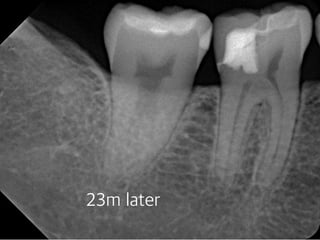
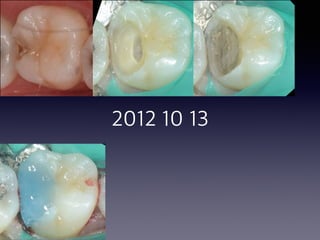
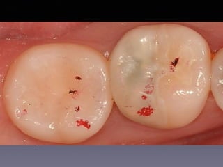
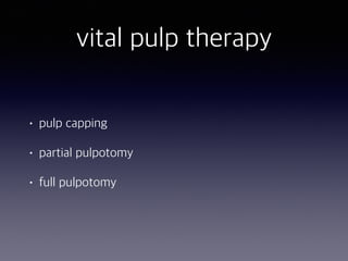
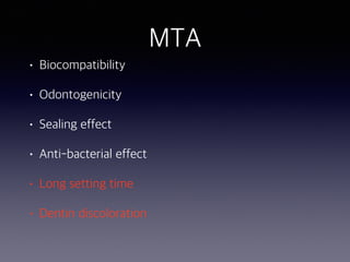
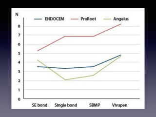
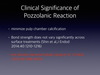
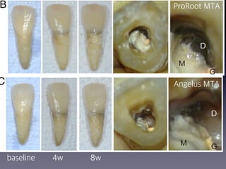
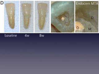
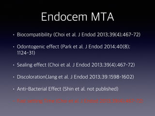
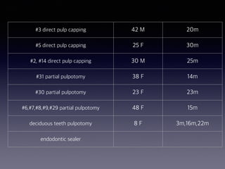
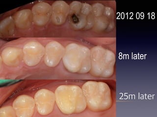
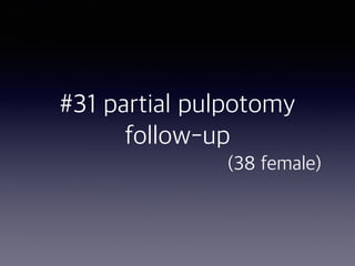
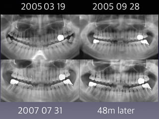
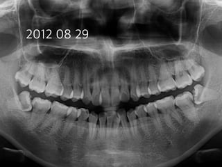
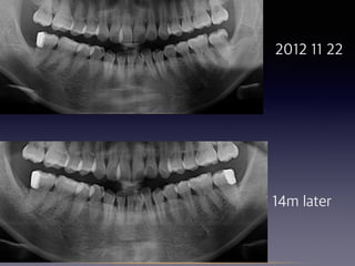
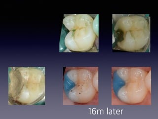
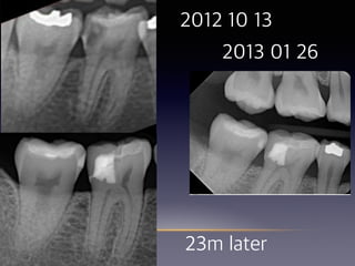
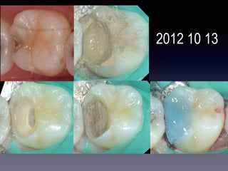
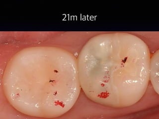
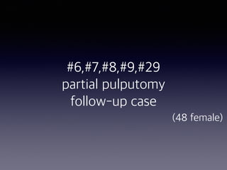
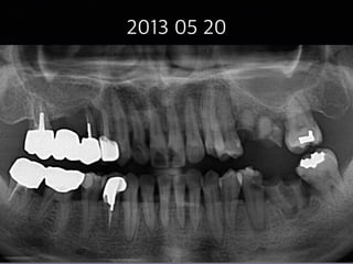
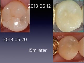
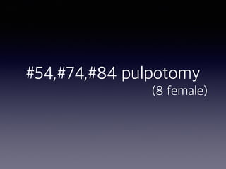
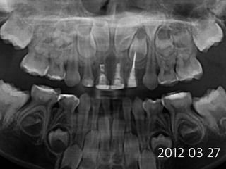
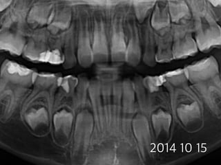
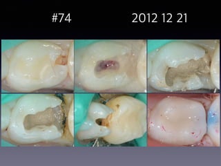
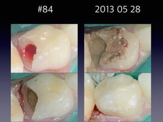
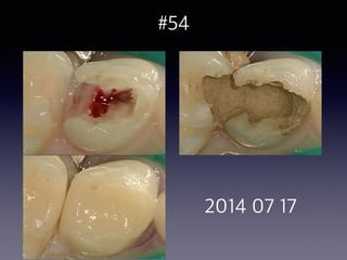
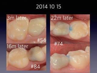
Prognosis of treatment
using Endocem MTA
case report about
direct pulp capping and partial pulpotomy
Seung Pil Jung DDS
• Seoul Pil Dental Clinic: Seoul
• Private Practice Mar.2012 ~
• Seoul Leaders Dental Clinic: Seo-San
• Private Practice Mar.2005~Sep.2010
• Korean Army Service: Seo-San
• Public Health Dentist Mar.2001~Apr.2004
• Graduated from Seoul National Univ. Feb.2000
• Vital pulp therapy
• Pulp capping using MTA
• Endocem MTA
• Pozzolanic reaction
• Characteristics
• Indication
• Directions for use
• Case
• Conclusion
• Discussion
2012 10 13
23m later
2012 10 13
21m later
vital pulp therapy
• pulp capping
• partial pulpotomy
• full pulpotomy
pulp capping,
partial pulpotomy, and
full pulptomy
pathways of the pulp - 10th edition 625~630
Vital pulp therapy:
Requirements for Success
• Treatment of a noninflammed pulp
• Bacteria-tight seal
• Pulp dressing
MTA as a capping agent
• high pH similar to calcium hydroxide when unset
• after setting, will create an excellent bacteria-tight seal
• hard enough to act as a base for a final restoration
• need a moist environment for at least 6 hours to set
properly—> two step procedure
• cause discoloration in the tooth crown
• high cost
Calcium
Hydroxide
success rate
pulp capping
without any removal of the
sort tissue
80%
partial
pulpotomy
the removal of coronal pulp
tissue to the level of healthy
pulp
95%
full
pulpotomy
the removal of the entire
coronal pulp to a level the root
orifices
75%
partial pulpotomy technique
• anesthesia,possibly without a vasoconstrictor
• rubber dam
• superficial disinfection
• 1-to 2-mm deep cavity into the pulp using a high-speed hand piece with a sterile diamond bur
• If bleeding is excessive, the pulp is amputated deeper until only moderate hemorrhage is seen
• excess blood is carefully removed by rinsing with sterile saline and the area is dried with a sterile cotton
pellet
• 5% NaOCl is recommended to rinse the plural wound
• chemical amputation of the blood coagulum
• remove damaged pulp cells, dentin chips, and other debris
• provide hemorrhage control with minimal damage the normal pulp tissue underneath
• do not allow blood clot to develop
partial pulpotomy technique
• 1-to 2-mm deep cavity into the pulp
• using a high-speed hand piece with a sterile diamond bur
• until only moderate hemorrhage is seen
• excess blood is carefully removed
• 5% NaOCl
• do not allow blood clot
• little or no history of pain
• absence of radiographic signs, percussion sensitivity,
swelling, or mobility
• exposures exceeding 2mm, bleeding could not
controlled within 1~2minutes excluded
• 93.5%, 91.4% healing
Partial pulpotomy on asymptomatic young
permanent posterior teeth(calcium hydroxide)
pulpotomy on symptomatic young
permanent (calcium hydroxide)
• 6 teeth
• temporary pain
• widened PDL ligament space
• condensing osteitis
• 66.7% healed
pulpotomy on symptomatic young
permanent teeth(calcium hydroxide)
• 26 permanent vital molars with caries pulp
exposures and apical periodontitis
• 16~ 72 months observed
• 24 teeth (92.3%)
methods
• 40 patients (7~45 years)
• pulp-capping treatment
• no more than reversible pulpits (cold test,
radiographic examination)
first visit
• remove caries using a caries detector
• hemostasis using NaOCl
• place Pro-root MTA over the exposures and all
surrounding dentin
• restore provisionally with un-bonded Clearfil
Photocore
second visit
• sensibility test
• confirm MTA curing
• restore with bonded composite
results
• observation period: 9 years
• followed : 49/53 teeth
• favorable outcome: 97.96%
• all teeth having open apexes showed completed
root formation(15/15)
MTA
• Biocompatibility
• Odontogenicity
• Sealing effect
• Anti-bacterial effect
• Long setting time
• Dentin discoloration
Endocem MTA
• mineral trioxide aggregate-derived pozzolan
cement
Pozzolanic Reaction
MTA surface after setting
calcium hydroxide
calcium silicate
hydrate
active silica
calcium silicate
hydrate
• minimize pulp chamber calcification
Clinical Significance of
Pozzolanic Reaction
17
2012 10 18
16m later
• minimize pulp chamber calcification
• Bond strength does not vary significantly across
surface treatments (Shin et al, J Endod
2014;40:1210–1216)
Clinical Significance of
Pozzolanic Reaction
• minimize pulp chamber calcification
• Bond strength does not vary significantly across
surface treatments (Shin et al,J Endod
2014;40:1210–1216)
• not discolor dentinal tubule (Jang et al. J Endod
2013;39:1598–1602)
Clinical Significance of
Pozzolanic Reaction
ProRoot MTA
Angelus MTA
baseline 4w 8w
Endocem MTA
baseline 4w 8w
Endocem MTA
• Biocompatibility (Choi et al. J Endod 2013;39(4);
467-72)
• MG63 cell
• 3days
• cytoplasmic extension
ProRoot
IRM
Endocem
Endocem MTA
• Biocompatibility (Choi et al. J Endod 2013;39(4);
467-72)
• Odontogenic effect (Park et al. J Endod 2014;40(8);
1124-31)
Odontogenic Effect
of a Fast-setting Pozzolan-based
Pulp Capping Material
Su-Jung Park, DDS, PhD,* Seok-Mo Heo, DDS, PhD,†
Sung-Ok Hong, DDS, MSD,*

Yun-Chan Hwang, DDS, PhD,jj
Kwang-Won Lee, DDS, PhD,‡
and Kyung-San Min, DDS, PhD‡§
Our results indicate that ProRoot and Endocem
have similar biocompatibility and odontogenic
effects. Therefore, Endocem is as effective a
pulp capping material as ProRoot. (J Endod
2014)
Endocem MTA
• Biocompatibility (Choi et al. J Endod 2013;39(4);
467-72)
• Odontogenic effect (Park et al. J Endod 2014;40(8);
1124-31)
• Sealing effect (Choi et al. J Endod 2013;39(4);
467-72)
Endocem MTA
ProRoot MTA IRM
Endocem MTA
• Biocompatibility (Choi et al. J Endod 2013;39(4);
467-72)
• Odontogenic effect (Park et al. J Endod 2014;40(8);
1124-31)
• Sealing effect (Choi et al. J Endod 2013;39(4);
467-72)
• Discoloration(Jang et al. J Endod 2013;39:1598–
1602)
Endocem MTA
• Biocompatibility (Choi et al. J Endod 2013;39(4);
467-72)
• Odontogenic effect (Park et al. J Endod 2014;40(8);
1124-31)
• Sealing effect (Choi et al. J Endod 2013;39(4);467-72)
• Discoloration(Jang et al. J Endod 2013;39:1598–1602)
• Anti-Bacterial Effect (Shin et al. not published)
•Shin et al. not published
Joo-Hee Shin, DDS, MSD, PhD


Department of Conservative Dentistry, Korea University
Medical Center, Korea University, Seoul, Korea
• Streptococcus mutans; dental caries
• Enterococcus faecalis; failed endodontic lesion
• porphyromonas gingivalis; periodontitis
Endocem MTA
• Biocompatibility (Choi et al. J Endod 2013;39(4);467-72)
• Odontogenic effect (Park et al. J Endod 2014;40(8);
1124-31)
• Sealing effect (Choi et al. J Endod 2013;39(4);467-72)
• Discoloration(Jang et al. J Endod 2013;39:1598–1602)
• Anti-Bacterial Effect (Shin et al. not published)
• Fast setting Time (Choi et al. J Endod 2013;39(4);467-72)
Indication
• Lining of cavity in pulp capping
• Lining of cavity in partial pulpotomy
• Lining of cavity after pulpotomy of deciduous teeth
• Canal filling for apical closure in apexogenesis
• Restoration of root canal perforation
• Restoration of internal resorption lesion
• Root end filling
• Endodontic sealer
Indication
• Lining of cavity in pulp capping
• Lining of cavity in partial pulpotomy
• Lining of cavity after pulpotomy of deciduous teeth
• Endodontic sealer
Directions for use
partial pulpotomy technique
• anesthesia,possibly without a vasoconstrictor
• rubber dam
• superficial disinfection
• 1-to 2-mm deep cavity into the pulp using a high-speed hand piece with a sterile diamond bur
• If bleeding is excessive, the pulp is amputated deeper until only moderate hemorrhage is seen
• excess blood is carefully removed by rinsing with sterile saline and the area is dried with a sterile cotton
pellet
• 5% NaOCl is recommended to rinse the plural wound
• chemical amputation of the blood coagulum
• remove damaged pulp cells, dentin chips, and other debris
• provide hemorrhage control with minimal damage the normal pulp tissue underneath
• do not allow blood clot to develop
partial pulpotomy technique
• 1-to 2-mm deep cavity into the pulp
• using a high-speed hand piece with a sterile diamond bur
• until only moderate hemorrhage is seen
• excess blood is carefully removed
• 5% NaOCl
• do not allow blood clot
• isolation
• remove decay closest to pulp tissue with a new
sterilized high speed diamond bur
Directions for use
• isolation
• remove decay closest to pulp tissue with a new
sterilized high speed diamond bur
• rinse thoroughly with 5.25% NaOCl until bleeding
stops
Directions for use
• isolation
• remove decay closest to pulp tissue with a new sterilized high speed
diamond bur
• rinse thoroughly with 5.25% NaOCl until bleeding stops
• mix Endocem with distilled water and apply a thin layer
• remove excess moisture with a sterile cotton pellet, and gently pack
it avoid dead space
• before Endocem hardens, add the rest of Endocem to filled the
cavity
Directions for use
• isolation
• remove decay closest to pulp tissue with a new sterilized high speed
diamond bur
• rinse thoroughly with 5.25% NaOCl until bleeding stops
• mix Endocem with distilled water and apply a thin layer
• remove excess moisture with a sterile cotton pellet, and gently pack it
avoid dead space
• before Endocem hardens, add the rest of Endocem to filled the cavity
• remove part of the exterior Endocem
Directions for use
• isolation
• remove decay closest to pulp tissue with a new sterilized high speed diamond bur
• rinse thoroughly with 5.25% NaOCl until bleeding stops
• mix Endocem with distilled water and apply a thin layer
• remove excess moisture with a sterile cotton pellet, and gently pack it avoid dead
space
• before Endocem hardens, add the rest of Endocem to filled the cavity
• remove part of the exterior Endocem
• apply resin
Directions for use
Precautions
Precautions
• depth and width is important
Precautions
• depth : 3 mm
• width: to cover all dentinal tubule
Precautions
• depth and width is important
• use undercut in narrow and shallow area
Precautions
• depth and width is important
• use undercut in narrow and shallow area
• After setting is complete, apply a strong stream of
water with a 3way syringe check for wash-out
Endocem Case
#3 direct pulp capping 42 M 20m
#5 direct pulp capping 25 F 30m
#2, #14 direct pulp capping 30 M 25m
#31 partial pulpotomy 38 F 14m
#30 partial pulpotomy 23 F 23m
#6,#7,#8,#9,#29 partial pulpotomy 48 F 15m
deciduous teeth pulpotomy 8 F 3m,16m,22m
endodontic sealer
#3 direct pulp capping
follow–up

(42 male)
2012 12 03
20m later
2013 01 19
2012 12 03
2012 12 03
20m later
#5 direct pulp capping follow–
up

(25 female)
2012 07 31
30m later
2012 07 31
2012 07 31
30m later
#2, #14 direct pulp capping
follow-up 

(30 male)
2012 09 18


2012 09 18
25m later
20
2012 09 20 2012 09 27
2012 09 18
25m later
8m later
2012 09 18
8m later
25m later
#31 partial pulpotomy
follow-up 

(38 female)
2005 03 19 2005 09 28
2007 07 31 48m later
2012 08 29
2012 11 22
14m later
16m later
#30 Partial pulpotomy
follow-up case 

(23 female)

2013 01 26
2012 10 13
23m later
2012 10 13
26
21m later


#6,#7,#8,#9,#29
partial pulputomy
follow-up case
(48 female)
2013 05 20
31
2013 05 20
2013 06 28
2013 05 20
15m later
2013 06 12
15m later
2013 05 20
2013 05 21 #9
2013 05 27 #7
2013 06 04 #6,#8
2013 06 13 #29
2013 05 20
15m later
38
2013 05 20
2013 06 12
15m later
#54,#74,#84 pulpotomy
(8 female)
2012 03 27
2014 10 15
#74 2012 12 21
#84 2013 05 28
2014 07 17
#54
2014 10 15
#54
#74
#84
22m later
16m later
3m later
Endodontic sealer
#30
#9, #10
#12,#14
#21
#20 #30
2014 11 10 2014 11 18
2014 11 19 2015 02 26
conclusion
• decrease the risk of endodontic treatment
• shorten chair time of removing infected dentin
• safer material in deciduous teeth pulpotomy than FC
• reduce treatment expenses
• improve treated tooth’s prognosis
discussion
• long-term follow-up
• need to examine cytotoxity and calcinogenicity over
longer duration
• need to study treating inflamed pulp tissue
THANK YOU

Vital pulp therapy

  • 1.
    Prognosis of treatment usingEndocem MTA case report about direct pulp capping and partial pulpotomy
  • 2.
    Seung Pil JungDDS • Seoul Pil Dental Clinic: Seoul • Private Practice Mar.2012 ~ • Seoul Leaders Dental Clinic: Seo-San • Private Practice Mar.2005~Sep.2010 • Korean Army Service: Seo-San • Public Health Dentist Mar.2001~Apr.2004 • Graduated from Seoul National Univ. Feb.2000
  • 3.
    • Vital pulptherapy • Pulp capping using MTA • Endocem MTA • Pozzolanic reaction • Characteristics • Indication • Directions for use • Case • Conclusion • Discussion
  • 4.
  • 5.
  • 6.
  • 7.
  • 9.
    vital pulp therapy •pulp capping • partial pulpotomy • full pulpotomy
  • 10.
    pulp capping, partial pulpotomy,and full pulptomy pathways of the pulp - 10th edition 625~630
  • 11.
    Vital pulp therapy: Requirementsfor Success • Treatment of a noninflammed pulp • Bacteria-tight seal • Pulp dressing
  • 12.
    MTA as acapping agent • high pH similar to calcium hydroxide when unset • after setting, will create an excellent bacteria-tight seal • hard enough to act as a base for a final restoration • need a moist environment for at least 6 hours to set properly—> two step procedure • cause discoloration in the tooth crown • high cost
  • 13.
    Calcium Hydroxide success rate pulp capping withoutany removal of the sort tissue 80% partial pulpotomy the removal of coronal pulp tissue to the level of healthy pulp 95% full pulpotomy the removal of the entire coronal pulp to a level the root orifices 75%
  • 14.
    partial pulpotomy technique •anesthesia,possibly without a vasoconstrictor • rubber dam • superficial disinfection • 1-to 2-mm deep cavity into the pulp using a high-speed hand piece with a sterile diamond bur • If bleeding is excessive, the pulp is amputated deeper until only moderate hemorrhage is seen • excess blood is carefully removed by rinsing with sterile saline and the area is dried with a sterile cotton pellet • 5% NaOCl is recommended to rinse the plural wound • chemical amputation of the blood coagulum • remove damaged pulp cells, dentin chips, and other debris • provide hemorrhage control with minimal damage the normal pulp tissue underneath • do not allow blood clot to develop
  • 15.
    partial pulpotomy technique •1-to 2-mm deep cavity into the pulp • using a high-speed hand piece with a sterile diamond bur • until only moderate hemorrhage is seen • excess blood is carefully removed • 5% NaOCl • do not allow blood clot
  • 18.
    • little orno history of pain • absence of radiographic signs, percussion sensitivity, swelling, or mobility • exposures exceeding 2mm, bleeding could not controlled within 1~2minutes excluded • 93.5%, 91.4% healing Partial pulpotomy on asymptomatic young permanent posterior teeth(calcium hydroxide)
  • 19.
    pulpotomy on symptomaticyoung permanent (calcium hydroxide) • 6 teeth • temporary pain • widened PDL ligament space • condensing osteitis • 66.7% healed
  • 20.
    pulpotomy on symptomaticyoung permanent teeth(calcium hydroxide) • 26 permanent vital molars with caries pulp exposures and apical periodontitis • 16~ 72 months observed • 24 teeth (92.3%)
  • 22.
    methods • 40 patients(7~45 years) • pulp-capping treatment • no more than reversible pulpits (cold test, radiographic examination)
  • 23.
    first visit • removecaries using a caries detector • hemostasis using NaOCl • place Pro-root MTA over the exposures and all surrounding dentin • restore provisionally with un-bonded Clearfil Photocore
  • 24.
    second visit • sensibilitytest • confirm MTA curing • restore with bonded composite
  • 25.
    results • observation period:9 years • followed : 49/53 teeth • favorable outcome: 97.96% • all teeth having open apexes showed completed root formation(15/15)
  • 26.
    MTA • Biocompatibility • Odontogenicity •Sealing effect • Anti-bacterial effect • Long setting time • Dentin discoloration
  • 27.
    Endocem MTA • mineraltrioxide aggregate-derived pozzolan cement
  • 28.
  • 29.
    MTA surface aftersetting calcium hydroxide calcium silicate hydrate active silica calcium silicate hydrate
  • 30.
    • minimize pulpchamber calcification Clinical Significance of Pozzolanic Reaction
  • 31.
  • 32.
    • minimize pulpchamber calcification • Bond strength does not vary significantly across surface treatments (Shin et al, J Endod 2014;40:1210–1216) Clinical Significance of Pozzolanic Reaction
  • 35.
    • minimize pulpchamber calcification • Bond strength does not vary significantly across surface treatments (Shin et al,J Endod 2014;40:1210–1216) • not discolor dentinal tubule (Jang et al. J Endod 2013;39:1598–1602) Clinical Significance of Pozzolanic Reaction
  • 36.
  • 37.
  • 38.
    Endocem MTA • Biocompatibility(Choi et al. J Endod 2013;39(4); 467-72)
  • 39.
    • MG63 cell •3days • cytoplasmic extension
  • 40.
  • 41.
    Endocem MTA • Biocompatibility(Choi et al. J Endod 2013;39(4); 467-72) • Odontogenic effect (Park et al. J Endod 2014;40(8); 1124-31)
  • 42.
    Odontogenic Effect of aFast-setting Pozzolan-based Pulp Capping Material Su-Jung Park, DDS, PhD,* Seok-Mo Heo, DDS, PhD,† Sung-Ok Hong, DDS, MSD,*
 Yun-Chan Hwang, DDS, PhD,jj Kwang-Won Lee, DDS, PhD,‡ and Kyung-San Min, DDS, PhD‡§
  • 44.
    Our results indicatethat ProRoot and Endocem have similar biocompatibility and odontogenic effects. Therefore, Endocem is as effective a pulp capping material as ProRoot. (J Endod 2014)
  • 45.
    Endocem MTA • Biocompatibility(Choi et al. J Endod 2013;39(4); 467-72) • Odontogenic effect (Park et al. J Endod 2014;40(8); 1124-31) • Sealing effect (Choi et al. J Endod 2013;39(4); 467-72)
  • 47.
  • 48.
    Endocem MTA • Biocompatibility(Choi et al. J Endod 2013;39(4); 467-72) • Odontogenic effect (Park et al. J Endod 2014;40(8); 1124-31) • Sealing effect (Choi et al. J Endod 2013;39(4); 467-72) • Discoloration(Jang et al. J Endod 2013;39:1598– 1602)
  • 49.
    Endocem MTA • Biocompatibility(Choi et al. J Endod 2013;39(4); 467-72) • Odontogenic effect (Park et al. J Endod 2014;40(8); 1124-31) • Sealing effect (Choi et al. J Endod 2013;39(4);467-72) • Discoloration(Jang et al. J Endod 2013;39:1598–1602) • Anti-Bacterial Effect (Shin et al. not published)
  • 50.
    •Shin et al.not published Joo-Hee Shin, DDS, MSD, PhD 
 Department of Conservative Dentistry, Korea University Medical Center, Korea University, Seoul, Korea
  • 51.
    • Streptococcus mutans;dental caries • Enterococcus faecalis; failed endodontic lesion • porphyromonas gingivalis; periodontitis
  • 53.
    Endocem MTA • Biocompatibility(Choi et al. J Endod 2013;39(4);467-72) • Odontogenic effect (Park et al. J Endod 2014;40(8); 1124-31) • Sealing effect (Choi et al. J Endod 2013;39(4);467-72) • Discoloration(Jang et al. J Endod 2013;39:1598–1602) • Anti-Bacterial Effect (Shin et al. not published) • Fast setting Time (Choi et al. J Endod 2013;39(4);467-72)
  • 55.
    Indication • Lining ofcavity in pulp capping • Lining of cavity in partial pulpotomy • Lining of cavity after pulpotomy of deciduous teeth • Canal filling for apical closure in apexogenesis • Restoration of root canal perforation • Restoration of internal resorption lesion • Root end filling • Endodontic sealer
  • 56.
    Indication • Lining ofcavity in pulp capping • Lining of cavity in partial pulpotomy • Lining of cavity after pulpotomy of deciduous teeth • Endodontic sealer
  • 57.
  • 58.
    partial pulpotomy technique •anesthesia,possibly without a vasoconstrictor • rubber dam • superficial disinfection • 1-to 2-mm deep cavity into the pulp using a high-speed hand piece with a sterile diamond bur • If bleeding is excessive, the pulp is amputated deeper until only moderate hemorrhage is seen • excess blood is carefully removed by rinsing with sterile saline and the area is dried with a sterile cotton pellet • 5% NaOCl is recommended to rinse the plural wound • chemical amputation of the blood coagulum • remove damaged pulp cells, dentin chips, and other debris • provide hemorrhage control with minimal damage the normal pulp tissue underneath • do not allow blood clot to develop
  • 59.
    partial pulpotomy technique •1-to 2-mm deep cavity into the pulp • using a high-speed hand piece with a sterile diamond bur • until only moderate hemorrhage is seen • excess blood is carefully removed • 5% NaOCl • do not allow blood clot
  • 60.
    • isolation • removedecay closest to pulp tissue with a new sterilized high speed diamond bur Directions for use
  • 62.
    • isolation • removedecay closest to pulp tissue with a new sterilized high speed diamond bur • rinse thoroughly with 5.25% NaOCl until bleeding stops Directions for use
  • 64.
    • isolation • removedecay closest to pulp tissue with a new sterilized high speed diamond bur • rinse thoroughly with 5.25% NaOCl until bleeding stops • mix Endocem with distilled water and apply a thin layer • remove excess moisture with a sterile cotton pellet, and gently pack it avoid dead space • before Endocem hardens, add the rest of Endocem to filled the cavity Directions for use
  • 66.
    • isolation • removedecay closest to pulp tissue with a new sterilized high speed diamond bur • rinse thoroughly with 5.25% NaOCl until bleeding stops • mix Endocem with distilled water and apply a thin layer • remove excess moisture with a sterile cotton pellet, and gently pack it avoid dead space • before Endocem hardens, add the rest of Endocem to filled the cavity • remove part of the exterior Endocem Directions for use
  • 68.
    • isolation • removedecay closest to pulp tissue with a new sterilized high speed diamond bur • rinse thoroughly with 5.25% NaOCl until bleeding stops • mix Endocem with distilled water and apply a thin layer • remove excess moisture with a sterile cotton pellet, and gently pack it avoid dead space • before Endocem hardens, add the rest of Endocem to filled the cavity • remove part of the exterior Endocem • apply resin Directions for use
  • 71.
  • 72.
    Precautions • depth andwidth is important
  • 73.
    Precautions • depth :3 mm • width: to cover all dentinal tubule
  • 75.
    Precautions • depth andwidth is important • use undercut in narrow and shallow area
  • 76.
    Precautions • depth andwidth is important • use undercut in narrow and shallow area • After setting is complete, apply a strong stream of water with a 3way syringe check for wash-out
  • 77.
  • 78.
    #3 direct pulpcapping 42 M 20m #5 direct pulp capping 25 F 30m #2, #14 direct pulp capping 30 M 25m #31 partial pulpotomy 38 F 14m #30 partial pulpotomy 23 F 23m #6,#7,#8,#9,#29 partial pulpotomy 48 F 15m deciduous teeth pulpotomy 8 F 3m,16m,22m endodontic sealer
  • 79.
    #3 direct pulpcapping follow–up
 (42 male)
  • 80.
    2012 12 03 20mlater 2013 01 19
  • 81.
  • 82.
  • 83.
    #5 direct pulpcapping follow– up
 (25 female)
  • 84.
  • 85.
  • 86.
  • 87.
    #2, #14 directpulp capping follow-up 
 (30 male)
  • 88.
  • 89.
    20 2012 09 202012 09 27
  • 90.
    2012 09 18 25mlater 8m later
  • 91.
    2012 09 18 8mlater 25m later
  • 92.
  • 93.
    2005 03 192005 09 28 2007 07 31 48m later
  • 94.
  • 95.
  • 96.
  • 97.
    #30 Partial pulpotomy follow-upcase 
 (23 female)

  • 98.
    2013 01 26 201210 13 23m later
  • 99.
  • 100.
  • 101.
  • 102.
  • 103.
    2013 06 28 201305 20 15m later
  • 104.
  • 105.
  • 106.
  • 107.
  • 108.
  • 109.
  • 110.
  • 111.
    38 2013 05 20 201306 12 15m later
  • 112.
  • 113.
  • 114.
  • 115.
  • 116.
  • 117.
  • 118.
    2014 10 15 #54 #74 #84 22mlater 16m later 3m later
  • 119.
  • 120.
  • 121.
  • 122.
    2014 11 102014 11 18 2014 11 19 2015 02 26
  • 123.
    conclusion • decrease therisk of endodontic treatment • shorten chair time of removing infected dentin • safer material in deciduous teeth pulpotomy than FC • reduce treatment expenses • improve treated tooth’s prognosis
  • 124.
    discussion • long-term follow-up •need to examine cytotoxity and calcinogenicity over longer duration • need to study treating inflamed pulp tissue
  • 125.