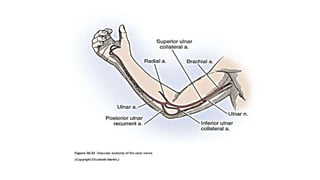
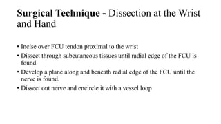
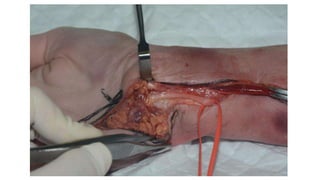
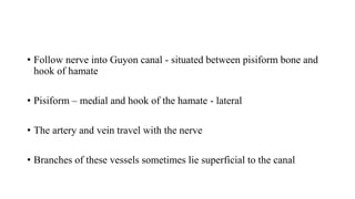
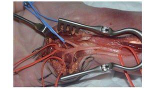
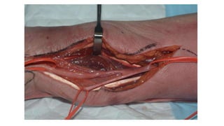
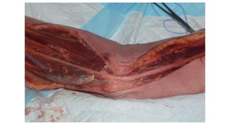
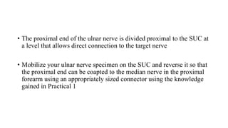
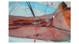
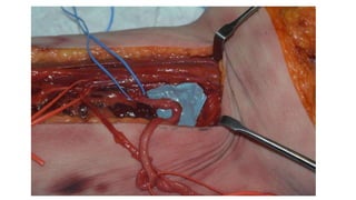
Vascularised Ulnar Nerve
Harvest Technique
Cadaveric dissection
First Nerve transfer workshop,
KTPH, Singapore
Practical 6
Surgical Technique - Dissection at the Wrist
and Hand
• Incise over FCU tendon proximal to the wrist
• Dissect through subcutaneous tissues until radial edge of the FCU is
found
• Develop a plane along and beneath radial edge of the FCU until the
nerve is found.
• Dissect out nerve and encircle it with a vessel loop
• Follow nerve into Guyon canal - situated between pisiform bone and
hook of hamate
• Pisiform – medial and hook of the hamate - lateral
• The artery and vein travel with the nerve
• Branches of these vessels sometimes lie superficial to the canal
• The deep branch winds around the hook of the hamate and is indented
by the fibrous margins of the overlying hypothenar muscles
• It then runs deep to the flexor tendons and the deep palmar arterial
arch
Dissection in the Olecranon Notch and
Proximal Forearm
• Follow the ulnar nerve proximally to the olecranon notch
• Split the two heads of the FCU to expose the nerve at the proximal
forearm level
Dissection in the Distal Arm
• Open the investing fascia to expose the ulnar nerve
• Branches to the underlying FCU and elbow joint and collateral vessels
may tether the nerve just proximal to the olecranon notch
• Divide the ulnar nerve at the wrist – divide dorsal cutaneous branch as
well as the motor branches to the extrinsic finger and wrist flexors
• Bleeding should be seen from the end of the vascularized nerve
• The proximal end of the ulnar nerve is divided proximal to the SUC at
a level that allows direct connection to the target nerve
• Mobilize your ulnar nerve specimen on the SUC and reverse it so that
the proximal end can be coapted to the median nerve in the proximal
forearm using an appropriately sized connector using the knowledge
gained in Practical 1
Thank you

Vascularised Ulnar Nerve Graft

  • 1.
    Vascularised Ulnar Nerve HarvestTechnique Cadaveric dissection First Nerve transfer workshop, KTPH, Singapore Practical 6
  • 3.
    Surgical Technique -Dissection at the Wrist and Hand • Incise over FCU tendon proximal to the wrist • Dissect through subcutaneous tissues until radial edge of the FCU is found • Develop a plane along and beneath radial edge of the FCU until the nerve is found. • Dissect out nerve and encircle it with a vessel loop
  • 5.
    • Follow nerveinto Guyon canal - situated between pisiform bone and hook of hamate • Pisiform – medial and hook of the hamate - lateral • The artery and vein travel with the nerve • Branches of these vessels sometimes lie superficial to the canal
  • 7.
    • The deepbranch winds around the hook of the hamate and is indented by the fibrous margins of the overlying hypothenar muscles • It then runs deep to the flexor tendons and the deep palmar arterial arch
  • 9.
    Dissection in theOlecranon Notch and Proximal Forearm • Follow the ulnar nerve proximally to the olecranon notch • Split the two heads of the FCU to expose the nerve at the proximal forearm level
  • 11.
    Dissection in theDistal Arm • Open the investing fascia to expose the ulnar nerve • Branches to the underlying FCU and elbow joint and collateral vessels may tether the nerve just proximal to the olecranon notch
  • 12.
    • Divide theulnar nerve at the wrist – divide dorsal cutaneous branch as well as the motor branches to the extrinsic finger and wrist flexors • Bleeding should be seen from the end of the vascularized nerve
  • 13.
    • The proximalend of the ulnar nerve is divided proximal to the SUC at a level that allows direct connection to the target nerve • Mobilize your ulnar nerve specimen on the SUC and reverse it so that the proximal end can be coapted to the median nerve in the proximal forearm using an appropriately sized connector using the knowledge gained in Practical 1
  • 16.