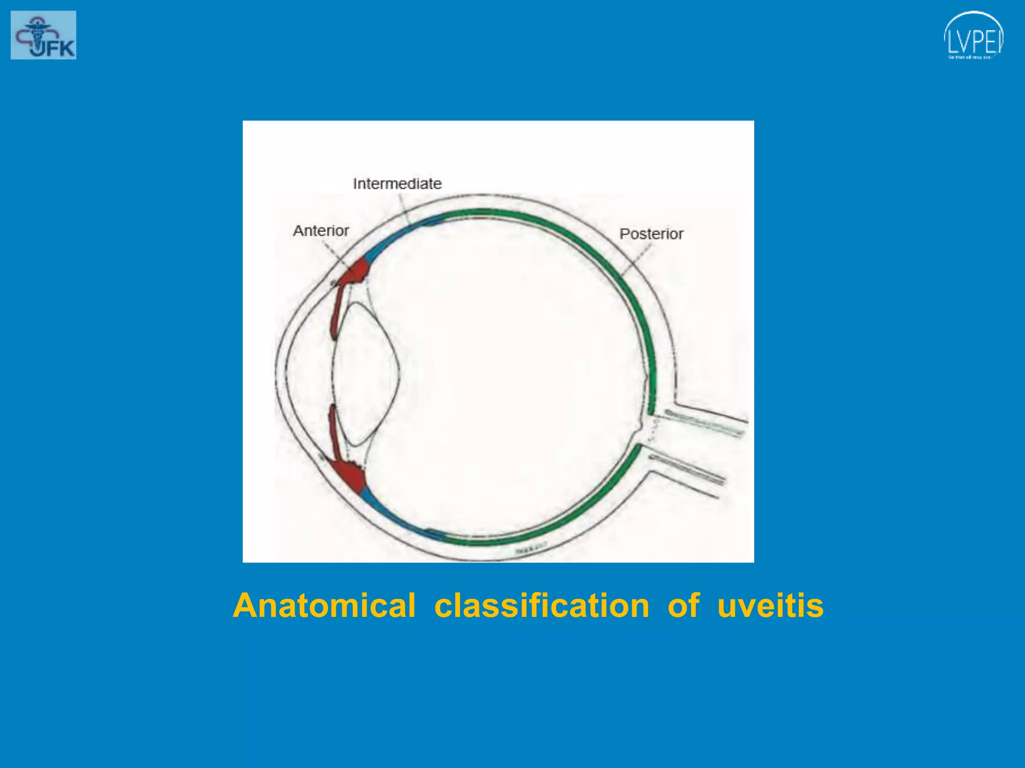
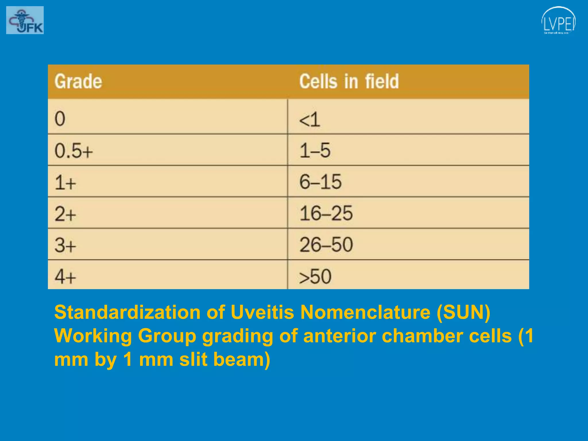
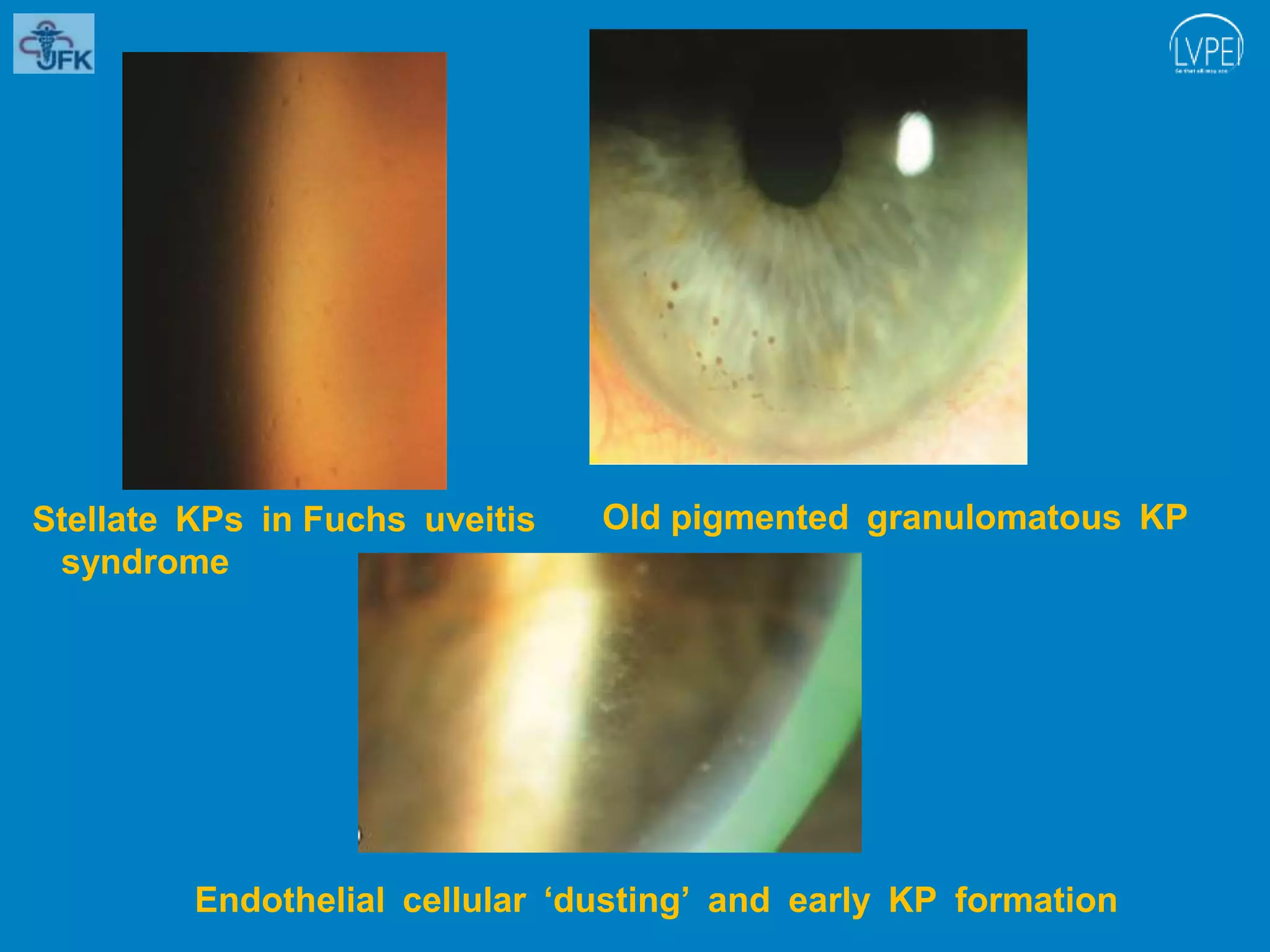
Uvea consists of the middle, pigmented, vascular structures of the eye and includes the iris, ciliary body, and choroid
Uveitis is broadly defined as inflammation (ie, -itis)of the uvea (from the Latin uva, meaning "grape")
It emphasizes the close relationship between the anatomically distinct parts of the uveal tract, for inflammatory processes tend to involve the uvea as a whole and are generally not limited to a single region
Congenital third ( oculomotor )nerve palsyVinitkumar MJ
Ask about pregnancy & birth history due to the association with birth trauma & perinatal complications.
Inquire whether the patient is meeting their developmental milestones or if they are exhibiting any other neurologic signs or symptoms. Although additional focal neurologic abnormalities or generalized delays in development have been described in these patients, their presence may increase concerns of other neurologic etiologies.
Determine whether they have signs of oculomotor synkinesis, such as asking the parents if they notice eye or eyelid movement during feeding.
Question if they have any family history of strabismus.
Heritability may suggest other forms of strabismus with known genetic associations.
Vitreous (Attachments, age changes, vitreous hemorrhage, Vitreous Detachment)Maryam Fida
Vitreous
Vitreous is an inert, avascular, transparent, jelly like structure.
Serve as one of refractive media of the eye and has optical functions.
It gives structural integrity to eye and provide nutrients to the lens, ciliary body and retina.
Constitute 80% volume of the eye.
Contain collagen fibrils, mucopoly-saccharides and hyaluronic acid.
It’s a hydrophilic gel which become ‘’fluid’’ when protein coagulates.
Reasons for coagulation of proteins could be,
• Advancing senile age
• Degenerations, e.g. as in high myopia
• Chemical and mechanical trauma
Internal limiting membrane on inner surface of retina separate it from vitreous. There is potential space ‘subhyaloid space’ between two.
Figure 1 structures of vitreous
Vitreous attachments
1. Anteriorly to the lens and ciliary epithelium in front of ora serrata. Part of vitreous about 4mm across ora serrata is called as ‘base of vitreous’. Here, attachment is strongest.
2. Posteriorly to the edge of optic disc and macula lutea (foveal region) forming ring shaped structure around them.
Figure 2 vitreous attachments
Age changes in Vitreous
Vitreous undergo certain physical and biochemical changes with aging.
1. At birth_ the Cloquet’s canal runs straight from lens to optic disc. It contains primary vitreous.
2. In young persons_ vitreous gel is homogenous but its fibers become coarse with process of advancing age.
3. In old age and high myopes_ secondary vitreous liquified (syneresis) and shrinks, producing a vitreous detachment, vitreous and retinal hemorrhage and retinal break.
Figure 3 Vitreous at birth Figure 4 Vitreous in young adults.
Figure 5 vitreous detachment in old age
Vitreous Hemorrhage
Vitreous hemorrhage is the extravasation, or leakage, of blood into the areas in and around the vitreous humor of the eye.
TYPES
There are two types of vitreous hemorrhage.
1. Peripheral or subhyaloid hemorrhage _ occurs between retina and vitreous.
Blood remains fluid, red in color
Blood moves with gravity forming boat-shaped figure in macular area.
Figure 3 subhyaloid hemorrhage
2. Intravitreal hemorrhage_ the hemorrhage may get absorbed or degenerate to form a white fibrous tissue mass.
Etiology
Common causes of vitreous hemorrhage are;
i. Trauma_ by contusion or penetrating injury
ii. Vitreous retraction_ vitreous fibrous bands or membrane retraction
iii. Eale’s disease_ due to retinal vasculitis and periphlebitis
iv. Blood dyscrasias_ leukemia, sickle cell anemia, purpura.
v. Diabetes mellitus_ common in diabetic proliferative retinopathy
vi. Central retinal vein thrombosis
vii. Malignant hypertension_ often results in large intravitreal hemorrhage.
Symptoms
I. Black spots or clouds maybe seen in front of eye.
II. Impaired vision maybe up to perception of light.
Signs
1. Fundus examination
a) Faint or no red reflex seen
b) Grey opacities maybe present in vitreous
Figure 4 (A) Fundus exami
Uveitis
• Inflammation of uveal tissue.
• Associated inflammation of adjacent structures, such as Retina, Vitreous, Sclera and Cornea.
Figure 1 uveitis
Anatomical classification
Clinical classification
Pathological classification
Etiological classification
(Duke Elder’s)
1. Anterior uveitis
Can be divided as follow;
1) Iritis_ inflammation mainly the iris
2) Iridocyclitis _iris and pars plicata involved
3) Cyclitis_ pars plicata is affected
Acute uveitis
Onset is sudden,
Last for less than 3 weeks Granulomatous uveitis
Infective nature
Inflammation is insidious in onset
Chronic in nature with minimum clinical features Infective uveitis
2. Intermediate uveitis
Inflammation of pars plana, peripheral retina and choroid.
Also called as “pars planitis”. Chronic uveitis
Onset is insidious
Duration is more than 3 weeks
Non-granulomatous uveitis
due to allergic or immune related reaction
acute onset
short duration
Allergic uveitis or immune related uveitis
3. Posterior uveitis
Inflammation of choroid(choroiditis)
Associated inflammation of retina (chorioretinitis) Recurrent uveitis
uveitis keeps reoccurring periodically
Toxic uveitis
4. Panuveitis
Inflammation of whole uveal tract Traumatic uveitis
5. Uveitis associated with non-infective systemic diseases
6. Idiopathic uveitis
7. Neoplastic
Figure 2 anatomical classification of uveitis
Panuveitis
Endophthalmitis
Panophthalmitis
Inflammation of all layers of uvea of eye
Can also affect lens, retina, optic nerve and vitreous causing reduced vision or blindness. Inflammation of internal structures of the eye, I;e choroid, retina and vitreous Purulent inflammation of all structures of eye
Including all the three coats and Tenon’s capsule as well.
Etiology
1. Idiopathic
After ruling out other causes
2. Infectious
Tuberculosis
Syphilis
Lyme disease
Leptospirosis
Infectious endophthalmitis
3. Immune related
Sarcoidosis
Vogt-koyanagi-Harada syndrome
Sympathetic ophthalmitis
Behcet syndrome
Etiology
Acute process 1-7 days following intraocular surgery such as Cataract surgery and filtering operation
Commonly caused by Bacteria-staphylococcus, pseudomonas, pneumococcus, streptococcus, E. coli,
Fungus -aspergillus fumigatus, candida albicans, fusarium,
Etiology
1.Exogenous
Due to infected wounds
Common pathogens are pneumococcus, staphylococcus, pseudomonas, pneumococcus, streptococcus, E. coli.
2.Endogenous
Due to metastasis of infected embolus in retinal artery and choroidal vessels.
Clinical Features
• Sudden onset of unilateral pain, redness, photophobia
• Maybe associated with lacrimation
• Visual acuity is usually good at presentation except in eyes with severe hypopyon.
• Low IOP
• Fibrinous exudate
• Posterior synechiae
• Miosis
• Aqueous flare and cells
• Endothelial dusting
Clinical Features
Bacterial endophthalmitis
• Sudden onset with severe pain
• Redness
• Visual loss
• Lid oedema, chemosis, corneal haze
• Low
Eyelid laceration repair with defects.pptxSHAYRI PILLAI
PRINCIPLES OF EYELID REPAIR
Wounds should be copiously irrigated and explored, with the removal of any foreign material after local anesthesia
Reconstruction should be done in layers as per correct anatomical orientation
Wounds should not be extended to explore structures unless the exploration is for suspected foreign body
The orbital septum if damaged should never be repaired-result incompromised eyelid excursion and even lagophthalmos
Vernal keratoconjunctivitis (VKC) is a chronic, bilateral, at times asymmetrical, seasonally exacerbated, allergic inflammation of the ocular surface, involving tarsal and⁄or bulbar conjunctiva
More common in children and young adults
Has an atopic background
First mentioned in the ophthalmic literature as conjunctiva lymphatica more than 150 years ago
More common in the summers than in the winters
Pseudoexfoliative material is a grey-white fibrillary substance
Age related multifactorial, progressive disorder
Material is deposited on various ocular structures includes-
- Lens capsule
- Zonular fibres
- Iris
- Trabeculum
- Conjunctiva
Deposits in blood vessels,skin, gall bladder, kidneys,lungs,heart and meninges
Ocular surface squamous neoplasia (OSSN) includes spectrum of disease from mild dysplasia to carcinoma in situ to invasive squamous cell carcinoma
Involving conjunctiva and cornea
Usually in the interpalpebral area, mostly at the limbus
Retinoscope is an objective refraction instrument used to
determine the spherocylindrical refractive error, as well as
observe optical aberrations, irregularities, and opacities.
The technique is called Retinoscopy/Skiascopy/Shadow Test
PCR- breach in the posterior capsule of the crystalline lens during cataract surgery
Common complication
Lead to sub-optimal visual outcomes if not recognized early or managed appropriately
Complication varies with the stage at which it occurred or was recognized
Consequent Vitreous loss -major determinant of post-operative outcomes
TASS
Toxic solution enters the anterior chamber
Produce severe intraocular inflammation as well as corneal edema
Form of sterile, noninfectious endophthalmitis
Endophthalmitis
Present in an acute form or in a more indolent or chronic form the latter is associated with organisms of lower pathogenicity
Since de Wecker first described the “filtering cicatrix”, as a surgical treatment for glaucoma in 1882
Glaucoma filtering surgery (GFS) differs from most surgical procedures in that inhibition of wound healing is desirable to achieve surgical success
Ophthalmoscopy
A clinical examination of the posterior segment by the means of an ophthalmoscope.
It is primarily done to assess the state of fundus and detect the opacities of ocular media.
Ophthalmoscope
An instrument that allows the ophthalmologist to look inside a person’s eye and see the details of the living retina
Testing colour vision requires elaborate apparatus for scientific investigation. There are two objectives in testing for colour blindness
(i) the exact nature of the defect and
(ii) whether the subject is likely to be a source of danger to the community.
After the tunnel has been dissected with crescent knife
Entry is made into the anterior chamber at 10 O’ Clock
position with MVR/V-Lance 20G blade
Viscoelastic is injected to make the eyeball hypertensive
A special property of the cone system is color vision.
Perceiving color allows humans (and many other animals) to discriminate objects on the basis of the distribution of the wavelengths of light that they reflect to the eye.
The perception of color is a response to electromagnetic energy at wavelengths between 400 and 700 nm, which is absorbed by cone outer segment visual pigments.
The degree to which a beam of light is deflected as it
passes through the lens depends on the focal and prismatic power of the lens and the distance from its optical centre.
TEST BANK for Operations Management, 14th Edition by William J. Stevenson, Ve...kevinkariuki227
TEST BANK for Operations Management, 14th Edition by William J. Stevenson, Verified Chapters 1 - 19, Complete Newest Version.pdf
Ethanol (CH3CH2OH), or beverage alcohol, is a two-carbon alcohol
that is rapidly distributed in the body and brain. Ethanol alters many
neurochemical systems and has rewarding and addictive properties. It
is the oldest recreational drug and likely contributes to more morbidity,
mortality, and public health costs than all illicit drugs combined. The
5th edition of the Diagnostic and Statistical Manual of Mental Disorders
(DSM-5) integrates alcohol abuse and alcohol dependence into a single
disorder called alcohol use disorder (AUD), with mild, moderate,
and severe subclassifications (American Psychiatric Association, 2013).
In the DSM-5, all types of substance abuse and dependence have been
combined into a single substance use disorder (SUD) on a continuum
from mild to severe. A diagnosis of AUD requires that at least two of
the 11 DSM-5 behaviors be present within a 12-month period (mild
AUD: 2–3 criteria; moderate AUD: 4–5 criteria; severe AUD: 6–11 criteria).
The four main behavioral effects of AUD are impaired control over
drinking, negative social consequences, risky use, and altered physiological
effects (tolerance, withdrawal). This chapter presents an overview
of the prevalence and harmful consequences of AUD in the U.S.,
the systemic nature of the disease, neurocircuitry and stages of AUD,
comorbidities, fetal alcohol spectrum disorders, genetic risk factors, and
pharmacotherapies for AUD.
Factory Supply Best Quality Pmk Oil CAS 28578–16–7 PMK Powder in Stockrebeccabio
Factory Supply Best Quality Pmk Oil CAS 28578–16–7 PMK Powder in Stock
Telegram: bmksupplier
signal: +85264872720
threema: TUD4A6YC
You can contact me on Telegram or Threema
Communicate promptly and reply
Free of customs clearance, Double Clearance 100% pass delivery to USA, Canada, Spain, Germany, Netherland, Poland, Italy, Sweden, UK, Czech Republic, Australia, Mexico, Russia, Ukraine, Kazakhstan.Door to door service
Hot Selling Organic intermediates
- Video recording of this lecture in English language: https://youtu.be/lK81BzxMqdo
- Video recording of this lecture in Arabic language: https://youtu.be/Ve4P0COk9OI
- Link to download the book free: https://nephrotube.blogspot.com/p/nephrotube-nephrology-books.html
- Link to NephroTube website: www.NephroTube.com
- Link to NephroTube social media accounts: https://nephrotube.blogspot.com/p/join-nephrotube-on-social-media.html
New Directions in Targeted Therapeutic Approaches for Older Adults With Mantl...i3 Health
i3 Health is pleased to make the speaker slides from this activity available for use as a non-accredited self-study or teaching resource.
This slide deck presented by Dr. Kami Maddocks, Professor-Clinical in the Division of Hematology and
Associate Division Director for Ambulatory Operations
The Ohio State University Comprehensive Cancer Center, will provide insight into new directions in targeted therapeutic approaches for older adults with mantle cell lymphoma.
STATEMENT OF NEED
Mantle cell lymphoma (MCL) is a rare, aggressive B-cell non-Hodgkin lymphoma (NHL) accounting for 5% to 7% of all lymphomas. Its prognosis ranges from indolent disease that does not require treatment for years to very aggressive disease, which is associated with poor survival (Silkenstedt et al, 2021). Typically, MCL is diagnosed at advanced stage and in older patients who cannot tolerate intensive therapy (NCCN, 2022). Although recent advances have slightly increased remission rates, recurrence and relapse remain very common, leading to a median overall survival between 3 and 6 years (LLS, 2021). Though there are several effective options, progress is still needed towards establishing an accepted frontline approach for MCL (Castellino et al, 2022). Treatment selection and management of MCL are complicated by the heterogeneity of prognosis, advanced age and comorbidities of patients, and lack of an established standard approach for treatment, making it vital that clinicians be familiar with the latest research and advances in this area. In this activity chaired by Michael Wang, MD, Professor in the Department of Lymphoma & Myeloma at MD Anderson Cancer Center, expert faculty will discuss prognostic factors informing treatment, the promising results of recent trials in new therapeutic approaches, and the implications of treatment resistance in therapeutic selection for MCL.
Target Audience
Hematology/oncology fellows, attending faculty, and other health care professionals involved in the treatment of patients with mantle cell lymphoma (MCL).
Learning Objectives
1.) Identify clinical and biological prognostic factors that can guide treatment decision making for older adults with MCL
2.) Evaluate emerging data on targeted therapeutic approaches for treatment-naive and relapsed/refractory MCL and their applicability to older adults
3.) Assess mechanisms of resistance to targeted therapies for MCL and their implications for treatment selection
These simplified slides by Dr. Sidra Arshad present an overview of the non-respiratory functions of the respiratory tract.
Learning objectives:
1. Enlist the non-respiratory functions of the respiratory tract
2. Briefly explain how these functions are carried out
3. Discuss the significance of dead space
4. Differentiate between minute ventilation and alveolar ventilation
5. Describe the cough and sneeze reflexes
Study Resources:
1. Chapter 39, Guyton and Hall Textbook of Medical Physiology, 14th edition
2. Chapter 34, Ganong’s Review of Medical Physiology, 26th edition
3. Chapter 17, Human Physiology by Lauralee Sherwood, 9th edition
4. Non-respiratory functions of the lungs https://academic.oup.com/bjaed/article/13/3/98/278874
These lecture slides, by Dr Sidra Arshad, offer a quick overview of physiological basis of a normal electrocardiogram.
Learning objectives:
1. Define an electrocardiogram (ECG) and electrocardiography
2. Describe how dipoles generated by the heart produce the waveforms of the ECG
3. Describe the components of a normal electrocardiogram of a typical bipolar leads (limb II)
4. Differentiate between intervals and segments
5. Enlist some common indications for obtaining an ECG
Study Resources:
1. Chapter 11, Guyton and Hall Textbook of Medical Physiology, 14th edition
2. Chapter 9, Human Physiology - From Cells to Systems, Lauralee Sherwood, 9th edition
3. Chapter 29, Ganong’s Review of Medical Physiology, 26th edition
4. Electrocardiogram, StatPearls - https://www.ncbi.nlm.nih.gov/books/NBK549803/
5. ECG in Medical Practice by ABM Abdullah, 4th edition
6. ECG Basics, http://www.nataliescasebook.com/tag/e-c-g-basics
Pulmonary Thromboembolism - etilogy, types, medical- Surgical and nursing man...VarunMahajani
Disruption of blood supply to lung alveoli due to blockage of one or more pulmonary blood vessels is called as Pulmonary thromboembolism. In this presentation we will discuss its causes, types and its management in depth.
Couples presenting to the infertility clinic- Do they really have infertility...Sujoy Dasgupta
Dr Sujoy Dasgupta presented the study on "Couples presenting to the infertility clinic- Do they really have infertility? – The unexplored stories of non-consummation" in the 13th Congress of the Asia Pacific Initiative on Reproduction (ASPIRE 2024) at Manila on 24 May, 2024.
Ozempic: Preoperative Management of Patients on GLP-1 Receptor Agonists Saeid Safari
Preoperative Management of Patients on GLP-1 Receptor Agonists like Ozempic and Semiglutide
ASA GUIDELINE
NYSORA Guideline
2 Case Reports of Gastric Ultrasound
Lung Cancer: Artificial Intelligence, Synergetics, Complex System Analysis, S...Oleg Kshivets
RESULTS: Overall life span (LS) was 2252.1±1742.5 days and cumulative 5-year survival (5YS) reached 73.2%, 10 years – 64.8%, 20 years – 42.5%. 513 LCP lived more than 5 years (LS=3124.6±1525.6 days), 148 LCP – more than 10 years (LS=5054.4±1504.1 days).199 LCP died because of LC (LS=562.7±374.5 days). 5YS of LCP after bi/lobectomies was significantly superior in comparison with LCP after pneumonectomies (78.1% vs.63.7%, P=0.00001 by log-rank test). AT significantly improved 5YS (66.3% vs. 34.8%) (P=0.00000 by log-rank test) only for LCP with N1-2. Cox modeling displayed that 5YS of LCP significantly depended on: phase transition (PT) early-invasive LC in terms of synergetics, PT N0—N12, cell ratio factors (ratio between cancer cells- CC and blood cells subpopulations), G1-3, histology, glucose, AT, blood cell circuit, prothrombin index, heparin tolerance, recalcification time (P=0.000-0.038). Neural networks, genetic algorithm selection and bootstrap simulation revealed relationships between 5YS and PT early-invasive LC (rank=1), PT N0—N12 (rank=2), thrombocytes/CC (3), erythrocytes/CC (4), eosinophils/CC (5), healthy cells/CC (6), lymphocytes/CC (7), segmented neutrophils/CC (8), stick neutrophils/CC (9), monocytes/CC (10); leucocytes/CC (11). Correct prediction of 5YS was 100% by neural networks computing (area under ROC curve=1.0; error=0.0).
CONCLUSIONS: 5YS of LCP after radical procedures significantly depended on: 1) PT early-invasive cancer; 2) PT N0--N12; 3) cell ratio factors; 4) blood cell circuit; 5) biochemical factors; 6) hemostasis system; 7) AT; 8) LC characteristics; 9) LC cell dynamics; 10) surgery type: lobectomy/pneumonectomy; 11) anthropometric data. Optimal diagnosis and treatment strategies for LC are: 1) screening and early detection of LC; 2) availability of experienced thoracic surgeons because of complexity of radical procedures; 3) aggressive en block surgery and adequate lymph node dissection for completeness; 4) precise prediction; 5) adjuvant chemoimmunoradiotherapy for LCP with unfavorable prognosis.
Ocular injury ppt Upendra pal optometrist upums saifai etawah
Uveitis signs and symptoms.pptx
1. Uveitis
Signs and Symptoms
January 8 2020
Dr Shayri Pillai
Ist Year Ophthalmology Resident
Liberia Eye Centre
JFK Memorial Medical Center
L V Prasad Eye Institute
3. INTRODUCTION
Uvea consists of the middle, pigmented, vascular structures
of the eye and includes the iris, ciliary body, and choroid
Uveitis is broadly defined as inflammation (ie, -itis)of the
uvea (from the Latin uva, meaning "grape")
It emphasizes the close relationship between the
anatomically distinct parts of the uveal tract, for
inflammatory processes tend to involve the uvea as a
whole and are generally not limited to a single region
5. Standardization of Uveitis Nomenclature
The Standardization of Uveitis Nomenclature (SUN)
Working Group guidance on uveitis terminology,
endorsed by the International Uveitis Study Group
(IUSG)
SUN Working Group in 2005 develop an anatomical
classification system, descriptors, standardized grading
systems, and terminology
The system was adopted by leading uveitis specialists
from all over the world
6. Based on uveitic entities into etiologic categories
(infectious or noninfectious) and then follows this basic
anatomical classification into 4 groups:
Anterior- Anterior chamber is the primary site of
inflammation
Intermediate- Primarily vitreous inflammation; includes
pars planitis
Posterior- Retina and/or choroid
Pan uveitis- All uveal structures are involved
12. Apart from the classifications based on the anatomical
site of involvement, uveitis can also be categorized by
clinical course as acute, chronic, recurrent and by
pathology
They are of two types—
Granulomatous
Non-granulomatous
13. Granulomatous iridocyclitis is characterized by the
presence of large, greasy ‘mutton fat’ keratic
precipitates
They are deposits of white blood cells (mainly
lymphocytes) derived from the aqueous, on the corneal
endothelium and dense posterior synechiae with
clusters of inflammatory cells on the pupillary border
Koeppe nodules or on the peripheral part of the
anterior surface of the iris (Busacca nodules)
17. Non-granulomatous uveitis is characterized by the
presence of fine keratic precipitates, acute onset and
short duration, diffuse in extension and without focal
lesions in the iris
There is a considerable flare and cells in the anterior
chamber
Keratic precipitates are few and composed of lymphoid
cells and polymorphs
19. Anterior uveitis is inflammation involving the anterior uveal
tract – the iris and the anterior part (pars plicata) of the
ciliary
body – and is the most common form of uveitis
Iritis refers to inflammation primarily involving the iris, and
iridocyclitis to involvement of both the iris and anterior
ciliary body; in practice these are interchangeable as they
cannot be distinguished clinically
Acute anterior uveitis (AAU) is the most common
presentation, of which HLA-B27-related and idiopathic
forms make up the largest proportion
21. Symptoms
AAU consist of the rapid onset of unilateral pain,
photophobia, redness and watery discharge, sometimes
preceded by mild ocular discomfort for a few days
Blurring of vision is related to severity
As recurrent disease is very common, especially with
the idiopathic and HLA-B27related types, there will often
be a history of previous similar episodes
CAU may be of insidious or acute onset, and can be
asymptomatic until the development of complications
such as cataract
22. Visual acuity is variably impaired depending on the
severity of inflammation and the presence of
complications
It is frequently only mildly reduced in AAU
23. Ciliary injection (perilimbal injection, ciliary flush or just
‘injection’) is circumcorneal conjunctival hyperaemia
with a violaceous (purplish) hue due to involvement of
deeper blood vessels , and is typically seen in anterior
uveitis of acute onset
Ciliary injection is characteristically absent in some
forms of CAU, and occasionally AAU
Ciliary injection
24. Miosis due to pupillary sphincter spasm predisposes to
the formation of posterior synechiae
Miosis
25. Anterior chamber cells are a dependable indicator of
inflammatory activity
Grading (SUN Working Group) is performed by
estimating the number of cells in a 1 mm by 1 mm slit
beam field, employing adequate light intensity and
magnification
This must be performed before pupillary dilatation,
which can lead to shedding of pigment cells into the
aqueous
Inflammatory cells are commonly also seen in the
anterior vitreous
26. Standardization of Uveitis Nomenclature (SUN)
Working Group grading of anterior chamber cells (1
mm by 1 mm slit beam)
27. Hypopyon refers to a whitish purulent exudate
composed of myriad inflammatory cells in the
inferior part of the anterior chamber (AC), forming a
horizontal level under the influence of gravity
Hypopyon is common in HLA-B27-associated AAU
when a high fibrin content makes it immobile and
slow to absorb
Hypopyon
28. In patients with Behçet disease the hypopyon
contains minimal fibrin and so characteristically
shifts according to the patient’s head position
29. Keratic precipitates (KP) are deposits on the corneal
endothelium composed of inflammatory cells such as
lymphocytes, plasma cells and macrophages
Fresh KP in early anterior uveitis
30. They are usually concentrated inferiorly, often in a
triangular pattern with the apex pointing up (Arlt
triangle) under the influence of gravity and aqueous
convection currents; a notable exception is Fuchs
uveitis syndrome (FUS), in which they are diffusely
distributed
31. KP characteristics indicate the probable type of
uveitis:
Typically smaller in the non-granulomatous
inflammation typical of AAU
Medium to large in (classically chronic) granulomatous
inflammation in which cell types may include epithelioid
and multinucleated cells
Large greasy appearing granulomatous KP are in a
‘mutton fat’ appearance
33. KP are small to medium and adopt a star-shaped
(‘stellate’) or filamentous morphology in FUS
KP usually resolve as acute inflammation subsides:
long-standing non-granulomatous KP may become
pigmented; granulomatous KP may become
pigmented and/or assume a ‘ground glass’
appearance
Endothelial dusting by numerous individual cells
precedes the formation of true KP aggregates
34. Stellate KPs in Fuchs uveitis
syndrome
Old pigmented granulomatous KP
Endothelial cellular ‘dusting’ and early KP formation
35. Aqueous flare is haziness of the normally clear fluid in
the anterior chamber, reflecting the presence of protein
due to breakdown of the blood–aqueous barrier
Presence of flare indicates active inflammation with a
resultant higher risk of complications over the longer term
Flare may be graded clinically using a slit lamp to assess
the degree of interference with visualization of iris and
lens
When available, laser flare photometry gives greater
objectivity
37. SUN Working Group slit lamp grading scheme for
anterior chamber flare
38. Fibrinous exudate in the anterior chamber is
common in severe AAU, and as with hypopyon is
often seen with HLA-B27-related inflammation
Fibrinous exudate
39. Iris nodules: Koeppe nodules are located on the
pupillary margin and may be the site of posterior
synechiae formation They can occur in both
granulomatous and non-granulomatous anterior uveitis
Koeppe nodules in Fuchs uveitis syndrome
40. Busacca nodules involve the iris stroma and are a
feature of granulomatous uveitis
Yellowish nodules can develop from dilated iris vessels
(roseolae) in syphilitic uveitis.
Busacca and Koeppe nodules Very large nodule in sarcoid uveiti
41. Iris ‘pearls’ may be seen in lepromatous chronic anterior
uveitis
Iris crystals (Russell bodies), thought to consist of
immunoglobulin deposits, are a rare finding in some
cases of chronic uveitis including FUS
Iris crystals (Russell bodies) in chronic syphilitic uveitis
42. Posterior synechiae (PS) are inflammatory adhesions
between the pupil margin and the anterior lens capsule
and may be particularly likely to form at the location of a
Koeppe nodule
They can develop rapidly, and to prevent their formation
initial prophylaxis with a mydriatic agent is routine in all but
very mild AAU
Once established, every attempt must be made to break
PS before they become permanent
43. Extensive synechiae and
pigment on the lens following
severe acute anterior uveitis
Adhesions in active acute anterior
uveitis
45. Iris atrophy may offer useful diagnostic clues
Diffuse stromal atrophy is seen in FUS, and patchy or
sectoral atrophy can occur in herpetic uveitis both patterns
may be seen in both simplex and zoster-related
inflammation, though the latter is said to more commonly
give a sectoral pattern
Extensive iris atrophy following herpes zoster
ophthalmicus – predominantly sectoral pattern
46. Iris neovascularization (rubeosis iridis) can occur,
particularly in chronic inflammation
The process tends to be less acute than with a
primary vascular cause such as central retinal vein
occlusion
Abnormal iris vessels are very common in FUS, but
do not cause synechial angle closure
48. Intermediate uveitis (IU) is a chronic, relapsing
disease of insidious onset in which, according to the
SUN Working Group, the vitreous is the primary site
of inflammation as determined clinically
It incorporates pars planitis, posterior cyclitis and
hyalitis
50. Symptoms
Presentation is with the insidious onset of blurred
vision, often accompanied by vitreous floaters; there is
usually no pain or redness
Though initial symptoms are often unilateral, objective
findings are typically present asymmetrically in both
eyes
51. Visual acuity is variably affected depending on
inflammatory activity and complications, particularly
CMO
The disease may last as long as 15 years and
preservation of vision will depend largely on control of
macular disease
In follow-up of up to 4 years, 75% of patients maintain
a visual acuity of 6/12 or better
52. Anterior uveitis- In PP there may be a few cells and
small scattered KP which occasionally have an
inferior linear distribution
In other forms of IU, anterior uveitis and its
associated findings such as PS can be more
prominent, especially in children, and in sarcoidosis
and Lyme disease
53. Vitreous Vitreous cells with anterior predominance
are universal, with vitreous condensation and haze
in more severe cases
Snowballs are whitish focal collections of
inflammatory cells and exudate, usually most
numerous in the inferior vitreous
56. Peripheral periphlebitis is also very common
Careful examination of a normal fellow eye in
apparently unilateral disease may reveal mild vascular
sheathing
57. Snowbanking is characterized by a grey–white
fibrovascular and/or exudative plaque that may occur
in any or all quadrants
Most frequently found inferiorly
58. Intermediate uveitis. (A) Snowballs; (B) peripheral periphlebitis
and snowballs; (C) inferior snowbanking and snowballs
59. Neovascularization may occur, particularly in the
retinal periphery (often associated with snowbanks)
and on the optic nerve head; the latter usually
resolves when activity is controlled
This can sometimes lead to vitreous haemorrhage,
retinal detachment and cyclitic membrane formation
60. Optic disc swelling is common, especially in younger
patients
CMO occurs in up to half of patients and is the major
cause of impaired visual acuity
Macular Epiretinal Membrane, Retinal Detachment,
Cataract, Glaucoma can also be seen
62. Inflammations of the posterior uvea exhibit the
general characteristics of those affecting the anterior
part of the uveal tract
They may appear either in the form of isolated foci of
inflammation or they may be diffuse
63. Typical symptoms of posterior uveitis include
the presence of ‘floaters’ with or without a
diminution of vision, which is often painless
Occasionally accompanied by pain, photophobia
and some redness if there is associated
involvement of the anterior segment
64. Signs include detectable inflammatory cells and
opacities in the vitreous (vitritis), exudates or infiltration
in the retina or choroid, oedema of the retina and choroid,
and sheathing of vessels
Other less frequent features include:
Disc oedema, retinal haemorrhages, associated signs of
anterior segment inflammation such as posterior
synechiae, anterior aqueous flare and cells, i.e. ‘spill-over’
uveitis
Late changes such as a complicated cataract,
glaucoma, retinal detachment or choroidal
neovascularization may occur
70. REFERENCES
Skuta,G.L. et. al. American Academy of Ophthalmology
Intra ocular inflammation and Uveitis 2018 Edition. USA
Pg. 83-89
Bowling , B. Kanski’s Clinical Ophthalmology: A Systemic
Approach, 8th Ed., 2016. Australia. P.399-455
Sihota, R. et al. Parsons’ Diseases of the Eye, 22nd Ed.,
2015 p.232-239
Anterior- Anterior chamber is the primary site of inflammation
Intermediate- involving vitreous Primarily vitreous inflammation; includes pars planitis
Posterior- Retina and/or choroid
Pan uveitis- All uveal structures are involved.
Rrayitis.. HLA-B27(human leukocyte antigen B27) are the protein found on white blood cells. HLA-27 is a genetic marker in people with inflammatory arthritis of the spine and joints.
Illumination – Cone of light 45- 60 angle brightest to observe flares
Behsat disease is a rare disorder causes blood vessels inflammation throughout the body. Include recurrent oral and genital ulcers, uveitis.
ART TRIANGLE Histology showing typical aggregate of inflammatory cells on the corneal endothelium
EPITHELOID
STEEL LATE, FILAMENTOUS
BLOOD AQUEOUS BARRIER.IS A BLLOD OCULAR BARRIER FORMED BY THE TIGHT JUNCTIONS(ZONULA OCCLUDENS AND ZONULA ADHERANS) BETWEEN THE INNER NON PIGMENTED EPITHELIUM OF CILIARY BODYAND NON FENESTRATED ENDOTHELIUM OF IRIS CAPILLARIES.THE ENDOTHELIAL. CELLS OF IRIS CAPILLARIES ARE NOT JOINED BY TIGHT JUNCTIONS AND IN INFLAMMATORY CONDITION THEY BECOME LEAKY ,CAUSING AQUEOUS FLARES.
ROCEOLE. SYPHILITIC
LEPROMATOUS.RUSELL
MYDRIATIC. STABLISED
Posterior synechiae. (A) Adhesions in active acute anterior uveitis; (B) extensive synechiae and pigment on the lens following severe acute anterior uveitis; (C) recently broken synechiae in a patient with HLA-B27-associated acute anterior uveitis
CYSTOID MACULAR EDEMA (CMO)
PARS PLANITIS(PP), INTERMEDIATE UVEITIS(IU), POSTERIOR SYNECHIAE(PS)
perifillbuytis
Periphlebitis is inflammation of the outer coat of vein or tissues around the vein. Eales disease: characterized by periphelbitis , occlusion and retinal neovascularization. Eales affects peripheral retina of young males characterized by recurrent vitreous hemorrhages.
Ocular sarcoidosis. (A) Very large granulomatous ‘mutton fat’ keratic precipitates; (B) large iris nodules; (C) nodular involvement of the trabecular meshwork; (D) snowballs
Active Toxoplasma retinitis. (A) Typical ‘satellite’ lesion adjacent to an old scar; (B) two small foci; (C) severe vitreous haze and ‘headlight in the fog’ appearance of lesion
Acute retinal necrosis. (A) Peripheral infiltrates with well-defined borders – there is vitreous haze, and a few perivascular haemorrhages; (B) advanced disease reaching the posterior pole; (C) full-thickness retinal necrosis
HIV infection of the retinal vascular endothelium, and abnormalities of flow. It manifests with cotton-wool spots and/or retinal haemorrhages and sometimes capillary abnormalities such as microaneurysms.