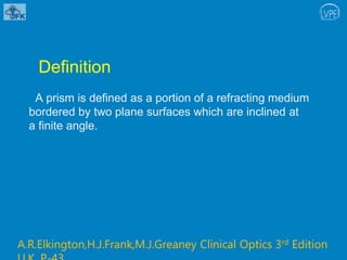
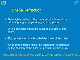
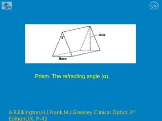
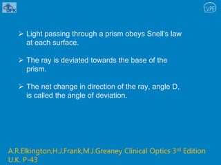
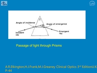
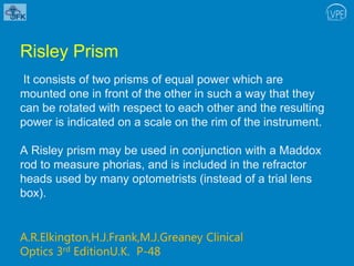
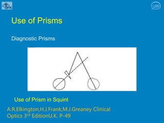
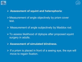
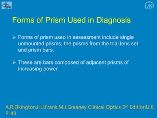
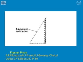
Presenter- Shayri Pillai
Liberia Eye Centre
JFK Memorial Medical Centre
LV Prasad Eye Institute
India
PRISMS
AUGUST 28 2019
Definition
A prism is defined as a portion of a refracting medium
bordered by two plane surfaces which are inclined at
a finite angle.
A.R.Elkington,H.J.Frank,M.J.Greaney Clinical Optics 3rd Edition
 The angle α between the two surfaces is called the
refracting angle or apical angle of the prism.
 A line bisecting the angle is called the axis of the
prism.
 The opposite surface is called the base of the prism.
 When prescribing prisms, the orientation is indicated
by the position of the base, e.g. 'base-in', 'base-up'.
Prism-Refraction
A.R.Elkington,H.J.Frank,M.J.Greaney Clinical Optics 3rd Edition U.K.
Prism. The refracting angle (α).
A.R.Elkington,H.J.Frank,M.J.Greaney Clinical Optics 3rd
EditionU.K. P-43
 Light passing through a prism obeys Snell's law
at each surface.
 The ray is deviated towards the base of the
prism.
 The net change in direction of the ray, angle D,
is called the angle of deviation.
A.R.Elkington,H.J.Frank,M.J.Greaney Clinical Optics 3rd Edition
U.K. P-43
 For any particular prism, the angle of deviation
D is least when the angle of incidence equals
the angle of emergence.
 Refraction is then said to be symmetrical and
the angle is called the angle of minimum
deviation.
A.R.Elkington,H.J.Frank,M.J.Greaney Clinical Optics 3rd
EditionU.K. P-43
Passage of light through Prisms
A.R.Elkington,H.J.Frank,M.J.Greaney Clinical Optics 3rd EditionU.K.
P-44
 The angle of deviation is given by the formula:
D= (n-1) α
Thus, for a glass prism of refractive index 1.5
D= (1.5-1) α
= α/2
In other words, the angle of deviation equals half the
refracting angle for a glass prism.
A.R.Elkington,H.J.Frank,M.J.Greaney Clinical Optics 3rd
EditionU.K. P-44
 The image formed by a prism is erect, virtual and
displaced towards the apex of the prism.
 Deviation is reduced to a minimum when light
passes through the prism symmetrically.
Image formation by the Prism
A.R.Elkington,H.J.Frank,M.J.Greaney Clinical Optics 3rd EditionU.K.
P-44
 There are two primary positions in which the power of
a prism may be specified, the position of minimum
deviation and the Prentice position.
 In the Prentice position one surface of the prism is
normal to the ray of light so that all the deviation takes
place at the other surface of the prism
A.R.Elkington,H.J.Frank,M.J.Greaney Clinical Optics 3rd
EditionU.K. P-44
The Prentice position of a prism
A.R.Elkington,H.J.Frank,M.J.Greaney Clinical Optics 3rd
EditionU.K. P-45
 The deviation of light in the Prentice position is
greater than that in the position of minimum deviation,
because in the Prentice position the angle of
incidence does not equal the angle of emergence.
 Therefore the Prentice position power of any prism is
greater than its power in the position of minimum
deviation.
A.R.Elkington,H.J.Frank,M.J.Greaney Clinical Optics 3rd
EditionU.K. P-45
 It is the Prentice position power which is normally
specified for glass ophthalmic prisms, e.g. trial lens
prisms.
 While it is the power in the position of minimum
deviation which is specified for plastic ophthalmic
prisms, e.g. prism bars.
A.R.Elkington,H.J.Frank,M.J.Greaney Clinical Optics 3rd
EditionU.K. P-45
Notation of Prisms
The power of any prism can be expressed in various
units.
The Prism Diopter (∆)
A prism of one prism diopter power (1∆) produces a
linear apparent displacement of 1 cm, of an object O,
situated at 1 m.
A.R.Elkington,H.J.Frank,M.J.Greaney Clinical Optics 3rd EditionU.K. P
The prism diopter and angle of apparent deviation ( θ ).
A.R.Elkington,H.J.Frank,M.J.Greaney Clinical Optics 3rd
EditionU.K. P-46
 Angle of Apparent Deviation
The apparent displacement of the object O can also
be measured in terms of the angle θ , the angle of
apparent deviation .
Under conditions of ophthalmic usage a prism of 1
prism diopter power produces an angle of apparent
deviation of 1/2°. Thus 1 prism diopter = 1/2°.
 The Centrad (∇)
 This unit differs from the prism diopter only in that the
image displacement is measured along an arc 1 m
from the prism .
 The centrad produces a very slightly greater angle of
deviation than the prism diopter, but the difference, in
practice, is negligible.
The Centrad
A.R.Elkington,H.J.Frank,M.J.Greaney Clinical Optics 3rd
EditionU.K. P-46
 Refracting Angle
 A prism may also be described by its refracting
angle . However, unless the refractive index of
the prism material is also known, the prism
power cannot be deduced.
Risley Prism
It consists of two prisms of equal power which are
mounted one in front of the other in such a way that they
can be rotated with respect to each other and the resulting
power is indicated on a scale on the rim of the instrument.
A Risley prism may be used in conjunction with a Maddox
rod to measure phorias, and is included in the refractor
heads used by many optometrists (instead of a trial lens
box).
A.R.Elkington,H.J.Frank,M.J.Greaney Clinical
Optics 3rd EditionU.K. P-48
Use of Prisms
Diagnostic Prisms
Use of Prism in Squint
A.R.Elkington,H.J.Frank,M.J.Greaney Clinical
Optics 3rd EditionU.K. P-49
 Assessment of squint and heterophoria:
Measurement of angle objectively by prism cover
test.
 Measurement of angle subjectively by Maddox rod.
To assess likelihood of diplopia after proposed squint
surgery in adults.
 Assessment of simulated blindness.
 If a prism is placed in front of a seeing eye, the eye will
move to regain fixation.
Forms of Prism Used in Diagnosis
 Forms of prism used in assessment include single
unmounted prisms, the prisms from the trial lens set
and prism bars.
 These are bars composed of adjacent prisms of
increasing power.
A.R.Elkington,H.J.Frank,M.J.Greaney Clinical Optics 3rd EditionU.K.
P-49
Therapeutic Prisms
 Convergence insufficiency
The commonest therapeutic use of prisms in the orthoptic
department is in building up the fusional reserve of patients
with convergence insufficiency.
 The prisms are used baseout during the patients'
exercise periods. They are not worn constantly.
A.R.Elkington,H.J.Frank,M.J.Greaney Clinical Optics 3rd
EditionU.K. P-50
 To relieve diplopia in certain cases of squint.
 These include decompensated heterophorias,
small vertical squints and some paralytic squints
with diplopia in the primary position.
 Prisms are reserved for those patients for whom
surgery is not indicated.
Forms of Therapeutic Prism
 Temporary wear-
 Prisms used in treatment include clip-on spectacle
prisms for trial wear.
 An improvement on these are Fresnel prisms, which
are available in all powers employed clinically.
A.R.Elkington,H.J.Frank,M.J.Greaney Clinical Optics 3rd
EditionU.K. P-49
 A Fresnel prism consists of a plastic sheet of parallel
tiny prisms of identical refracting angle.
 The overall prismatic effect is the same as that of a
single large prism.
 The sheets are lighter than a glass prism and can be
stuck on to the patient's glasses.
Fresnel Prism
A.R.Elkington,H.J.Frank,M.J.Greaney Clinical
Optics 3rd EditionU.K. P-50
Permanent wear
Permanent incorporation of a prism into a patient's
spectacles can be achieved by decentring the
spherical lens already present. Alternatively, prisms
can be mounted in spectacles.
 Prisms in Optical Instruments
 Prisms are commonly used in ophthalmic
instruments as reflectors of light.
 The prism is designed and orientated so that total
internal reflection occurs within it.
 It can be seen that prisms give greater flexibility in
dealing with an image than do mirrors.
 There are many possible systems available
Instruments in which prisms are used include the
slit lamp microscope, the applanation tonometer
and the keratometer.
A.R.Elkington,H.J.Frank,M.J.Greaney Clinical Optics 3rd
EditionU.K P-50
Thank you!
Excellence Equity Efficiency
L V Prasad Eye Institute

Prisms.pptx

  • 1.
    Presenter- Shayri Pillai LiberiaEye Centre JFK Memorial Medical Centre LV Prasad Eye Institute India PRISMS AUGUST 28 2019
  • 2.
    Definition A prism isdefined as a portion of a refracting medium bordered by two plane surfaces which are inclined at a finite angle. A.R.Elkington,H.J.Frank,M.J.Greaney Clinical Optics 3rd Edition
  • 3.
     The angleα between the two surfaces is called the refracting angle or apical angle of the prism.  A line bisecting the angle is called the axis of the prism.  The opposite surface is called the base of the prism.  When prescribing prisms, the orientation is indicated by the position of the base, e.g. 'base-in', 'base-up'. Prism-Refraction A.R.Elkington,H.J.Frank,M.J.Greaney Clinical Optics 3rd Edition U.K.
  • 4.
    Prism. The refractingangle (α). A.R.Elkington,H.J.Frank,M.J.Greaney Clinical Optics 3rd EditionU.K. P-43
  • 5.
     Light passingthrough a prism obeys Snell's law at each surface.  The ray is deviated towards the base of the prism.  The net change in direction of the ray, angle D, is called the angle of deviation. A.R.Elkington,H.J.Frank,M.J.Greaney Clinical Optics 3rd Edition U.K. P-43
  • 6.
     For anyparticular prism, the angle of deviation D is least when the angle of incidence equals the angle of emergence.  Refraction is then said to be symmetrical and the angle is called the angle of minimum deviation. A.R.Elkington,H.J.Frank,M.J.Greaney Clinical Optics 3rd EditionU.K. P-43
  • 7.
    Passage of lightthrough Prisms A.R.Elkington,H.J.Frank,M.J.Greaney Clinical Optics 3rd EditionU.K. P-44
  • 8.
     The angleof deviation is given by the formula: D= (n-1) α Thus, for a glass prism of refractive index 1.5 D= (1.5-1) α = α/2 In other words, the angle of deviation equals half the refracting angle for a glass prism. A.R.Elkington,H.J.Frank,M.J.Greaney Clinical Optics 3rd EditionU.K. P-44
  • 9.
     The imageformed by a prism is erect, virtual and displaced towards the apex of the prism.  Deviation is reduced to a minimum when light passes through the prism symmetrically. Image formation by the Prism A.R.Elkington,H.J.Frank,M.J.Greaney Clinical Optics 3rd EditionU.K. P-44
  • 10.
     There aretwo primary positions in which the power of a prism may be specified, the position of minimum deviation and the Prentice position.  In the Prentice position one surface of the prism is normal to the ray of light so that all the deviation takes place at the other surface of the prism A.R.Elkington,H.J.Frank,M.J.Greaney Clinical Optics 3rd EditionU.K. P-44
  • 11.
    The Prentice positionof a prism A.R.Elkington,H.J.Frank,M.J.Greaney Clinical Optics 3rd EditionU.K. P-45
  • 12.
     The deviationof light in the Prentice position is greater than that in the position of minimum deviation, because in the Prentice position the angle of incidence does not equal the angle of emergence.  Therefore the Prentice position power of any prism is greater than its power in the position of minimum deviation. A.R.Elkington,H.J.Frank,M.J.Greaney Clinical Optics 3rd EditionU.K. P-45
  • 13.
     It isthe Prentice position power which is normally specified for glass ophthalmic prisms, e.g. trial lens prisms.  While it is the power in the position of minimum deviation which is specified for plastic ophthalmic prisms, e.g. prism bars. A.R.Elkington,H.J.Frank,M.J.Greaney Clinical Optics 3rd EditionU.K. P-45
  • 14.
    Notation of Prisms Thepower of any prism can be expressed in various units. The Prism Diopter (∆) A prism of one prism diopter power (1∆) produces a linear apparent displacement of 1 cm, of an object O, situated at 1 m. A.R.Elkington,H.J.Frank,M.J.Greaney Clinical Optics 3rd EditionU.K. P
  • 15.
    The prism diopterand angle of apparent deviation ( θ ). A.R.Elkington,H.J.Frank,M.J.Greaney Clinical Optics 3rd EditionU.K. P-46
  • 16.
     Angle ofApparent Deviation The apparent displacement of the object O can also be measured in terms of the angle θ , the angle of apparent deviation . Under conditions of ophthalmic usage a prism of 1 prism diopter power produces an angle of apparent deviation of 1/2°. Thus 1 prism diopter = 1/2°.
  • 17.
     The Centrad(∇)  This unit differs from the prism diopter only in that the image displacement is measured along an arc 1 m from the prism .  The centrad produces a very slightly greater angle of deviation than the prism diopter, but the difference, in practice, is negligible.
  • 18.
  • 19.
     Refracting Angle A prism may also be described by its refracting angle . However, unless the refractive index of the prism material is also known, the prism power cannot be deduced.
  • 20.
    Risley Prism It consistsof two prisms of equal power which are mounted one in front of the other in such a way that they can be rotated with respect to each other and the resulting power is indicated on a scale on the rim of the instrument. A Risley prism may be used in conjunction with a Maddox rod to measure phorias, and is included in the refractor heads used by many optometrists (instead of a trial lens box). A.R.Elkington,H.J.Frank,M.J.Greaney Clinical Optics 3rd EditionU.K. P-48
  • 21.
    Use of Prisms DiagnosticPrisms Use of Prism in Squint A.R.Elkington,H.J.Frank,M.J.Greaney Clinical Optics 3rd EditionU.K. P-49
  • 22.
     Assessment ofsquint and heterophoria: Measurement of angle objectively by prism cover test.  Measurement of angle subjectively by Maddox rod. To assess likelihood of diplopia after proposed squint surgery in adults.  Assessment of simulated blindness.  If a prism is placed in front of a seeing eye, the eye will move to regain fixation.
  • 23.
    Forms of PrismUsed in Diagnosis  Forms of prism used in assessment include single unmounted prisms, the prisms from the trial lens set and prism bars.  These are bars composed of adjacent prisms of increasing power. A.R.Elkington,H.J.Frank,M.J.Greaney Clinical Optics 3rd EditionU.K. P-49
  • 24.
    Therapeutic Prisms  Convergenceinsufficiency The commonest therapeutic use of prisms in the orthoptic department is in building up the fusional reserve of patients with convergence insufficiency.  The prisms are used baseout during the patients' exercise periods. They are not worn constantly. A.R.Elkington,H.J.Frank,M.J.Greaney Clinical Optics 3rd EditionU.K. P-50
  • 25.
     To relievediplopia in certain cases of squint.  These include decompensated heterophorias, small vertical squints and some paralytic squints with diplopia in the primary position.  Prisms are reserved for those patients for whom surgery is not indicated.
  • 26.
    Forms of TherapeuticPrism  Temporary wear-  Prisms used in treatment include clip-on spectacle prisms for trial wear.  An improvement on these are Fresnel prisms, which are available in all powers employed clinically. A.R.Elkington,H.J.Frank,M.J.Greaney Clinical Optics 3rd EditionU.K. P-49
  • 27.
     A Fresnelprism consists of a plastic sheet of parallel tiny prisms of identical refracting angle.  The overall prismatic effect is the same as that of a single large prism.  The sheets are lighter than a glass prism and can be stuck on to the patient's glasses.
  • 28.
  • 29.
    Permanent wear Permanent incorporationof a prism into a patient's spectacles can be achieved by decentring the spherical lens already present. Alternatively, prisms can be mounted in spectacles.
  • 30.
     Prisms inOptical Instruments  Prisms are commonly used in ophthalmic instruments as reflectors of light.  The prism is designed and orientated so that total internal reflection occurs within it.  It can be seen that prisms give greater flexibility in dealing with an image than do mirrors.
  • 31.
     There aremany possible systems available Instruments in which prisms are used include the slit lamp microscope, the applanation tonometer and the keratometer. A.R.Elkington,H.J.Frank,M.J.Greaney Clinical Optics 3rd EditionU.K P-50
  • 32.
    Thank you! Excellence EquityEfficiency L V Prasad Eye Institute