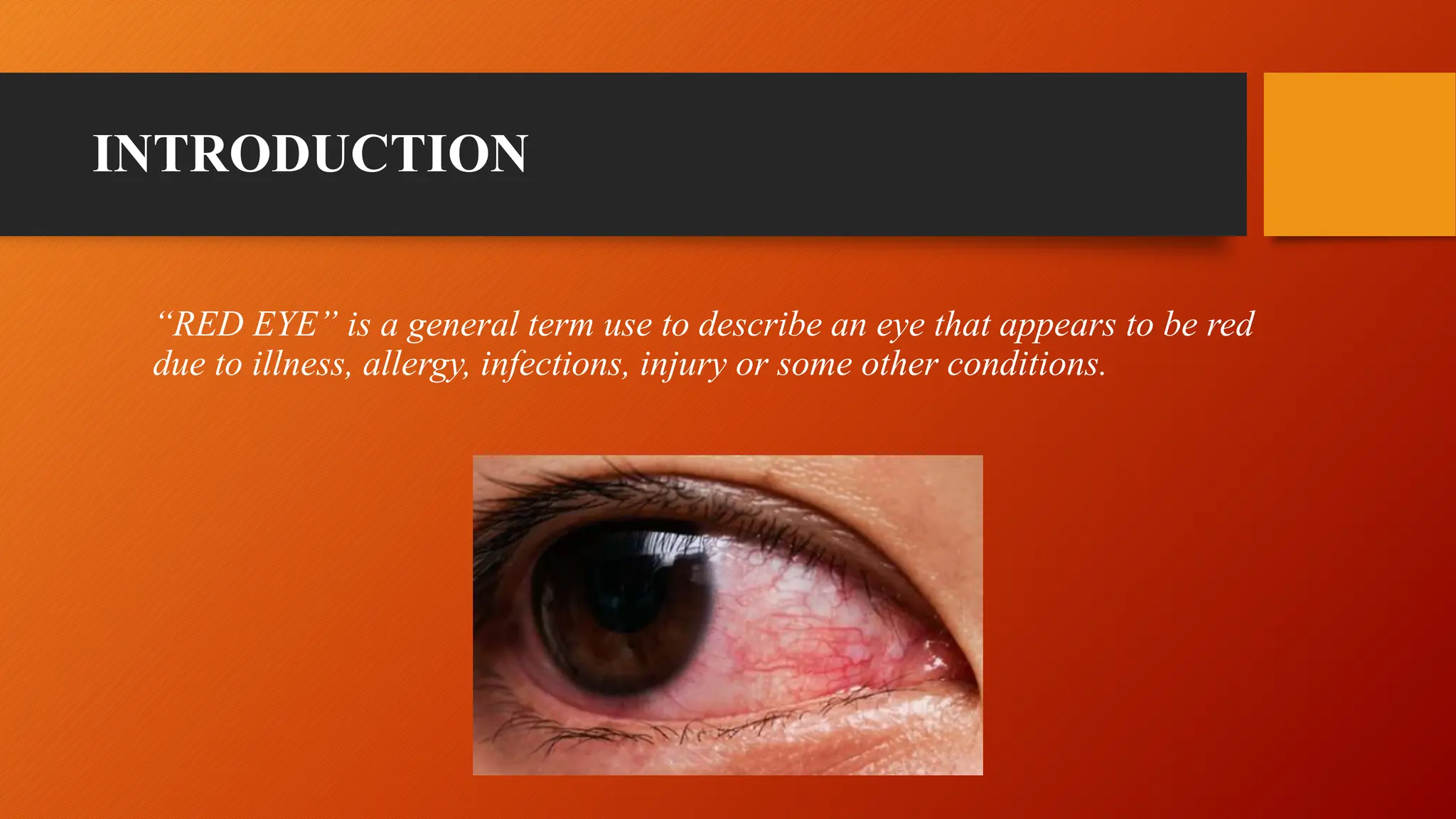
The document discusses signs of intraocular inflammation and distinguishes between granulomatous and non-granulomatous uveitis. It describes key signs of acute anterior uveitis including redness, pain, photophobia, aqueous cells and flare, hypopyon, iris signs like synechiae, and lens complications. Granulomatous uveitis is characterized by mutton fat KPs, iris nodules, and thick synechiae, while non-granulomatous uveitis presents with fine KPs, no iris nodules, and fine synechiae with more cells and flare. The document also notes the distinction between acute versus chronic conditions as causes of red eye
Red Eye - Common Causes, Diagnosis and Treatment.pptxMedinfopedia Blog
Red eye is a non-specific term that is used to describe an eye that appears red due to intraocular or extra-ocular pathologies which can be as a result of infections, inflammations, allergies or trauma.
It is usually as a result of vasodilation in the anterior portion of the eye. It is a sign of an underlying disease, not a diagnosis.
Uveal tissue is the middle vascular coat of the eyeball.
From anterior to posterior, it can be divided into 3 parts –
IRIS, CILIARY BODY CHOROID.
Iris is the anterior most part of the uveal tract.
The iris consists of four layers,
Anterior limiting layer
Iris stroma
Anterior epithelial layer
Posterior epithelial layer
The colour of iris depends on Anterior limiting layer.
Flu Vaccine Alert in Bangalore Karnatakaaddon Scans
As flu season approaches, health officials in Bangalore, Karnataka, are urging residents to get their flu vaccinations. The seasonal flu, while common, can lead to severe health complications, particularly for vulnerable populations such as young children, the elderly, and those with underlying health conditions.
Dr. Vidisha Kumari, a leading epidemiologist in Bangalore, emphasizes the importance of getting vaccinated. "The flu vaccine is our best defense against the influenza virus. It not only protects individuals but also helps prevent the spread of the virus in our communities," he says.
This year, the flu season is expected to coincide with a potential increase in other respiratory illnesses. The Karnataka Health Department has launched an awareness campaign highlighting the significance of flu vaccinations. They have set up multiple vaccination centers across Bangalore, making it convenient for residents to receive their shots.
To encourage widespread vaccination, the government is also collaborating with local schools, workplaces, and community centers to facilitate vaccination drives. Special attention is being given to ensuring that the vaccine is accessible to all, including marginalized communities who may have limited access to healthcare.
Residents are reminded that the flu vaccine is safe and effective. Common side effects are mild and may include soreness at the injection site, mild fever, or muscle aches. These side effects are generally short-lived and far less severe than the flu itself.
Healthcare providers are also stressing the importance of continuing COVID-19 precautions. Wearing masks, practicing good hand hygiene, and maintaining social distancing are still crucial, especially in crowded places.
Protect yourself and your loved ones by getting vaccinated. Together, we can help keep Bangalore healthy and safe this flu season. For more information on vaccination centers and schedules, residents can visit the Karnataka Health Department’s official website or follow their social media pages.
Stay informed, stay safe, and get your flu shot today!
Couples presenting to the infertility clinic- Do they really have infertility...Sujoy Dasgupta
Dr Sujoy Dasgupta presented the study on "Couples presenting to the infertility clinic- Do they really have infertility? – The unexplored stories of non-consummation" in the 13th Congress of the Asia Pacific Initiative on Reproduction (ASPIRE 2024) at Manila on 24 May, 2024.
Pulmonary Thromboembolism - etilogy, types, medical- Surgical and nursing man...VarunMahajani
Disruption of blood supply to lung alveoli due to blockage of one or more pulmonary blood vessels is called as Pulmonary thromboembolism. In this presentation we will discuss its causes, types and its management in depth.
Explore natural remedies for syphilis treatment in Singapore. Discover alternative therapies, herbal remedies, and lifestyle changes that may complement conventional treatments. Learn about holistic approaches to managing syphilis symptoms and supporting overall health.
These simplified slides by Dr. Sidra Arshad present an overview of the non-respiratory functions of the respiratory tract.
Learning objectives:
1. Enlist the non-respiratory functions of the respiratory tract
2. Briefly explain how these functions are carried out
3. Discuss the significance of dead space
4. Differentiate between minute ventilation and alveolar ventilation
5. Describe the cough and sneeze reflexes
Study Resources:
1. Chapter 39, Guyton and Hall Textbook of Medical Physiology, 14th edition
2. Chapter 34, Ganong’s Review of Medical Physiology, 26th edition
3. Chapter 17, Human Physiology by Lauralee Sherwood, 9th edition
4. Non-respiratory functions of the lungs https://academic.oup.com/bjaed/article/13/3/98/278874
New Drug Discovery and Development .....NEHA GUPTA
The "New Drug Discovery and Development" process involves the identification, design, testing, and manufacturing of novel pharmaceutical compounds with the aim of introducing new and improved treatments for various medical conditions. This comprehensive endeavor encompasses various stages, including target identification, preclinical studies, clinical trials, regulatory approval, and post-market surveillance. It involves multidisciplinary collaboration among scientists, researchers, clinicians, regulatory experts, and pharmaceutical companies to bring innovative therapies to market and address unmet medical needs.
- Video recording of this lecture in English language: https://youtu.be/lK81BzxMqdo
- Video recording of this lecture in Arabic language: https://youtu.be/Ve4P0COk9OI
- Link to download the book free: https://nephrotube.blogspot.com/p/nephrotube-nephrology-books.html
- Link to NephroTube website: www.NephroTube.com
- Link to NephroTube social media accounts: https://nephrotube.blogspot.com/p/join-nephrotube-on-social-media.html
Title: Sense of Smell
Presenter: Dr. Faiza, Assistant Professor of Physiology
Qualifications:
MBBS (Best Graduate, AIMC Lahore)
FCPS Physiology
ICMT, CHPE, DHPE (STMU)
MPH (GC University, Faisalabad)
MBA (Virtual University of Pakistan)
Learning Objectives:
Describe the primary categories of smells and the concept of odor blindness.
Explain the structure and location of the olfactory membrane and mucosa, including the types and roles of cells involved in olfaction.
Describe the pathway and mechanisms of olfactory signal transmission from the olfactory receptors to the brain.
Illustrate the biochemical cascade triggered by odorant binding to olfactory receptors, including the role of G-proteins and second messengers in generating an action potential.
Identify different types of olfactory disorders such as anosmia, hyposmia, hyperosmia, and dysosmia, including their potential causes.
Key Topics:
Olfactory Genes:
3% of the human genome accounts for olfactory genes.
400 genes for odorant receptors.
Olfactory Membrane:
Located in the superior part of the nasal cavity.
Medially: Folds downward along the superior septum.
Laterally: Folds over the superior turbinate and upper surface of the middle turbinate.
Total surface area: 5-10 square centimeters.
Olfactory Mucosa:
Olfactory Cells: Bipolar nerve cells derived from the CNS (100 million), with 4-25 olfactory cilia per cell.
Sustentacular Cells: Produce mucus and maintain ionic and molecular environment.
Basal Cells: Replace worn-out olfactory cells with an average lifespan of 1-2 months.
Bowman’s Gland: Secretes mucus.
Stimulation of Olfactory Cells:
Odorant dissolves in mucus and attaches to receptors on olfactory cilia.
Involves a cascade effect through G-proteins and second messengers, leading to depolarization and action potential generation in the olfactory nerve.
Quality of a Good Odorant:
Small (3-20 Carbon atoms), volatile, water-soluble, and lipid-soluble.
Facilitated by odorant-binding proteins in mucus.
Membrane Potential and Action Potential:
Resting membrane potential: -55mV.
Action potential frequency in the olfactory nerve increases with odorant strength.
Adaptation Towards the Sense of Smell:
Rapid adaptation within the first second, with further slow adaptation.
Psychological adaptation greater than receptor adaptation, involving feedback inhibition from the central nervous system.
Primary Sensations of Smell:
Camphoraceous, Musky, Floral, Pepperminty, Ethereal, Pungent, Putrid.
Odor Detection Threshold:
Examples: Hydrogen sulfide (0.0005 ppm), Methyl-mercaptan (0.002 ppm).
Some toxic substances are odorless at lethal concentrations.
Characteristics of Smell:
Odor blindness for single substances due to lack of appropriate receptor protein.
Behavioral and emotional influences of smell.
Transmission of Olfactory Signals:
From olfactory cells to glomeruli in the olfactory bulb, involving lateral inhibition.
Primitive, less old, and new olfactory systems with different path
TEST BANK for Operations Management, 14th Edition by William J. Stevenson, Ve...kevinkariuki227
TEST BANK for Operations Management, 14th Edition by William J. Stevenson, Verified Chapters 1 - 19, Complete Newest Version.pdf
Report Back from SGO 2024: What’s the Latest in Cervical Cancer?bkling
Are you curious about what’s new in cervical cancer research or unsure what the findings mean? Join Dr. Emily Ko, a gynecologic oncologist at Penn Medicine, to learn about the latest updates from the Society of Gynecologic Oncology (SGO) 2024 Annual Meeting on Women’s Cancer. Dr. Ko will discuss what the research presented at the conference means for you and answer your questions about the new developments.
For Better Surat #ℂall #Girl Service ❤85270-49040❤ Surat #ℂall #Girls
Red Eye.pptx
1. RED EYE (UVEITIS V/S OTHER
CAUSES OF RED EYE)
Dr. Gariyashee Lahkar
2. • Describe the signs of intraocular inflammation
• Granulomatous v/s non-granulomatous
• Acute iridocyclitis from chronic condition
THINGS TO LEARN
3. INTRODUCTION
“RED EYE” is a general term use to describe an eye that appears to be red
due to illness, allergy, infections, injury or some other conditions.
7. SIGNS:
• LID OEDEMA: usually mild.
• CIRCUMCORNEAL HYPERAEMIA/INJECTION:
due to involvement of deeper blood vessels.
• MIOSIS due to pupillary sphincter spasm predisposes to
the formation of posterior synechiae.
• CORNEAL OEDEMA : due to toxic rise in IOP.
Circumcorneal injection
Miosis
8. • KERATIC PRECIPITATES (KP)
Small KP
Mutton fat KPs
•Keratic precipitates (KP) are deposits
on the corneal endothelium composed
of inflammatory cells such as
lymphocytes, plasma cells and
macrophages. They are usually
concentrated inferiorly, often in a
triangular pattern with the apex
pointing up (Arlt triangle) under the
influence of gravity and aqueous
convection currents.
Types:
1. Mutton fat KPs
2. Small & medium KP (granular)
3. Fine KPs
4. Old KPs
9. AQUEOUS CELLS and AQUEOUS FLARE
They are a dependable indicator of
inflammatory activity. Grading (SUN
Working Group) is performed by
estimating the number of cells in a 1
mm by 1 mm slit beam .
AQUEOUS FLARE
It is the haziness of the normally
clear fluid in the anterior chamber,
reflecting the presence of protein
due to breakdown of the blood–
aqueous barrier.
AQUEOUS CELLS
10.
11. HYPOPYON:
Whitish purulent exudate composed of
inflammatory cells in the inferior part of the
anterior chamber (AC)
Hypopyon Hyphema
HYPHEMA:
Presence of blood in anterior chamber.
May be seen in traumatic cases.
13. • IRIS NODULES :
It can occur in both
granulomatous and
non-granulomatous
anterior uveitis.
Busacca nodules
involve the iris stroma
and are a feature of
granulomatous uveitis.
Koeppe nodules are
located on the
pupillary margin and
may be the site of
posterior synechiae
formation
14. • POSTERIOR SYNECHIAE (PS) : Inflammatory adhesions between the pupil margin and the anterior lens capsule.
Posterior synechiae
Posterior synechiae can be:
1. Segmental : Part of pupillary margin
attached to lens capsule.
2. Annular(ring) synechiae : Whole circle
of pupillary margin attached to lens
capsule.
3. Total: The posterior chamber is filled
with exudates which ties down the iris to
the lens capsule.
15. Sluggish or absent pupillary reaction
Irregular pupil due to segmental posterior synechiae
Festooned Pupil
Seclusio Pupillae: Due
to presence of annular
synechiae, Whole
circle of pupillary
margin attached to lens
capsule.
Occlusio pupillae:
Exudates organize
across the pupillary
area, forming a film of
opaque fibrous
tissue,leading to a
blocked pupil or
occlusio pupillae.
Seclusio Pupillae
PUPILLARY SIGNS:
20. Once diagnosis of iridocyclitis is established, an
attempt should be made to know whether the
condition is of granulomatous or non-granulomatous
type.
PATHOLOGICAL CLASSIFICATION:
• Granulomatous.
• Non- Granulomatous.
Granulomatous versus non-granulomatous uveitis:
21. Granulomatous uveitis
• Caused by invasion of living organisms or Autoimmune etiology.
• Type IV Hypersensitivity reaction.
• Onset is usually insidious but prolonged due to quiescent inflammatory reaction.
• Inflammation is mild.
• Commonly involves choroid, retina with vitreous exudates and nodular lesions.
22. Characterised by :
• Dense nodular infiltration of the tissues.
• Mutton fat greasy KPs are seen.
• Iris nodules seen.
• Synechiae are broad based and thick.
Granulomatous uveitis
23. Non-granulomatous uveitis:
• Onset is acute.
• Short duration
• Diffuse in extension.
• Inflammation is marked.
• Choroid/ retina / vitreous opacities are absent or fine.
24. • Multiple fine dispersed KP’s are seen, composed of lymphoid cells and polymorphs.
• Iris nodules are absent.
• Synechiae are fine and filamentous.
• Considerable flare and cells present in Anterior Chamber.
Non-granulomatous uveitis: