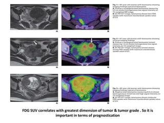
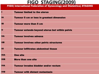
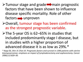
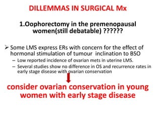
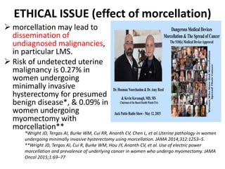
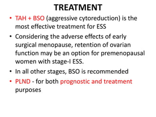
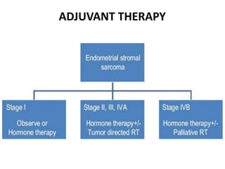
This document summarizes information about uterine sarcomas, with a focus on leiomyosarcomas and endometrial stromal sarcomas. It discusses the clinical presentation, diagnostic challenges, classification, staging, prognostic factors, surgical management, and adjuvant therapies for these rare but aggressive uterine cancers. Key points include the difficulty of pre-operative diagnosis, the importance of surgical staging and cytoreduction, and the limited but emerging role of adjuvant therapies like radiation and chemotherapy.
Recurrent pregnancy loss is a significant redroductive medical problem, influencing 2%–5% of couples. ... Throughout the years, proof based medications, for example, surgical correction of uterine abnormalities or asprin and heparin for antiphospholipid syndrome have improved the results for couples with repetitive pregnancy loss.
4 cases of pelvic mass are discussed .Adnexal mass invilves masses arisinf from ovary,fallopian tube,uterus,bowel and some miscellenious masses.USG is used to detect its size and the origin.Histopathological findings are diagnostic.
Primary Endometrial Stromal Sarcoma arising from Cervixiosrjce
IOSR Journal of Dental and Medical Sciences is one of the speciality Journal in Dental Science and Medical Science published by International Organization of Scientific Research (IOSR). The Journal publishes papers of the highest scientific merit and widest possible scope work in all areas related to medical and dental science. The Journal welcome review articles, leading medical and clinical research articles, technical notes, case reports and others.
Diagnostic Biomarker Candidates Including NT5DC2 for Human Uterine Mesenchyma...YogeshIJTSRD
Unfortunately, uterine leiomyosarcoma still has a poor prognosis. The National Cancer Institute reported that the median overall survival mOS at stage I to stage IV of leiomyosarcoma was 31 months. Takuma Hayashi | Kenji Sano | Tomoyuki Ichimura | Susumu Tonegawa | Nobuo Yaegashi | Ikuo Konishi "Diagnostic Biomarker Candidates Including NT5DC2 for Human Uterine Mesenchymal Tumors" Published in International Journal of Trend in Scientific Research and Development (ijtsrd), ISSN: 2456-6470, Volume-5 | Issue-3 , April 2021, URL: https://www.ijtsrd.com/papers/ijtsrd38686.pdf Paper URL: https://www.ijtsrd.com/medicine/other/38686/diagnostic-biomarker-candidates-including-nt5dc2-for-human-uterine-mesenchymal-tumors/takuma-hayashi
Tubular Adenoma of the Breast: A Rare Case Presentation and Review of Literat...Crimsonpublisherssmoaj
Tubular Adenoma of the Breast: A Rare Case Presentation and Review of Literature by Emmanuel Owusu Ofori* in Crimson Publishers: Surgery Open Access Journal
Tubular adenomas are a rare breast entity constituting 0.3-1.7% of all benign tumours first described in 1968 typically affecting women in their reproductive ages (15-49 years). Few cases have been reported in the literature. They are rarely found before menarche or after menopause. Preoperatively, tubular adenomas are difficult to differentiate from fibroadenoma, phyllodes and tubular carcinoma. We report the case of a 25-year-old female who reported to our clinic with 4-month history of painless left breast mass which had not seen any significant changes in size. Histological findings were consistent with tubular adenoma.
https://crimsonpublishers.com/smoaj/fulltext/SMOAJ.000541.php
For more open access journals in Crimson Publishers
Please click on: https://crimsonpublishers.com/
For more articles on Surgery Open Access Journal
Please click on link: https://crimsonpublishers.com/smoaj/index.php
Please follow the below link for our LinkedIn page
https://www.linkedin.com/company/crimsonpublishers
Evaluation of Breast Cancer in Reference to Skin ChangesQUESTJOURNAL
Introduction:- Breast cancer is the most commonly occurring female cancer in the world which is more than double that of the second ranked cancer i.e. cervical cancer. Breast cancer accounts for 23% of all cancer deaths. It is the most frequent cancer death in developing countries of the world. Mammary skin changed in breast carcinoma is categorized as advance stage in breast cancer classification. In the present study we evaluated the correlation of macroscopic, microscopic and no skin changes with axillary lymph node using histologic factor dermal lymphatic involvement. Materials and methods: prospective study was conducted on 42 breast cancer admitted patients of different age groups. Based on degree of skin involvement patients were placed into four groups, i.e. clinical stageT1 toT4. All groups were compared on the basis of percentage of patients involvement according to T1, T2, T3 and T4 stage, tumor size, histopathological dermis and epidermis involvement, dermal lymphatic invasion, tumor size and tumor subtype. Results: Majority of the patients with skin (dermis and epidermis) infiltration by the tumor (94.4%) were of T4 stage (along with dermal lymphatic invasion). Majority of the patients with only dermal lymphatic involvement (87.5%) without dermis and epidermis infiltration were of T2 stage. Conclusion: In our study, most of the patients of stage T2 and T3 with dermal lymphatic invasion had involved node when studied by routine histologic technique even though they were not clinically palpable. The identification of characteristics of the primary tumor like dermal lymphatic invasion that are associated with nodal metastases should encourage the surgeon to perform a more extensive axillary lymph node dissection and the pathologist to use methods of examining the nodes that increase the likelihood of finding metastatic disease. From this we can conclude that patients in early stage breast cancer may also have metastatic axillary lymph nodes. Dermal lymphatic invasion may be regarded as the precursor of nodal involvement, and all patients with nodal involvement can be assumed to have lymphatic invasion in the primary tumors. However the converse may not be true, not all patients with lymphatic invasion have nodal involvement.
Poly-cystic ovarian syndrome is am emerging problem in an adolescent age group which needs to be addressed because of different diagnostic criteria in this age group.
Prix Galien International 2024 Forum ProgramLevi Shapiro
June 20, 2024, Prix Galien International and Jerusalem Ethics Forum in ROME. Detailed agenda including panels:
- ADVANCES IN CARDIOLOGY: A NEW PARADIGM IS COMING
- WOMEN’S HEALTH: FERTILITY PRESERVATION
- WHAT’S NEW IN THE TREATMENT OF INFECTIOUS,
ONCOLOGICAL AND INFLAMMATORY SKIN DISEASES?
- ARTIFICIAL INTELLIGENCE AND ETHICS
- GENE THERAPY
- BEYOND BORDERS: GLOBAL INITIATIVES FOR DEMOCRATIZING LIFE SCIENCE TECHNOLOGIES AND PROMOTING ACCESS TO HEALTHCARE
- ETHICAL CHALLENGES IN LIFE SCIENCES
- Prix Galien International Awards Ceremony
Lung Cancer: Artificial Intelligence, Synergetics, Complex System Analysis, S...Oleg Kshivets
RESULTS: Overall life span (LS) was 2252.1±1742.5 days and cumulative 5-year survival (5YS) reached 73.2%, 10 years – 64.8%, 20 years – 42.5%. 513 LCP lived more than 5 years (LS=3124.6±1525.6 days), 148 LCP – more than 10 years (LS=5054.4±1504.1 days).199 LCP died because of LC (LS=562.7±374.5 days). 5YS of LCP after bi/lobectomies was significantly superior in comparison with LCP after pneumonectomies (78.1% vs.63.7%, P=0.00001 by log-rank test). AT significantly improved 5YS (66.3% vs. 34.8%) (P=0.00000 by log-rank test) only for LCP with N1-2. Cox modeling displayed that 5YS of LCP significantly depended on: phase transition (PT) early-invasive LC in terms of synergetics, PT N0—N12, cell ratio factors (ratio between cancer cells- CC and blood cells subpopulations), G1-3, histology, glucose, AT, blood cell circuit, prothrombin index, heparin tolerance, recalcification time (P=0.000-0.038). Neural networks, genetic algorithm selection and bootstrap simulation revealed relationships between 5YS and PT early-invasive LC (rank=1), PT N0—N12 (rank=2), thrombocytes/CC (3), erythrocytes/CC (4), eosinophils/CC (5), healthy cells/CC (6), lymphocytes/CC (7), segmented neutrophils/CC (8), stick neutrophils/CC (9), monocytes/CC (10); leucocytes/CC (11). Correct prediction of 5YS was 100% by neural networks computing (area under ROC curve=1.0; error=0.0).
CONCLUSIONS: 5YS of LCP after radical procedures significantly depended on: 1) PT early-invasive cancer; 2) PT N0--N12; 3) cell ratio factors; 4) blood cell circuit; 5) biochemical factors; 6) hemostasis system; 7) AT; 8) LC characteristics; 9) LC cell dynamics; 10) surgery type: lobectomy/pneumonectomy; 11) anthropometric data. Optimal diagnosis and treatment strategies for LC are: 1) screening and early detection of LC; 2) availability of experienced thoracic surgeons because of complexity of radical procedures; 3) aggressive en block surgery and adequate lymph node dissection for completeness; 4) precise prediction; 5) adjuvant chemoimmunoradiotherapy for LCP with unfavorable prognosis.
These simplified slides by Dr. Sidra Arshad present an overview of the non-respiratory functions of the respiratory tract.
Learning objectives:
1. Enlist the non-respiratory functions of the respiratory tract
2. Briefly explain how these functions are carried out
3. Discuss the significance of dead space
4. Differentiate between minute ventilation and alveolar ventilation
5. Describe the cough and sneeze reflexes
Study Resources:
1. Chapter 39, Guyton and Hall Textbook of Medical Physiology, 14th edition
2. Chapter 34, Ganong’s Review of Medical Physiology, 26th edition
3. Chapter 17, Human Physiology by Lauralee Sherwood, 9th edition
4. Non-respiratory functions of the lungs https://academic.oup.com/bjaed/article/13/3/98/278874
Explore natural remedies for syphilis treatment in Singapore. Discover alternative therapies, herbal remedies, and lifestyle changes that may complement conventional treatments. Learn about holistic approaches to managing syphilis symptoms and supporting overall health.
Couples presenting to the infertility clinic- Do they really have infertility...Sujoy Dasgupta
Dr Sujoy Dasgupta presented the study on "Couples presenting to the infertility clinic- Do they really have infertility? – The unexplored stories of non-consummation" in the 13th Congress of the Asia Pacific Initiative on Reproduction (ASPIRE 2024) at Manila on 24 May, 2024.
Ethanol (CH3CH2OH), or beverage alcohol, is a two-carbon alcohol
that is rapidly distributed in the body and brain. Ethanol alters many
neurochemical systems and has rewarding and addictive properties. It
is the oldest recreational drug and likely contributes to more morbidity,
mortality, and public health costs than all illicit drugs combined. The
5th edition of the Diagnostic and Statistical Manual of Mental Disorders
(DSM-5) integrates alcohol abuse and alcohol dependence into a single
disorder called alcohol use disorder (AUD), with mild, moderate,
and severe subclassifications (American Psychiatric Association, 2013).
In the DSM-5, all types of substance abuse and dependence have been
combined into a single substance use disorder (SUD) on a continuum
from mild to severe. A diagnosis of AUD requires that at least two of
the 11 DSM-5 behaviors be present within a 12-month period (mild
AUD: 2–3 criteria; moderate AUD: 4–5 criteria; severe AUD: 6–11 criteria).
The four main behavioral effects of AUD are impaired control over
drinking, negative social consequences, risky use, and altered physiological
effects (tolerance, withdrawal). This chapter presents an overview
of the prevalence and harmful consequences of AUD in the U.S.,
the systemic nature of the disease, neurocircuitry and stages of AUD,
comorbidities, fetal alcohol spectrum disorders, genetic risk factors, and
pharmacotherapies for AUD.
Report Back from SGO 2024: What’s the Latest in Cervical Cancer?bkling
Are you curious about what’s new in cervical cancer research or unsure what the findings mean? Join Dr. Emily Ko, a gynecologic oncologist at Penn Medicine, to learn about the latest updates from the Society of Gynecologic Oncology (SGO) 2024 Annual Meeting on Women’s Cancer. Dr. Ko will discuss what the research presented at the conference means for you and answer your questions about the new developments.
Report Back from SGO 2024: What’s the Latest in Cervical Cancer?
UTERINE LEIOMYOSARCOMA
1. UTERINE
SARCOMAS
Presented by:
DR KIRAN PANDEY
MBBS MS FRCOG FIMSA FICMCH MAMS
•PROF & HOD, DEPT OF Obs & Gyne, GSVM
MC
•PRESIDENT KANPUR OB&GYN SOCIETY
2016-18
DR PAVIKA LAL
ASSISTANT PROFESSOR
DEPT OF Obs & Gyne, GSVM MC
2. Prof. & Head. Department of OB/GY., G.S.V.M Medical College, Kanpur
Head of the Department since 10 years
DR. KIRAN PANDEY
M.D., FICOG, FICMCH, FIMSA, MAMS
ACADEMIC ACTIVITIES & RESEARCH ACTIVITIES
Published > 100 research Papers in National & International Journals, and presented 45 Research Presentation
in National Conferences. Best prize for 3 papers in AICOG.
As Secretary KOGS & Scientific Secretary IMA organized several CMEs & workshop.
•Important Project for in vivo detection of cervical pre cancers with
IIT Kanpur.
•Evaluation of Progesterone vaginal ring (PVR) in India with ICMR
• Active participation in Govt. programs like ICMR, SIFPSA, PPTCT ,IYCF, NRHM.
Special Areas Of Interest-Menopausal problems, Oncology, Infertility, Adolescent health, Gynae plastic surg.
Contributed a chapter – Gestational diabetes in “Current trends” in Obst. & Gynae.
AWARDS ACHIEVED
Received 9 National, 6 State level & 8 District level Awards & more than 25 awards at IMA.
Felicitated on World women’s day for being a “WOMEN OF SUBSTANCE”.
MEDICO SOCIAL ACTIVITIES
Received 8 awards from various Social Organization & is Actively participating in Health campus, Awareness seminars on
female feticide ,organ donation and Adolescent Health.
Actively involved in Kanpur Obstetrics & Gynaecological Society in various capacities & is presently working as Honorary
President, KOGS.
Nominated as central zone coordinator 2013-14 by National Adolescent Health Committee of FOGSI.
1995-AICOG: Chairman poster committee, 2006- UPCON: Chairman Scientific committee, 2011- FORCE: Organizing se
5. INTRODUCTION
Uterine sarcomas accounts for 3–7% of all
malignant diseases of the uterine corpus
Incidence of LMS is <2/100,000women
Notorious for their aggressive nature and poor prognosis,
because of their location in the vascular myometrium of
the uterus
early invasion and widespread metastases, particularly to
the lungs.
6. Learning objectives-
To appreciate the diagnostic challenges
faced with LMS especially in view of the
similarities with uterine fibroids.
To learn about the current views on
surgical treatment and adjuvant therapy.
To have an understanding of the novel
therapies currently under investigation.
7. CLINCIAL PRESENTATION
Median Age – 47-56 years
Risk factors:-
i) Nulliparity.
ii) Increasing age
iii) Obesity.
iv) History of pelvic radiation.
v) Exposure to tamoxifen.
8. Symptoms:-
No symptoms specific for LMS
Abnormal vaginal bleeding &
Pelvic/abdominal pain are m/c c/p but
similar to fibroids further
compounds the difficulty in diagnosis
Incidental finding of LMS at surgery
remains the m/c form of presentation
as preop dx remains inadequate.
9. PITFALLS IN DIAGNOSIS
As conservative non-surgical techniques (eg.-
Uterine artery embolisation & use of GnRH )of
treating fibroids are becoming more popular, non-
detection of LMS is a major concern delay in
diagnosis delay in instituting the appropriate
treatment.
• Mostly detected at time of HP evaluation of a
hysterectomy or myomectomy
specimen(incidence of LMS in women operated
for uterine fibroids is about 0.5%*)
• *Leibsohn S, d’Ablaing G, Mishell DR Jr, Schlaerth JB. Leiomyosarcoma in a series of
hysterectomies performed for presumed uterine leiomyomas. Am J Obstet Gynecol
1990;162:968–74;discussion 974–6.
10. DIAGNOSIS
MRI&CECT are not specific difficult to distinguish LMS
from degenerating uterine fibroids.
Combined use of dynamic MR(gd enhanced)I and serum
measurement of LDH(3) isozyme the diagnostic value
to 100 %.
T1 T2
Fibroid homogeneous and
hypointense
homogeneous and
hypointense
LMS heterogeneous
hyperintense
irregular borders
and areas of
haemorrhage or
necrosis
11. Dynamic MRI Image(gadolinium enhanced)
T2-weighted imaging in preop pelvic MRI (A) Sagittal slice showing an irregular tumor
with high-signal intensity extending from the cavity to the right posterior wall of the
uterus (arrow 1). (B) Axial slice showing extension of the tumor to the right adnexa and
vessels (arrow 2) and the right internal iliac vein (arrow 3).
12. MRI image of uterine fibroid
A degenerating fibroidMRI showing normal fibroid
13. .
LMS LEIOMYOMA
CYTOLOGICAL ATYPIA
+
Moderate to severe
-
HIGH MITOTIC INDEX
+
≥10 per 10 HPF
+/-
COAGULATIVE TUMOUR
CELL NECROSIS + -
Leiomyoma showing spindle shaped cells
with elongated nuclei uniform in size with
Varying amount of connective tissue
HISTOLOGY
15. STUMPs(smooth muscle tumours of unknown
malignant potential) have some characteristic
features of LMS, but do not meet full criteria
The absence of coagulative necrosis and atypia
suggest a fibroid, even if the mitotic count is as
high as 20
Histological diagnosis by endometrial sampling
is unreliable as it cannot provide an accurate
result unless the tumour has reached the
surface of the endometrial cavity which gives a
low sensitivity of approximately 30%
16. IMMUNOHISTOCHEMISTRY
LMS stain +ive for SMA(smooth muscle actin),
desmin and caldesmon.
ER and PR expression is significantly lower in
LMS than leiomyoma (ER:40%vs78%; PR: 38% vs
88%).
Immunopositivity for p16 and p53 with a high
Ki-67 proliferation index also has been shown to
have high sensitivity and specificity for
differentiating LMS and leiomyomas.
(Differentiates LMS from leiomyoma)
17. A&B- IHC staining of diagnosed LMS for antibodies Against smooth muscle Actin and Desmin.
C&D- Staining negative for SMA and Desmin (leiomyoma)
18. FDG SUV correlates with greatest dimension of tumor & tumor grade . So it is
important in terms of prognostication
19.
20. FIGO( International Federation of Gynecology and Obstetrics) STAGING
I Tumour limited to the uterus
IA Tumour 5 cm or less in greatest dimension
IB Tumour more than 5 cm
II Tumour extends beyond uterus but within pelvis
IIA Tumour involves adnexa
IIB Tumour involves other pelvic structures
III Tumour infiltrates abdominal tissue
IIIA One site
IIIB More than one site
IVA Tumour invades bladder and/or rectum
IVB Tumour with distant metastasis
FIGO STAGING(2009)
21. Tumour stage and grade main prognostic
factors that have been shown to influence
disease specific mortality. Role of other
factors unproven
Overall, tumour stage has been confirmed
as the strongest prognostic variable.
The 5-year OS is 62–65% in studies that
included predominantly stage I disease, but
in studies with a higher proportion of
advanced disease it is as low as 29%.*
* Kapp DS, Shin JY, Chan JK. Prognostic factors and survival in 1396 patients with uterine
leiomyosarcomas: emphasis on impact of lymphadenectomy and oophorectomy. Cancer
2008;112:820–30.
23. PRINCIPLES OF MANAGEMENT
It is universally accepted that surgery is primary
treatment for LMS.
A TAH and BSO and appropriate surgical staging,
including peritoneal washings and sampling of
suspicious nodules, should be carried out .
It is recommended that aggressive surgical
cytoreduction at the time of initial diagnosis offers
the best possibility of prolonged survival.
24. SURGICAL MANAGEMENT
Three areas of contention with regard to
surgery are:-
i) Oophorectomy in the premenopausal
women.
II) Incidental finding of LMS in myomectomy
specimen.
iii) Role of Pelvic lymphadenectomy(PLND) in
early LMS
25. DILLEMMAS IN SURGICAL Mx
1.Oophorectomy in the premenopausal
women(still debatable) ??????
Some LMS express ERs with concern for the effect of
hormonal stimulation of tumour inclination to BSO
– Low reported incidence of ovarian mets in uterine LMS.
– Several studies show no difference in OS and recurrence rates in
early stage disease with ovarian conservation
consider ovarian conservation in young
women with early stage disease
26. 2.Incidental finding of LMS in myomectomy
specimen?
a completion procedure with total hysterectomy,
trachelectomy and/or bilateral salpingo-
oophorectomy is recommended.
A conservative approach following myomectomy
should only be taken for specific and accurately
selected women who strongly desire pregnancy
and well counselled about the risks involved.
27. ETHICAL ISSUE (effect of morcellation)
morcellation may lead to
dissemination of
undiagnosed malignancies,
in particular LMS.
Risk of undetected uterine
malignancy is 0.27% in
women undergoing
minimally invasive
hysterectomy for presumed
benign disease*, & 0.09% in
women undergoing
myomectomy with
morcellation**
*Wright JD, Tergas AI, Burke WM, Cui RR, Ananth CV, Chen L, et al.Uterine pathology in women
undergoing minimally invasive hysterectomy using morcellation. JAMA 2014;312:1253–5.
**Wright JD, Tergas AI, Cui R, Burke WM, Hou JY, Ananth CV, et al. Use of electric power
morcellation and prevalence of underlying cancer in women who undergo myomectomy. JAMA
Oncol 2015;1:69–77
28. THE MORCELLATION CONTROVERSY
• Amy Reed (an anesthesiologist) was diagnosed with fibroids and was
advised TLH with morcellation but things didn't go as expected. Dr.
Reed learned that she had a uterine leiomyosarcoma and THE
MORCELLATION MAY HAVE WORSENED HER PROGNOSIS
• Since then, Dr. Reed's husband, has led a campaign calling for a ban
on morcellation.
• FDA on April 2014 announced that it discourages the use of
laparoscopic power morcellators in most hysterectomy and
myomectomy procedures especially in suspected or confirmed
uterine malignancies/ peri or post menopausal women with fibroid
because of the risk of spreading unsuspected cancerous tissue.
29. Role of Pelvic lymphadenectomy(PLND) in
early LMS
Incidence of LN mets from uLMS is very low &
unlikely in the absence of extrauterine disease.
Routine PLND is not usually done for women with
the disease confined to the uterus and with
normal LNs on observation and palpation
LN status may have a role as a staging procedure
& in determining the need for adjuvant pelvic RT
but its therapeutic benefit is still to be proven
30. Management of Early uterine LMS
SURGERY
ADJUVANT
PELVIC RT
SYSTEMIC CT
EORTC trail(55874) – 13 years study -224
patients- 2 groups – observation & RT(51Gy
in 28# over 5 weeks)
No beneficial impact on OS
Premenopausal women:- TAH
+/- PLND ( in presence/absence
of extrauterine disease
respectively)
Postmenopausal female:- TAH +
BSO
31. Limited studies available- ongoing phase III
trial(NCTO1533207*)
Observation 4 cycles of adjuvant GEMCITABINE+ DOXORUBICIN
additional 4 cycles of DOXORUBICIN( regimen well tolerated)
This trial is still underway
*A RCT of adjuvant CT vs observation to determine
if adjuvant CT can improve OS in early uLMS
CT
32. ADVANCED OR METASTATIC UTERINE LMS
SURGERY
ADJUVANT
PELVIC RT
No role
LMS recurs only at
Metastatic sites
Secondary cytoreduction
surgery
Pulmonary mets
resection
Improved OS
SYSTEMIC CT
met. disease beyond the
Pelvis- not surgical candidates
OTHER MODALITIES
gemcitabine + docetaxel
- best regimen
34. Conclusion-
m/c uterine sarcoma
Notoriously aggressive in nature.
Preop dx is difficult & usually detected as an
incidental finding at surgery.
Tumour stage is the most important prognostic
factor.
Primary treatment is surgical(in early/
advanced/ recurrent ) while role of adjuvant
therapy(RT/CT) is still to be clearly defined.
Gemcitabine + docetaxel- best regimen for
adjuvant CT.
36. • ESS - very rare(.2%) of all uterine malignancies
• Annual incidence - 1–2 per million women
• An indolent tumor with local recurrences and
distant metastasis can occur even 20 years
after initial diagnosis. Median age-36-48yrs
37. ENDOMETRIAL STROMAL NODULE:
Well circumscribed, non encapsulated
Finger like projections into the myometrium <3mm & <3 in no.
No vascular invasion
LOW GRADE ESS:
irregular nodular growth in endometrium
Infiltrates(worm like) myometrium, myometrial & parametrial
veins
Cellular atypia & pleomorphism absent
Mitotic activity <5/10HPF
UNDIFFERENTIATED ESS(earlier k/a high grade ESS)
Resemble sarcomatous component of carcinosarcomas
Lack typical growth patterns and vascularity
Displaces myometrium(in contrast to infiltration in low gr ESS)
Abundant mitotic activity & marked cellular atypia
Shows prominent hemorrhages & necrosis within polypoidal
fleshy endometrial masses.
38. CLINICAL FEATURES
abnormal uterine bleeding(90%)
pelvic pain
dysmenorrhoea
• asymptomatic in 25% individuals
• 30 to 50% cases - extra uterine spread at the
time of the diagnosis
• Rarely ESS is initially present at an extra
uterine site(m/c ovary) – primary/ metastatic
lesion from an occult tumor of endometrium
or from a previously undiagnosed case where
a hysterectomy was done for a benign
leiomyoma of the uterus.
39. DIAGNOSIS
• similarity of ESS with normal endometrium+ the
lesion is almost always intra-myometrial
impossible to diagnose it with certainty on
curettage fragments
definitive diagnosis can be made only on a
hysterectomy specimen
• RADIOLOGY: USG –unreliable;
MRI
useful in preop diagnosis.
Bands of low signal intensity within myometrium.
Continuous extension of lesions into adjascent
structures(vessels & ligaments)
40. Endometrial stromal nodule-
Multiple collagen bundles are
interspersed among the
neoplastic stromal cells
high-grade endometrial stromal
sarcoma. Tumor cells with an
epithelioid morphology (left)
are juxtaposed to areas of
endometrial stromal neoplasia
with a fibroblastic appearance.
HISTOLOGY
41. IMMUNOHISTOCHEMICAL FEATURES
Low-grade endometrial stromal sarcoma
with smooth muscle differentiation.
Desmin immunoreaction highlights the
smooth muscle component (‘starburst’)
with minimal reaction of the background
endometrial stroma
high-grade endometrial stromal sarcoma.
CD10(cell surface neutral endopeptidase)
Positivity- helps in differentiating from
leiomyoma .H-ESS is almost always + for
ER & PR
IMMUNOHISTOCHEMISTRY: Strong and/or diffuse positivity for CD10 ; ESS is almost
always positive for both ER & PR
42. MRI image of ESS showing
increased intensity of the tumor
In comparison to normal
endometrium. It also shows
Continuity of tumor into broad
and round ligaments
43. stage Definition
Stage 1 CONFINED TO UTERUS
1A Tumor limited to endometrium/endocervix. No myometrial invasion
1B <= half of myometrial invasion
1C >half of myometrial invasion
Stage 2 EXTENDS TO PELVIS
2A Adnexal involvement
2B Extra uterine pelvic tissues
Stage 3 INVADES ABDOMINAL TISSUES
3A One site
3B > One site
3C Metastasis to pelvic & para aortic lymph nodes
Stage 4
4A Invades bladder &/or rectum
4B Distant metastasis
FIGO STAGING OF ESS
44. PROGNOSTIC FACTORS
• older patients (>50 yrs)
• black race
• advanced stage
• nodal metastasis
• high mitotic count(>5/10HPF)
• CD10 negative
• lack of ER&PR
• Lack of primary surgery
INDEPENDENT
POOR
PROGNOSIS
ESS has a better life expectancy than other sarcomas
45. TREATMENT
• TAH + BSO (aggressive cytoreduction) is the
most effective treatment for ESS
• Considering the adverse effects of early
surgical menopause, retention of ovarian
function may be an option for premenopausal
women with stage-I ESS.
• In all other stages, BSO is recommended
• PLND - for both prognostic and treatment
purposes
47. HORMONAL & TUMOR DIRECTED
THERAPY
The mechanism of action of progestins is to bind
progesterone receptors and down regulate gene
transcription leading to decreased endometrial
gland and stromal proliferation. GnRH agonists
down regulate GnRH receptors in the anterior
pituitary leading to a hypoestrogenic state.
megestrol/medroxy progesterone(160mg/d)
gonadotropin releasing hormone (GnRH)
analogues(7.5mg/month IM)
aromatase inhibitors
50. • MMT is a rare(0.6/1,00,000 women), highly
aggressive, rapidly progressing neoplasm
associated with a poor prognosis
• MMT is a biphasic tumour of the female genital
tract, composed of epithelial and mesenchymal
tissues. Median age- 62yrs.
• two categories: homologous and heterologous.
The homologous type: has tissues native to the
uterus(endometrium or smooth muscle)
The heterologous type: cartilage, skeletal muscle, or bone
is present which is not native to the uterus.
51. RISK FACTORS
• Nulliparity
• Advanced age
• Obesity
• Exposure to exogenous estrogens
• Long-term use of tamoxifen
• Pelvic RT
52. Stage Extent
Stage 1 Tumor confined to corpus uterii
1A <half of myometrium
1B >= half of myometrium
Stage 2 Invasion of cervical stroma but does not extend beyond uterus
Stage3 Loco-regional spread
3A Invades serosa of uterus / adnexa
3B Vaginal or parametrial involvement
3C Mets to pelvic &/or para aortic nodes
3C1 Pelvic nodes +
3C2 Para aortic nodes +
Stage4 Distant metastasis
4A Invasion of bladder orbowel mucosa
4B Intra abdominal &/ or inguinal nodes
CLASSIFICATION- MMTs are staged like CA endometrium
53. TYPICAL PATIENT PROFILE PRESENTING WITH UTERINE
CARCINOSARCOMA
Elderly female (usually 60–70 years), usually
postmenopausal
Presents with pyometra with vagina bleeding,
bloody/watery discharge, abdominal pain, and/or
mass (symptom triad)
Often past history of tamoxifen use
May be obese, hypertensive, nulliparous, and/or
diabetic
No previous history of uterine problems
54. DIAGNOSIS
• HISTOLOGY: unique biphasic morphology-both
epithelial and mesenchymal elements
• IHC: express epithelial membrane antigen (EMA),
pancytokeratin and stromal lineage markers in
relation to their histological appearances such as
desmin in muscle differentiation or S100 in areas
with chondroid or lipomatous differentiation
• Overexpression of tyrosine kinase receptors such
as HER-2(human epidermal growth factor
receptor ), EGFR(Epidermal growth factor
receptor), and KIT(a part of tyrosine kinase
receptor)
55. A-F:Homogeneous hyperintense intrauterine mass, enhancing with gadolinium,invades the myometrium
Lymphadenopathies are present, Surgery : large irregular mass Gross specimen: endometrial cavity
completely occupied by a polypoid lobulated tumor gray and dun with white areas (4,5x3,5 cm). It
invades the whole thickness of the uterine wall on the left side. On microscopic exam poorly
differentiated areas predominate over areas of adenocarcinoma with squamous spots mixed with
sarcomatous areas with muscular differentiation
56. • on MRI uterine MMTs may be indistinguishable
from endometrial carcinomas
• Enhancement equal to or greater than that of
the myometrium suggests the possibility of this
tumour-type
• CECT & 18F-Fluorodeoxyglucose Positron
Emission Tomography (18F-PDG PET) Scans are
other modalities useful in diagnosis
57. TREATMENT
• The primary treatment option is surgery
• High rates of relapse and metastases
postoperatively necessitate effective adjuvant
therapies
• surgical practice recommended for uterine
carcinosarcoma is surgical staging with TAH
with BSO, pelvic lymphadenectomy, and para-
aortic lymph-node sampling with peritoneal
washings.
58. • Radiotherapy contributes to decreased pelvic
recurrences
• Controversies still remain regarding the
techniques of radiation: localized pelvic radiation
by vaginal brachytherapy versus whole abdominal
radiation by external beam
• chemotherapy has a definitive role to minimize
both local and distal recurrence
• A combination of paclitaxal & carboplatin results
in RR of 80% and median disease free interval of
18months
59. SURGICAL STAGING(AGGRESSIVE CYTO-REDUCTION)
(TAH+BSO+PLND+PERITONEAL WASHING+OMENTAL BIOPSY)
STAGE 1&2 STAGE 3 STAGE 4
CYTO-REDUCTION SURGERY + RADIATION
(VAGINAL BRACHY/ EB RT)
? ROLE OF CYTO-REDUCTION SURGERY
ADJUVANT CHEMOTHERAPY
+HARMONAL THERAPY+MOLECULAR TARGETED
THERAPY