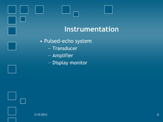
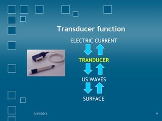
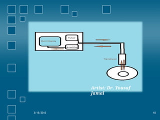
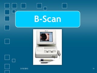
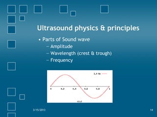
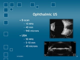
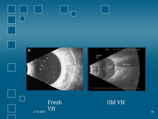
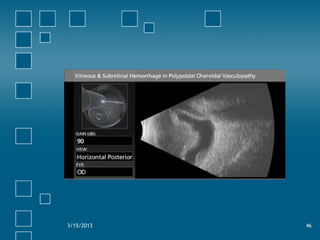
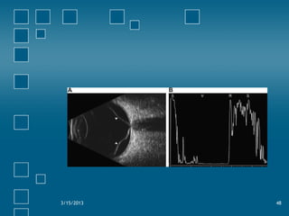
OPHTHALMIC ULTRASOUND
OPTOM RAJU GHOSH
Echolocation types
6
TYPES
A – SCAN
B – SCAN
Instrumentation
• Pulsed-echo system
− Transducer
− Amplifier
− Display monitor
3/15/2013 8
Transducer function
ELECTRIC CURRENT
TRANDUCER
US WAVES
SURFACE
3/15/2013 9
3/15/2013 10
Artist: Dr. Yousaf
Jamal
3/15/2013 11
B-Scan
B-scan
• Brightness mode scan
• OPD procedure
3/15/2013 12
Indications
• To examine intraocular structures with no
direct visualization of posterior segment
Or
• To confirm or differentiate between
pathologies in clear media
3/15/2013 13
Ultrasound physics & principles
• Parts of Sound wave
− Amplitude
− Wavelength (crest & trough)
− Frequency
3/15/2013 14
Frequency & its relations
With resolution
Image quality
With penetration
How much deep
3/15/2013 15
Medical Ultrasound frequencies
• Abdominal US
− 1-5 MHz
• Ophthalmic US (B-scan)
− 8-10 MHz
• Ultrasound Biomicroscopy (UBM)
− 20-50 MHz
Ophthalmic US
• B-scan
− 10 MHz
− 40 mm
− 940 microns
• UBM
− 50 MHz
− 5-10 mm
− 40 microns
3/15/2013 21
Principles of US
• Velocity
• Reflectivity
• Angle of incidence
• Absorption
3/15/2013 22
• Higher gain for weaker echoes
• Low gain for stronger echoes
3/15/2013 25
Angle of incidence
3/15/2013 26
Transduc
er
Probe positioning
• Trans-ocular approach
− Transverse
− Longitudinal
− Axial
• Para-ocular approach
3/15/2013 28
Transverse position
− Most commonly used position
− Shows about 6 clock hours
− Used for basic screening
− Detects lateral extent of pathology
− Probe is placed opposite to the examined
meridian
3/15/2013 29
Longitudinal positions
− Detects axial (AP) extent of pathology
− Useful for retinal tears detection
− Shows only 1 clock hour scan
3/15/2013 31
Axial positions
• Probe direct over the cornea
• Pt looks in primary gaze
• US waves pass thru center of lens and hit
optic nerve rather than macula
• Lens density affects the quality of image
3/15/2013 33
Basic screening technique
• Done for screening purpose in opaque
media
• Highest gain settings are used so weaker
signals shouldn’t be missed
• Any pathology found…further scanning is
required
3/15/2013 35
Technique
• 05 scans in different positions will detect
gross pathology
• Transverse position with limbus-to-fornix
approach in…
− 12, 3, 6 and 9 o'clock
• Horizontal axial scan …shows optic nerve
& macula in one image
• Print out of each position is taken with
labels
3/15/2013 36
Common examples
Vitreoretinal disorders
• Most common indication for B-scan
− Vitreous hemorrhage
− Retinal detachment
− Intraocular tumors
− Intraocular foreign bodies
3/15/2013 43
Vitreous hemorrhage
• Fresh:
− Dot-like…Echolucent or low reflectivity
• Old:
− Membrane-like…varying reflectivity &
dense inferiorly
3/15/2013 44
3/15/2013 45
Fresh
VH
Old VH
3/15/2013 46
Retinal detachment
• Rhegmatogenous R/D:
− Thin, continuous membrane anteriorly
separated from globe wall
− Echoes are of high amplitue-100% of
scleral spike
− Retinal cysts, subretinal hemorrhages may
be seen
3/15/2013 47
3/15/2013 48
• Tractional R/D:
− Traction membranes are seen
− R/D usually doesn’t extend to ora serrata
− Lower mobility in contrast to Rheg. R/D
3/15/2013 49
3/15/2013 50
3/15/2013 51
• Exudative R/D:
− Smooth elevation of retina
− Shifting fluid
3/15/2013 52
3/15/2013 53
Supine
position
Erect
position
Intraocular tumors
• Retinoblastoma:
− Single or multiple mass lesions arising
from retina
− Highly refractile calcium seeding in
vitreous +/- orbital shadowing
− R/D may be found
3/15/2013 54
3/15/2013 55
Intraocular foreign body
• Echodense signals with shadowing
• Persistence of signals at low gains
• Glass…reverberations
• Air bubble…may simulate IOFB
3/15/2013 59
3/15/2013 60
Dislocated lens
• Signals depend on clarity of lens
• Clear lens…Echolucent globular structure
• Brunescent lens…highly reflective with
shadowing
3/15/2013 61
3/15/2013 62

USG B SCAN OPHTHALMOLOGY