This study aims to use morphometric analysis of nuclear and nucleolar structures to distinguish between epithelial (EPI) and epithelial-to-mesenchymal (EMT) states of prostate cancer cell lines. Images of PC3 cell nuclei and nucleoli stained with fluorescent probes are analyzed using software to measure 3D characteristics. Preliminary results show a difference between EPI and EMT conditions. Further analysis of additional cell lines as well as correlating gene expression with nuclear positioning may help characterize differences and improve early cancer detection.
An understanding towards genetics and epigenetics is essential to cope up with the paradigm shift which is underway. Personalized medicine and gene therapy will confluence the days to come.
This review highlights traditional approaches as well as current advancements in the analysis of the gene expression data from cancer perspective.
Due to improvements in biometric instrumentation and automation, it has become easier to collect a lot of experimental data in molecular biology.
Analysis of such data is extremely important as it leads to knowledge discovery that can be validated by experiments. Previously, the diagnosis of complex genetic diseases has conventionally been done based on the non-molecular characteristics like kind of tumor tissue, pathological characteristics, and clinical phase.
The microarray data can be well accounted for high dimensional space and noise. Same were the reasons for ineffective and imprecise results. Several machine learning and data mining techniques are presently applied for identifying cancer using gene expression data.
While differences in efficiency do exist, none of the well-established approaches is uniformly superior to others. The quality of algorithm is important, but is not in itself a guarantee of the quality of a specific data analysis.
http://kaashivinfotech.com/
http://inplanttrainingchennai.com/
http://inplanttraining-in-chennai.com/
http://internshipinchennai.in/
http://inplant-training.org/
http://kernelmind.com/
http://inplanttraining-in-chennai.com/
http://inplanttrainingchennai.com/
Molecular imaging has revolutionized our perceptions of imaging. This high impact field is finding transformative applications in the understanding, detection, and treatment of nearly all diseases.
The field of molecular imaging is a an exciting fusion and integration of many different disciplines including molecular biology, chemistry and probe design, imaging technologies, visualization, and image analysis, that are focused on understanding, detecting, and treating oncological, neurological, cardiovascular, inflammatory, metabolic, and infectious diseases. Based on their strengths, different imaging modalities provide different but equally valuable information that can be integrated in advancing our understanding of these diseases.
As the era of ‘omics’ progresses towards personalized medicine, the field of molecular imaging is finding multiple uses in noninvasive characterization of the molecular features of diseases and their impact on function. In complex diseases such as cancer, with its tremendous genetic diversity, it is becoming increasingly important to develop molecularly-targeted treatment strategies that combine detection with treatment.
Magnetic Resonance Spectroscopy and MetabolomicsUzay Emir
Magnetic Resonance Spectroscopy, MRI, Human Connectome, 2-HG, 2-hydroxyglutarate, zoom, zoom MRSI, reduced field of View, rFOV, Cerebellum, High-resolution, IDH, Isocitrate, IDH1, IDH2, Cancer, Glioma, Parcellation, Macro Anatomical
Functional
Myeloarchitectonic
CYTO- AND RECEPTOR ARCHITECTONIC MAPPING OF THE HUMAN BRAIN
MR Spectroscopy Study Group
Multimodality Molecular Imaging – An Overview With Special Focus on PET/CTApollo Hospitals
Imaging capabilities have evolved from those that provide anatomical pictures to those that capture functional information and, more recently, molecular information (nuclear medicine, PET, SPECT, PET/CT, SPECT/CT, MRS, contrast-enhanced ultrasound, fluorescence and bioluminescence imaging). Multimodality imaging has emerged as a technology that utilizes the strengths of different modalities and yields a hybrid imaging platform with benefits superior to those of any of its individual components, considered alone. Leading edge hybrid imaging (combining multiple, complementary imaging technologies such as PET and CT) offer unique opportunities to “view” the molecular biology of disease, and the use of this equipment is on the rise.
A Classification of Cancer Diagnostics based on Microarray Gene Expression Pr...IJTET Journal
inAbstract— Pattern Recognition (PR) plays an important role in field of Bioinformatics. PR is concerned with processing raw measurement data by a computer to arrive at a prediction that can be used to formulate a decision to be taken. The important problem in which pattern recognition are applied have common that they are too complex to model explicitly. Diverse methods of this PR are used to analyze, segment and manage the high dimensional microarray gene data for classification. PR is concerned with the development of systems that learn to solve a given problem using a set of instances, each instances represented by a number of features. The microarray expression technologies are possible to monitor the expression levels of thousands of genes simultaneously. The microarrays generated large amount of data has stimulate the development of various computational methods to different biological processes by gene expression profiling. Microarray Gene Expression Profiling (MGEP) is important in Bioinformatics, it yield various high dimensional data used in various clinical applications like cancer diagnostics and drug designing. In this work a new schema has developed for classification of unknown malignant tumors into known class. According to this work an new classification scheme includes the transformation of very high dimensional microarray data into mahalanobis space before classification. The eligibility of the proposed classification scheme has proved to 10 commonly available cancer gene datasets, this contains both the binary and multiclass data sets. To improve the performance of the classification gene selection method is applied to the datasets as a preprocessing and data extraction step.
The IOSR Journal of Pharmacy (IOSRPHR) is an open access online & offline peer reviewed international journal, which publishes innovative research papers, reviews, mini-reviews, short communications and notes dealing with Pharmaceutical Sciences( Pharmaceutical Technology, Pharmaceutics, Biopharmaceutics, Pharmacokinetics, Pharmaceutical/Medicinal Chemistry, Computational Chemistry and Molecular Drug Design, Pharmacognosy & Phytochemistry, Pharmacology, Pharmaceutical Analysis, Pharmacy Practice, Clinical and Hospital Pharmacy, Cell Biology, Genomics and Proteomics, Pharmacogenomics, Bioinformatics and Biotechnology of Pharmaceutical Interest........more details on Aim & Scope).
All manuscripts are subject to rapid peer review. Those of high quality (not previously published and not under consideration for publication in another journal) will be published without delay
Presentation by Scott Woodman, MD, PhD. Presented at the 2018 Eyes on a Cure: Patient & Caregiver Symposium, hosted by the Melanoma Research Foundation's CURE OM initiative.
Our understanding of life at the molecular level is highly dependent on the ability to map the molecular details of individual proteins and nucleic acids as well as their interactions with each other and with small molecules (inhibitors, cofactors, substrates, etc.). The determination of 3-D structures of proteins is crucial for the understanding of these interactions as well as their structure–function relationships, which also has many practical applications in drug design and protein engineering.
E1512 Trial Spotlight for May 2013 ECOG-ACRIN NewsletterSara Bucknam
E1512 Trial spotlight for ECOG-ACRIN Newsletter. E1512 is a randomized, three‐arm phase II trial investigating the clinical efficacy and safety of erlotinib alone, cabozantinib alone, or erlotinib plus cabozantinib as second‐ or third‐line therapy in patients with EGFR wild‐type NSCLC.
Basic structural and functional unit of life
Understanding of cell morphology is critical to the study of biochemistry.
Divided and classified in many ways.
One common classification method is absence or presence of a cell nucleus.
A physical sciences network characterization of non-tumorigenic and metastati...Shashaanka Ashili
To investigate the transition from non-cancerous to metastatic from a physical sciences perspective, the
Physical Sciences–Oncology Centers (PS-OC) Network performed molecular and biophysical comparative studies of the non-tumorigenic MCF-10A and metastatic DA-MB-231 breast epithelial cell lines, commonly used as models of cancer metastasis. Experiments were performed in 20 laboratories from 12 PS-OCs. Each laboratory was supplied with identical aliquots and common reagents and culture protocols. Analyses of these measurements revealed dramatic differences in their mechanics, migration, adhesion, oxygen response, and proteomic profiles. Model-based multi-omics approaches identified key differences between these cells’ regulatory networks involved in morphology and survival. These results provide a multifaceted description of cellular parameters of two widely used cell lines and demonstrate the value of the PS-OC Network approach for integration of diverse experimental observations to elucidate the phenotypes associated with cancer metastasis.
Precision Radiotherapy: Tailoring Treatment for Individualised Cancer Care.pptxDr. Rituparna Biswas
Precision radiotherapy, also known as precision radiation therapy or targeted radiotherapy, is a cutting-edge approach in the field of radiation oncology that aims to deliver highly focused and accurate doses of radiation to cancerous cells while minimizing damage to surrounding healthy tissues.
Non negative matrix factorization ofr tuor classificationSahil Prajapati
The PPT aware about you the concept of Non Negative Matrix Factorization and how theses techniques can be used to treat cancer by the use of the coding such as a MATLAB,LABVIEW software to locate the tumor or the cancer part with the different approaches and tachniques.
Go through the PPT to know and how one can improvise my work for better results??
Please help me if one come up with other techniques.
MEDICAL IMAGING MUTIFRACTAL ANALYSIS IN PREDICTION OF EFFICIENCY OF CANCER TH...cscpconf
Based on pressing need for predictive performance improvement, we explored the value of pretherapy
tumour histology image analysis to predict chemotherapy response. It was shown that
multifractal analysis of breast tumour tissue prior to chemotherapy indeed has the capacity to
distinguish between histological images of the different chemotherapy responder groups with
accuracies of 91.4% for pPR, 82.9% for pCR and 82.1% for PD/SD.
MEDICAL IMAGING MUTIFRACTAL ANALYSIS IN PREDICTION OF EFFICIENCY OF CANCER TH...csandit
Based on pressing need for predictive performance improvement, we explored the value of pretherapy
tumour histology image analysis to predict chemotherapy response. It was shown that
multifractal analysis of breast tumour tissue prior to chemotherapy indeed has the capacity to
distinguish between histological images of the different chemotherapy responder groups with
accuracies of 91.4% for pPR, 82.9% for pCR and 82.1% for PD/SD.
Titles with Abstracts_2023-2024_Digital Image processing.pdfinfo751436
Digital image processing (DIP) refers to the manipulation of digital images using various algorithms and techniques to enhance or extract information from the images. There are several advantages to using digital image processing:
Enhancement of Image Quality:
DIP allows for the improvement of image quality by reducing noise, correcting distortions, and enhancing details. This is particularly useful in medical imaging, satellite imagery, and surveillance.
Image Restoration:
It helps in restoring images that have been degraded due to factors such as noise, blurring, or compression. Restoration techniques can improve the visual quality of images.
Image Compression:
DIP plays a crucial role in image compression, allowing for the reduction of file sizes while maintaining an acceptable level of image quality. This is essential for efficient storage and transmission of images over networks.
Image Recognition and Computer Vision:
DIP is widely used in computer vision applications for tasks such as object recognition, face detection, and gesture recognition. It enables machines to interpret and understand visual information.
Medical Image Processing:
In medical imaging, DIP is used for tasks like tumor detection, organ segmentation, and image reconstruction. It assists healthcare professionals in diagnosis and treatment planning.
Remote Sensing:
In satellite imagery and remote sensing applications, DIP helps analyze and interpret data for various purposes, including environmental monitoring, agriculture, and disaster management.
Geographic Information Systems (GIS):
DIP is employed in GIS to process and analyze spatial data, enabling the extraction of meaningful information from satellite imagery and maps.
Video Processing:
DIP is used in video processing for tasks such as video compression, object tracking, and motion analysis. It is essential in surveillance systems and video editing.
Authentication and Security:
DIP is utilized in authentication systems, such as fingerprint recognition and iris scanning. It also plays a role in security applications, such as facial recognition for access control.
Automated Inspection and Quality Control:
In industrial settings, DIP is used for automated inspection and quality control. It helps identify defects and ensures the production of high-quality products.
Entertainment and Multimedia:
DIP is integral in various entertainment and multimedia applications, including image and video editing, special effects, and virtual reality.
Scientific Research:
Researchers use DIP in fields such as astronomy, biology, and physics for image analysis, data extraction, and visualization.
In summary, digital image processing offers a wide range of advantages across various fields, contributing to improvements in image quality, information extraction, and automated decision-making processes.
Early diagnosis of cancers is a major requirement for patients and a
complicated job for the oncologist. If it is diagnosed early, it could have made
the patient more likely to live. For a few decades, fuzzy logic emerged as an
emphatic technique in the identification of diseases like different types of
cancers. The recognition of cancer diseases mostly operated with inexactness,
inaccuracy, and vagueness. This paper aims to design the fuzzy expert system
(FES) and its implementation for the detection of prostate cancer. Specifically,
prostate-specific antigen (PSA), prostate volume (PV), age, and percentage
free PSA (%FPSA) are used to determine prostate cancer risk (PCR), while
PCR serves as an output parameter. Mamdani fuzzy inference method is used
to calculate a range of PCR. The system provides a scale of risk of prostate
cancer and clears the path for the oncologist to determine whether their
patients need a biopsy. This system is fast as it requires minimum calculation
and hence comparatively less time which reduces mortality and morbidity and
is more reliable than other economic systems and can be frequently used by
doctors.
GRADE CATEGORIZATION OF TUMOUR CELLS WITH STANDARD AND REFERENTIAL FRONTIER A...
UROP Final poster
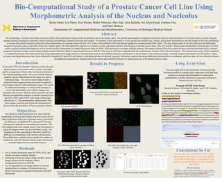
1. Bio-Computational Study of a Prostate Cancer Cell Line Using
Morphometric Analysis of the Nucleus and Nucleolus
The morphology of nuclear and cellular structures reflects tissue phenotype during normal development and in the disease states. The present gold standard of diagnosis of prostate cancer is characterization of 4um tissue biopsy sections using the
standard staining techniques for prostate clinical histology and pathology, hematoxylin/eosin and Feulgen. The purpose of this experiment is to use nuclear and nuceloli area, volume, and position information to provide insight on how the morphology
of nuclear and cellular structures reflect the cellular phenotypes of a prostate cancer cell line, PC3, when in the epithelial (EPI) and induced epithelial-to-mesenchymal (EMT) states. This may further assist in the early detection and differential
diagnosis of prostate cancer, especially in the early metastic phase. We will explore the extension of research on semi- automated machine classification of prostate cancer states. This will include 3 dimensional morphometric characteristics of whole
nuclei, nucleoli position information as well as nuclei/nuceloli relationships via simple fluorescent stains for DNA, RNA and nuclear envelope antibody staining. The images collected from these stains are then converted and analyzed by software
programs including Farsight, custom ImageJ plugins and the Statistics Online Computational Resource (SOCR). This analysis will lead to algorithms for the morphometric analysis of the collected imagery. 3D confocal microscopy will be compared
to standard fluorescent wide-field microscopy using segmentation and image analysis of characteristics such as size, number of nucleoli and distance to the perimeter of the nuclear envelope. Currently, imagery and statistics for 3D nuclear volume,
curvature, and fractal dimension have been collected for matched EPI and EMT cell cultures, with additional data on nucleoli expected. Preliminary analysis has shown differences between the two conditions, and more data will be collected to discern
the additional morphometric characteristics of the nuclei and nucleoli to increase the statistical power of the patterns shown to date.
Abstract
Brian Athey, Ivo Dinov, Ken Pienta, Walter Meixner, Alex Ade, Alex Kalinin, Ari Allyn-Feuer, Gordon Fon,
and John Mathew
Department of Computational Medicine and Bioinformatics, University of Michigan Medical School
Objectives
Conclusions So Far
Results in Progress
Methods
The next major goal in the experiment will be to develop
fluorescent probes to correlate gene expression levels and gene
locations in nuclear heterochromatin or euchromatin
compartments to further characterize expression differences
between the two cell lines.
Example of FISH Probe Design
Section of Intron 4 (Provided by Galaxy and UCSC Genome
Browser)
Probes by Bio-Search Technologies Stellaris:
actaattgttggtgctatctag
tttgaccaggctatttaaactt
gagtgtcagcatgttaaacatt
gctcactgcagccttgaactcctagactcaagccatcttcccacccagtagggctacggatgtacactaccatgc
ccagctgatttttttttaatttttgttttaattttttgtagagacaaaggggtcttgctatgttcccaggctggtgtctaac
tcctggccttaagtgatcctcccaacgtggcctcccaaagtgctggtattacaggtgtgagccactgcaactgac
ctatgtggttcttttgataggagagactaattgttggtgctatctagcacacactgtgtgtagacatcttgttaaat
agaaaatagatttatgggtatgactatgaagagtctaattccccaaaccacacacacaactctatctacgtttgac
caggctatttaaacttaactgcagagtgtcagcatgttaaacattgatttacataaaatgatagctgcccacttt
cttgtaaatgttataaaaactgtagagattaactaaaaaatgcacacagaagtttgctttcagttccacaagggtag
tttatttttgttataaaaacagtattccccactttcttagataccagatctctgcccagattttacccagtttcatcttgct
gctctctaatctcctatgtatgtaatatactttgaccatttaaatatgtattaagaca
Long Term GoalIntroduction
The objective of this experiment is to show that the
morphology of nuclear and cellular structures can be used to
make predictions of the type of prostate cancer, specifically
the differences in Epthelial PC3 cells and PC3 having
undergone the epithelial-to-mesenchymal transition. This
will expand on the work already done (1) but will consist of
using 3D imagery which may provide better results. The
hypothesis for this experiment is that there would be a
difference in morphology of the nucleus and nucleolus.
These differences could be used as another bio-marker in
combination with the Gleason scoring system, the present
standard diagnostic test in prostate biopsies.
• Use of simple fluorescent stains for DNA, RNA, and
Nuclear envelope antibody staining
• Analyzing of acquired images using Farsight, custom,
ImageJ plugins and the Statistics Online
Computational Resource (SOCR)
• Use of segmentation and image analysis of nuclear
characteristics to compare 3D confocal and two-
photon microscopy to standard fluorescent wide-field
microscopy.
From left to right: DAPI prolong Gold, Anti-
Fibrillarin, Ethidium Bromide
From left to right: DAPI, Nuclear Mask from
DAPI Channel, Image J Plugin to segment
connected and isolated nuclear masks
Results from plugin Segmentation
DAPI and FibrillarinNUP-98 and Fibrillarin
PC3 EMT from bottom left to top right:
Ethidium, DAPI, Fibrillarin
PC3 EMI from bottom left to top right: Ethidium,
DAPI, Fibrillarin
In the early 1970’s Dr. Donald F. Gleason and his lab used
stained sections of carcinoma cells to create a histologic
pattern arrangement of these cells, this became the basis of
the Gleason grading system. The score from the Gleason
gradient can give information on the tumor size and its
pathologic stage. This can be used to help to make a
prediction tumor aggressiveness and assist in prognosis
and treatment options.of the pathological stage. The search
for additional biomarkers to detect earlier changes in
tissue, and particularly early cellular changes, has
continued. One avenue which has shown promise has been
detecting morphometric changes in nuclear structure from
the Epi to EMT state of PC3 cells using 2-dimesional
microscopy (1). The morphometric measurements from
these images could be used to provide information to
doctors about the possibility of future metastatic disease.
(1) Verdone, J. E., Parsana, P., Veltri, R. W. and Pienta, K. J. (2015), “Epithelial–
mesenchymal transition in prostate cancer is associated with quantifiable changes in
nuclear structure.” Prostate, 75: 218–224. doi: 10.1002/pros.22908
Humphrey, Peter A. “Gleason Grading and prognostic factors in carcinoma of the
prostate.” Modern Pathology 17 (2004): 292-306. Web. 17 April 2015.
References
Currently the analysis of the PC3 Epi vs. MSC
nuclear/nucleolus images are not yet completed.
The initial results support the hypothesis: that
there is a statistical difference in the nucleolus
and nucleus.