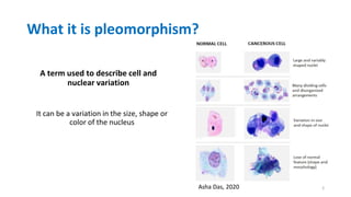
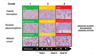
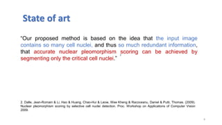
PLEOMORPHISM
Karla Margarita Muñoz Rico
Advisors: Eduardo Romero,
Ricardo Moncayo
08/09/2022
1
What it is pleomorphism?
A term used to describe cell and
nuclear variation
It can be a variation in the size, shape or
color of the nucleus
Asha Das, 2020 2
Nuclear atypia scoring
• It's a quantitative diagnostic
measure to assess the grade of
different cancers.
• Provides the degree of variation
in shape and size of cancer
nuclei compared to normal.
3
Nuclear atypia scoring
• The histological grading of cancer tissues, gives an estimate of patient
prognosis and is helpful in developing patient-related treatment
plans.
1. Das A, Nair MS, Peter SD. Computer-Aided Histopathological Image Analysis Techniques for Automated Nuclear Atypia Scoring
of Breast Cancer: a Review. J Digit Imaging. 2020 Oct;33(5):1091-1121
1
4
MODIFIED BLOOM-
RICHARDSON
GRADING SYSTEM
5
Problem
• These manual diagnoses require extensive training and experience
for the pathologist and even there will be great extent of
disagreement between regarding the grade, this is particularly true in
case of nuclear pleomorphism. This subjective of measurement and
poor reproducibility have resulted in great demand for an automated
grading system.
1. Das A, Nair MS, Peter SD. Computer-Aided Histopathological Image Analysis Techniques for Automated Nuclear Atypia Scoring
of Breast Cancer: a Review. J Digit Imaging. 2020 Oct;33(5):1091-1121
1
6
Research Question
How to reduce the subjectivity of measurement of nuclear
pleomorphism?
7
8
“Our proposed method is based on the idea that the input image
contains so many cell nuclei, and thus so much redundant information,
that accurate nuclear pleomorphism scoring can be achieved by
segmenting only the critical cell nuclei.”
State of art
2
2. Dalle, Jean-Romain & Li, Hao & Huang, Chao-Hui & Leow, Wee Kheng & Racoceanu, Daniel & Putti, Thomas. (2009).
Nuclear pleomorphism scoring by selective cell nuclei detection. Proc. Workshop on Applications of Computer Vision
2009.
State of art
3. Moncayo R., Romo-BucheliD., RomeroE. (2015) A Grading Strategy for Nuclear Pleomorphismin Histopathological BreastCancer Images Using a Bag of Features (BOF). In: Pardo A., Kittler J.
(eds) Progressin Pattern Recognition, Image Analysis, Computer Vision, and Applications. CIARP 2015. Lecture Notes in ComputerScience, vol 9423. Springer
“Was proposed a pleomorphism grading method that starts by a Grading Strategy for Nuclear
Pleomorphism detecting nuclei candidates (related to hematoxylin dye) using a color
deconvolution algorithm.”
3
State of art
4.Peregrina-Barreto H, Ramirez-Guatemala VY, Lopez-Armas GC, Cruz-Ramos JA. Characterization of Nuclear Pleomorphism and Tubules in Histopathological Images of Breast
Cancer. Sensors (Basel). 2022 Jul 28;22(15):5649. doi: 10.3390/s22155649. PMID: 35957203; PMCID: PMC9371191.
“Analysis of features depended on manual segmentation of the
centroid of nuclei, a time-consuming task. The study of healthy and
cancerous tissue may generate a large set of features, although not all
have the same relevance.”
10
4
11
State of art
“Due to annotated images from multiple sources and the CNN-models
ability to learn features that maps each pixel to its respective class, we
overcome challenges with anatomical variations and variations that
may arise from staining, different slide scanners and software
processing.”
5. Oskal, K.R.J., Risdal, M., Janssen, E.A.M. et al. A U-net based approach to epidermal tissue segmentation in whole
slide histopathological images. SN Appl. Sci. 1, 672 (2019). https://doi.org/10.1007/s42452-019-0694-y
5
Our proposal
Find a method to reduce the subjectivity of measurement of nuclear
pleomorphism in breast cancer images.
12
Dataset
13
Aperio Scanscope XT
Hamamatsu
Nanozoomer 2.0-HT.
MITOS-ATYPIA-14
Breast cancer
• 284 frames at X20
• 1,136 frames at X40
• List of mitosis given by two
different pathologists.
• The criteria for nuclear atypia
are provided as a number 1, 2
or 3
Pipeline
NORMALIZATION AND
NUCLEI DETECTION
FEATURES EXTRACTION
Statistical
analysis
VALIDATE THE METHOD
14
Pipeline
15
Preprocessing
NORMALIZATION AND
NUCLEI DETECTION
Processing
FEATURES EXTRACTION
Statistical
analysis
VALIDATE THE METHOD
16
FEATURES EXTRACTION CLASSIFIED
Pipeline
9. Hao Y, Qiao S, Zhang L, Xu T, Bai Y, Hu H, Zhang W, Zhang G. Breast Cancer Histopathological Images Recognition Based on Low Dimensional Three-
Channel Features. Front Oncol. 2021 Jun 14;11:657560.
10. Taken from Journal of pathology informatics,Ínternet, available in
https://www.jpathinformatics.org/viewimage.asp?img=JPatholInform_2019_10_1_32_270744_f22.jpg
NORMALIZATION AND NUCLEI DETECTION
Scheme 1
17
COLOR DECONVOLUTION NORMALIZATION NUCLEI
SEGMENTATION
6. Hao Y, Qiao S, Zhang L, Xu T, Bai Y, Hu H, Zhang W, Zhang G. Breast Cancer Histopathological Images Recognition Based on Low
Dimensional Three-Channel Features. Front Oncol. 2021 Jun 14;11:657560.
18
7..Michael E, Ma H, Li H, Kulwa F, Li J. Breast Cancer Segmentation Methods: Current Status and Future Potentials. Biomed Res Int. 2021 Jul
20;2021:9962109
8. Getao Du, Xu Cao, Jimin Liang, Xueli Chen, and Yonghua Zhan, Medical Image Segmentation based on U-Net: A Review. J Imaging Sci.
Technol. 2020, pp 20508 1:12
NORMALIZATION AND NUCLEI DETECTION
Scheme 2
Results
19
ORIGINAL
IMAGE
NORMALIZED
IMAGE
COLOR
DECONVOLUTION
SEGMENTED
NUCLEI
Scheme 1
Results
20
SEGMENTED
NUCLEI
ORIGINAL
IMAGE
NORMALIZED
IMAGE
COLOR
DECONVOLUTION
Scheme 1
Precision Recall F1 Score Jaccard
Watershed 0.89 0.83 0.86 0.85
Results: U-net
21
Jaccard Index F1 Score
0,72 0,82
Experiment: U-net
22
Precision Recall F1 Score Accuracy
U-net 0.8 0.82 0.81 0.8
Future work
• Features extraction.
• Correlate with genetic biomarkers (TP53)
• Validate.
23
Future work
• Can we relate a genetic marker to histopathology?
24
Future work
25
“TP53 mutations frequently occur in gastric cancer and
are associated with unfavorable clinical outcomes”
“TP53 may play an important role in activating tumor
immunity in GC and other cancer types and that the
TP53 mutation status could be useful in stratifying
cancer patients responsive to a certain
immunotherapy”
Correlate with genetic biomarkers (TP53)
11. Jiang, Z., Liu, Z., Li, M., Chen, C., & Wang, X. (2018). Immunogenomics Analysis Reveals that TP53 Mutations Inhibit Tumor
Immunity in Gastric Cancer. Translational oncology, 11(5), 1171–1187. https://doi.org/10.1016/j.tranon.2018.07.012
Thanks
26

pleomorphism (1) (1).pdf

  • 1.
    PLEOMORPHISM Karla Margarita MuñozRico Advisors: Eduardo Romero, Ricardo Moncayo 08/09/2022 1
  • 2.
    What it ispleomorphism? A term used to describe cell and nuclear variation It can be a variation in the size, shape or color of the nucleus Asha Das, 2020 2
  • 3.
    Nuclear atypia scoring •It's a quantitative diagnostic measure to assess the grade of different cancers. • Provides the degree of variation in shape and size of cancer nuclei compared to normal. 3
  • 4.
    Nuclear atypia scoring •The histological grading of cancer tissues, gives an estimate of patient prognosis and is helpful in developing patient-related treatment plans. 1. Das A, Nair MS, Peter SD. Computer-Aided Histopathological Image Analysis Techniques for Automated Nuclear Atypia Scoring of Breast Cancer: a Review. J Digit Imaging. 2020 Oct;33(5):1091-1121 1 4
  • 5.
  • 6.
    Problem • These manualdiagnoses require extensive training and experience for the pathologist and even there will be great extent of disagreement between regarding the grade, this is particularly true in case of nuclear pleomorphism. This subjective of measurement and poor reproducibility have resulted in great demand for an automated grading system. 1. Das A, Nair MS, Peter SD. Computer-Aided Histopathological Image Analysis Techniques for Automated Nuclear Atypia Scoring of Breast Cancer: a Review. J Digit Imaging. 2020 Oct;33(5):1091-1121 1 6
  • 7.
    Research Question How toreduce the subjectivity of measurement of nuclear pleomorphism? 7
  • 8.
    8 “Our proposed methodis based on the idea that the input image contains so many cell nuclei, and thus so much redundant information, that accurate nuclear pleomorphism scoring can be achieved by segmenting only the critical cell nuclei.” State of art 2 2. Dalle, Jean-Romain & Li, Hao & Huang, Chao-Hui & Leow, Wee Kheng & Racoceanu, Daniel & Putti, Thomas. (2009). Nuclear pleomorphism scoring by selective cell nuclei detection. Proc. Workshop on Applications of Computer Vision 2009.
  • 9.
    State of art 3.Moncayo R., Romo-BucheliD., RomeroE. (2015) A Grading Strategy for Nuclear Pleomorphismin Histopathological BreastCancer Images Using a Bag of Features (BOF). In: Pardo A., Kittler J. (eds) Progressin Pattern Recognition, Image Analysis, Computer Vision, and Applications. CIARP 2015. Lecture Notes in ComputerScience, vol 9423. Springer “Was proposed a pleomorphism grading method that starts by a Grading Strategy for Nuclear Pleomorphism detecting nuclei candidates (related to hematoxylin dye) using a color deconvolution algorithm.” 3
  • 10.
    State of art 4.Peregrina-BarretoH, Ramirez-Guatemala VY, Lopez-Armas GC, Cruz-Ramos JA. Characterization of Nuclear Pleomorphism and Tubules in Histopathological Images of Breast Cancer. Sensors (Basel). 2022 Jul 28;22(15):5649. doi: 10.3390/s22155649. PMID: 35957203; PMCID: PMC9371191. “Analysis of features depended on manual segmentation of the centroid of nuclei, a time-consuming task. The study of healthy and cancerous tissue may generate a large set of features, although not all have the same relevance.” 10 4
  • 11.
    11 State of art “Dueto annotated images from multiple sources and the CNN-models ability to learn features that maps each pixel to its respective class, we overcome challenges with anatomical variations and variations that may arise from staining, different slide scanners and software processing.” 5. Oskal, K.R.J., Risdal, M., Janssen, E.A.M. et al. A U-net based approach to epidermal tissue segmentation in whole slide histopathological images. SN Appl. Sci. 1, 672 (2019). https://doi.org/10.1007/s42452-019-0694-y 5
  • 12.
    Our proposal Find amethod to reduce the subjectivity of measurement of nuclear pleomorphism in breast cancer images. 12
  • 13.
    Dataset 13 Aperio Scanscope XT Hamamatsu Nanozoomer2.0-HT. MITOS-ATYPIA-14 Breast cancer • 284 frames at X20 • 1,136 frames at X40 • List of mitosis given by two different pathologists. • The criteria for nuclear atypia are provided as a number 1, 2 or 3
  • 14.
    Pipeline NORMALIZATION AND NUCLEI DETECTION FEATURESEXTRACTION Statistical analysis VALIDATE THE METHOD 14
  • 15.
  • 16.
    16 FEATURES EXTRACTION CLASSIFIED Pipeline 9.Hao Y, Qiao S, Zhang L, Xu T, Bai Y, Hu H, Zhang W, Zhang G. Breast Cancer Histopathological Images Recognition Based on Low Dimensional Three- Channel Features. Front Oncol. 2021 Jun 14;11:657560. 10. Taken from Journal of pathology informatics,Ínternet, available in https://www.jpathinformatics.org/viewimage.asp?img=JPatholInform_2019_10_1_32_270744_f22.jpg
  • 17.
    NORMALIZATION AND NUCLEIDETECTION Scheme 1 17 COLOR DECONVOLUTION NORMALIZATION NUCLEI SEGMENTATION 6. Hao Y, Qiao S, Zhang L, Xu T, Bai Y, Hu H, Zhang W, Zhang G. Breast Cancer Histopathological Images Recognition Based on Low Dimensional Three-Channel Features. Front Oncol. 2021 Jun 14;11:657560.
  • 18.
    18 7..Michael E, MaH, Li H, Kulwa F, Li J. Breast Cancer Segmentation Methods: Current Status and Future Potentials. Biomed Res Int. 2021 Jul 20;2021:9962109 8. Getao Du, Xu Cao, Jimin Liang, Xueli Chen, and Yonghua Zhan, Medical Image Segmentation based on U-Net: A Review. J Imaging Sci. Technol. 2020, pp 20508 1:12 NORMALIZATION AND NUCLEI DETECTION Scheme 2
  • 19.
  • 20.
  • 21.
  • 22.
    Experiment: U-net 22 Precision RecallF1 Score Accuracy U-net 0.8 0.82 0.81 0.8
  • 23.
    Future work • Featuresextraction. • Correlate with genetic biomarkers (TP53) • Validate. 23
  • 24.
    Future work • Canwe relate a genetic marker to histopathology? 24
  • 25.
    Future work 25 “TP53 mutationsfrequently occur in gastric cancer and are associated with unfavorable clinical outcomes” “TP53 may play an important role in activating tumor immunity in GC and other cancer types and that the TP53 mutation status could be useful in stratifying cancer patients responsive to a certain immunotherapy” Correlate with genetic biomarkers (TP53) 11. Jiang, Z., Liu, Z., Li, M., Chen, C., & Wang, X. (2018). Immunogenomics Analysis Reveals that TP53 Mutations Inhibit Tumor Immunity in Gastric Cancer. Translational oncology, 11(5), 1171–1187. https://doi.org/10.1016/j.tranon.2018.07.012
  • 26.