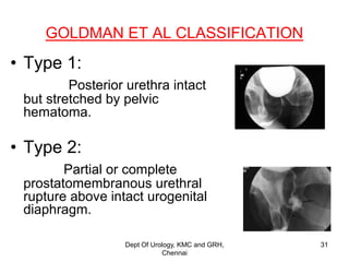
This document discusses the anatomy of the urogenital triangle and classification of urethral injuries. It begins with an overview of the bony pelvis and ligaments, followed by the muscles that support the pelvic floor including the levator ani and coccygeus. It then describes the urogenital diaphragm and its contents. Several classifications of urethral injuries are presented, including the McCallum & Col Pinto classification for posterior injuries and the AAST classification system for severity grading. Injuries can be anterior or posterior, with posterior injuries often associated with pelvic fractures from trauma.
ANATOMY OF UTERUS
ANATOMY OF OVARY
ANATOMY OF FALLOPIAN TUBES
ANATOMY OF UTERUS &ITS APPENDAGES
ANATOMY OF CERVIX
ANATOMY OF UTERUS PPT
BLOOD SUPPLY, NERVE SUPPLY, LYMPHATIC DRAINAGE
HISTOLOGY
ANATOMY OF UTERUS
ANATOMY OF OVARY
ANATOMY OF FALLOPIAN TUBES
ANATOMY OF UTERUS &ITS APPENDAGES
ANATOMY OF CERVIX
ANATOMY OF UTERUS PPT
BLOOD SUPPLY, NERVE SUPPLY, LYMPHATIC DRAINAGE
HISTOLOGY
HUMAN ANATOMY
regional anatomy
regional anatomy of pelvic
changsha medical university lecture
csmu lecture by an chen
uploaded by Prabesh raj jamkatel
pelvics
HUMAN ANATOMY
regional anatomy
regional anatomy of pelvic
changsha medical university lecture
csmu lecture by an chen
uploaded by Prabesh raj jamkatel
pelvics
Prix Galien International 2024 Forum ProgramLevi Shapiro
June 20, 2024, Prix Galien International and Jerusalem Ethics Forum in ROME. Detailed agenda including panels:
- ADVANCES IN CARDIOLOGY: A NEW PARADIGM IS COMING
- WOMEN’S HEALTH: FERTILITY PRESERVATION
- WHAT’S NEW IN THE TREATMENT OF INFECTIOUS,
ONCOLOGICAL AND INFLAMMATORY SKIN DISEASES?
- ARTIFICIAL INTELLIGENCE AND ETHICS
- GENE THERAPY
- BEYOND BORDERS: GLOBAL INITIATIVES FOR DEMOCRATIZING LIFE SCIENCE TECHNOLOGIES AND PROMOTING ACCESS TO HEALTHCARE
- ETHICAL CHALLENGES IN LIFE SCIENCES
- Prix Galien International Awards Ceremony
Lung Cancer: Artificial Intelligence, Synergetics, Complex System Analysis, S...Oleg Kshivets
RESULTS: Overall life span (LS) was 2252.1±1742.5 days and cumulative 5-year survival (5YS) reached 73.2%, 10 years – 64.8%, 20 years – 42.5%. 513 LCP lived more than 5 years (LS=3124.6±1525.6 days), 148 LCP – more than 10 years (LS=5054.4±1504.1 days).199 LCP died because of LC (LS=562.7±374.5 days). 5YS of LCP after bi/lobectomies was significantly superior in comparison with LCP after pneumonectomies (78.1% vs.63.7%, P=0.00001 by log-rank test). AT significantly improved 5YS (66.3% vs. 34.8%) (P=0.00000 by log-rank test) only for LCP with N1-2. Cox modeling displayed that 5YS of LCP significantly depended on: phase transition (PT) early-invasive LC in terms of synergetics, PT N0—N12, cell ratio factors (ratio between cancer cells- CC and blood cells subpopulations), G1-3, histology, glucose, AT, blood cell circuit, prothrombin index, heparin tolerance, recalcification time (P=0.000-0.038). Neural networks, genetic algorithm selection and bootstrap simulation revealed relationships between 5YS and PT early-invasive LC (rank=1), PT N0—N12 (rank=2), thrombocytes/CC (3), erythrocytes/CC (4), eosinophils/CC (5), healthy cells/CC (6), lymphocytes/CC (7), segmented neutrophils/CC (8), stick neutrophils/CC (9), monocytes/CC (10); leucocytes/CC (11). Correct prediction of 5YS was 100% by neural networks computing (area under ROC curve=1.0; error=0.0).
CONCLUSIONS: 5YS of LCP after radical procedures significantly depended on: 1) PT early-invasive cancer; 2) PT N0--N12; 3) cell ratio factors; 4) blood cell circuit; 5) biochemical factors; 6) hemostasis system; 7) AT; 8) LC characteristics; 9) LC cell dynamics; 10) surgery type: lobectomy/pneumonectomy; 11) anthropometric data. Optimal diagnosis and treatment strategies for LC are: 1) screening and early detection of LC; 2) availability of experienced thoracic surgeons because of complexity of radical procedures; 3) aggressive en block surgery and adequate lymph node dissection for completeness; 4) precise prediction; 5) adjuvant chemoimmunoradiotherapy for LCP with unfavorable prognosis.
Title: Sense of Taste
Presenter: Dr. Faiza, Assistant Professor of Physiology
Qualifications:
MBBS (Best Graduate, AIMC Lahore)
FCPS Physiology
ICMT, CHPE, DHPE (STMU)
MPH (GC University, Faisalabad)
MBA (Virtual University of Pakistan)
Learning Objectives:
Describe the structure and function of taste buds.
Describe the relationship between the taste threshold and taste index of common substances.
Explain the chemical basis and signal transduction of taste perception for each type of primary taste sensation.
Recognize different abnormalities of taste perception and their causes.
Key Topics:
Significance of Taste Sensation:
Differentiation between pleasant and harmful food
Influence on behavior
Selection of food based on metabolic needs
Receptors of Taste:
Taste buds on the tongue
Influence of sense of smell, texture of food, and pain stimulation (e.g., by pepper)
Primary and Secondary Taste Sensations:
Primary taste sensations: Sweet, Sour, Salty, Bitter, Umami
Chemical basis and signal transduction mechanisms for each taste
Taste Threshold and Index:
Taste threshold values for Sweet (sucrose), Salty (NaCl), Sour (HCl), and Bitter (Quinine)
Taste index relationship: Inversely proportional to taste threshold
Taste Blindness:
Inability to taste certain substances, particularly thiourea compounds
Example: Phenylthiocarbamide
Structure and Function of Taste Buds:
Composition: Epithelial cells, Sustentacular/Supporting cells, Taste cells, Basal cells
Features: Taste pores, Taste hairs/microvilli, and Taste nerve fibers
Location of Taste Buds:
Found in papillae of the tongue (Fungiform, Circumvallate, Foliate)
Also present on the palate, tonsillar pillars, epiglottis, and proximal esophagus
Mechanism of Taste Stimulation:
Interaction of taste substances with receptors on microvilli
Signal transduction pathways for Umami, Sweet, Bitter, Sour, and Salty tastes
Taste Sensitivity and Adaptation:
Decrease in sensitivity with age
Rapid adaptation of taste sensation
Role of Saliva in Taste:
Dissolution of tastants to reach receptors
Washing away the stimulus
Taste Preferences and Aversions:
Mechanisms behind taste preference and aversion
Influence of receptors and neural pathways
Impact of Sensory Nerve Damage:
Degeneration of taste buds if the sensory nerve fiber is cut
Abnormalities of Taste Detection:
Conditions: Ageusia, Hypogeusia, Dysgeusia (parageusia)
Causes: Nerve damage, neurological disorders, infections, poor oral hygiene, adverse drug effects, deficiencies, aging, tobacco use, altered neurotransmitter levels
Neurotransmitters and Taste Threshold:
Effects of serotonin (5-HT) and norepinephrine (NE) on taste sensitivity
Supertasters:
25% of the population with heightened sensitivity to taste, especially bitterness
Increased number of fungiform papillae
Couples presenting to the infertility clinic- Do they really have infertility...Sujoy Dasgupta
Dr Sujoy Dasgupta presented the study on "Couples presenting to the infertility clinic- Do they really have infertility? – The unexplored stories of non-consummation" in the 13th Congress of the Asia Pacific Initiative on Reproduction (ASPIRE 2024) at Manila on 24 May, 2024.
MANAGEMENT OF ATRIOVENTRICULAR CONDUCTION BLOCK.pdfJim Jacob Roy
Cardiac conduction defects can occur due to various causes.
Atrioventricular conduction blocks ( AV blocks ) are classified into 3 types.
This document describes the acute management of AV block.
These simplified slides by Dr. Sidra Arshad present an overview of the non-respiratory functions of the respiratory tract.
Learning objectives:
1. Enlist the non-respiratory functions of the respiratory tract
2. Briefly explain how these functions are carried out
3. Discuss the significance of dead space
4. Differentiate between minute ventilation and alveolar ventilation
5. Describe the cough and sneeze reflexes
Study Resources:
1. Chapter 39, Guyton and Hall Textbook of Medical Physiology, 14th edition
2. Chapter 34, Ganong’s Review of Medical Physiology, 26th edition
3. Chapter 17, Human Physiology by Lauralee Sherwood, 9th edition
4. Non-respiratory functions of the lungs https://academic.oup.com/bjaed/article/13/3/98/278874
Pulmonary Thromboembolism - etilogy, types, medical- Surgical and nursing man...VarunMahajani
Disruption of blood supply to lung alveoli due to blockage of one or more pulmonary blood vessels is called as Pulmonary thromboembolism. In this presentation we will discuss its causes, types and its management in depth.
Factory Supply Best Quality Pmk Oil CAS 28578–16–7 PMK Powder in Stockrebeccabio
Factory Supply Best Quality Pmk Oil CAS 28578–16–7 PMK Powder in Stock
Telegram: bmksupplier
signal: +85264872720
threema: TUD4A6YC
You can contact me on Telegram or Threema
Communicate promptly and reply
Free of customs clearance, Double Clearance 100% pass delivery to USA, Canada, Spain, Germany, Netherland, Poland, Italy, Sweden, UK, Czech Republic, Australia, Mexico, Russia, Ukraine, Kazakhstan.Door to door service
Hot Selling Organic intermediates
HOT NEW PRODUCT! BIG SALES FAST SHIPPING NOW FROM CHINA!! EU KU DB BK substit...GL Anaacs
Contact us if you are interested:
Email / Skype : kefaya1771@gmail.com
Threema: PXHY5PDH
New BATCH Ku !!! MUCH IN DEMAND FAST SALE EVERY BATCH HAPPY GOOD EFFECT BIG BATCH !
Contact me on Threema or skype to start big business!!
Hot-sale products:
NEW HOT EUTYLONE WHITE CRYSTAL!!
5cl-adba precursor (semi finished )
5cl-adba raw materials
ADBB precursor (semi finished )
ADBB raw materials
APVP powder
5fadb/4f-adb
Jwh018 / Jwh210
Eutylone crystal
Protonitazene (hydrochloride) CAS: 119276-01-6
Flubrotizolam CAS: 57801-95-3
Metonitazene CAS: 14680-51-4
Payment terms: Western Union,MoneyGram,Bitcoin or USDT.
Deliver Time: Usually 7-15days
Shipping method: FedEx, TNT, DHL,UPS etc.Our deliveries are 100% safe, fast, reliable and discreet.
Samples will be sent for your evaluation!If you are interested in, please contact me, let's talk details.
We specializes in exporting high quality Research chemical, medical intermediate, Pharmaceutical chemicals and so on. Products are exported to USA, Canada, France, Korea, Japan,Russia, Southeast Asia and other countries.
Anti ulcer drugs and their Advance pharmacology ||
Anti-ulcer drugs are medications used to prevent and treat ulcers in the stomach and upper part of the small intestine (duodenal ulcers). These ulcers are often caused by an imbalance between stomach acid and the mucosal lining, which protects the stomach lining.
||Scope: Overview of various classes of anti-ulcer drugs, their mechanisms of action, indications, side effects, and clinical considerations.
How to Give Better Lectures: Some Tips for Doctors
Urogenital triangle
1. ANATOMY OF UROGENITAL
TRIANGLE AND
CLASSIFICATION OF URETHRAL
INJURIES
Dept of Urology
Govt Royapettah Hospital and Kilpauk Medical College
Chennai
1
2. Moderators:
Professors:
• Prof. Dr. G. Sivasankar, M.S., M.Ch.,
• Prof. Dr. A. Senthilvel, M.S., M.Ch.,
Asst Professors:
• Dr. J. Sivabalan, M.S., M.Ch.,
• Dr. R. Bhargavi, M.S., M.Ch.,
• Dr. S. Raju, M.S., M.Ch.,
• Dr. K. Muthurathinam, M.S., M.Ch.,
• Dr. D. Tamilselvan, M.S., M.Ch.,
• Dr. K. Senthilkumar, M.S., M.Ch.
Dept Of Urology, KMC and GRH,
Chennai
2
3. BONY PELVIS
• Bony pelvis consists of two innominate bones-fused posteriorly to sacrum and
to each other anteriorly.
• Each innominate bone is composed of ilium,ischium and pubis.
• At the pelvic inlet,the true and false pelvis are seperated by the arcuate line.
• Arcuate line extends from the sacral promontory to the pectineal line of pubis.
• The inferior pelvic outlet is closed by the pelvic floor.
Dept Of Urology, KMC and GRH,
Chennai
3
4. LIGAMENTS
• Sacrospinous ligament:
Extends from the ischial spine to the lateral margins of the sacrum
and coccyx.The greater and lesser sciatic foramen are above and
below the ligament.
• Sacrotuberous ligament:
Extends from the ischial tuberosity to the coccyx.
Dept Of Urology, KMC and GRH,
Chennai
4
5. MUSCULAR SUPPORTS OF THE PELVIC
FLOOR
• Muscles that form the pelvic diaphragm include the
Levator ani and the coccygeus.
• Levator ani is composed of two major muscles from
medial to lateral-Pubococcygeus and Iliococcygeus.
Dept Of Urology, KMC and GRH,
Chennai
5
6. PUBOCOCCYGEUS
• Bulkier medial part that arises from the back of body of pubis
and anterior portion of arcus tendineus.
• The inner border forms the margin of the urogenital hiatus
,through which pass the urethra ,vagina and anorectum.
• Portions of the Pubococcygeus include Pubovaginalis,
Puboanalis and Puborectalis.
Dept Of Urology, KMC and GRH,
Chennai
6
7. • Iliococcygeus:
Thin lateral part of the levator ani that
arises from the arcusa tendineus of the levator ani to the
ischial spine.
• Coccygeus:
Extends from the ischial spine to coccyx
and lower sacrum and forms the posterior part of the
pelvic diaphragm.It sits on the pelvic surface of the
sacrospinous ligament.
Dept Of Urology, KMC and GRH,
Chennai
7
8. ARCUS TENDINEUS
• Arcus tendineus of the Levator ani is a dense
connective tissue structure
• Extends from the pubic ramus to the ischial
spine and courses along the surface of the
obturator internus.
Dept Of Urology, KMC and GRH,
Chennai
8
9. UROGENITAL DIAPHRAGM(TRIANGLE)
• The weakest point in the pelvic floor ,Urogenital hiatus,is
bridged by the Urogenital diaphragm – a structure
unique to humans.
• It is triangular and bridges the gap between the inferior
pubic rami bilaterally and the perineal body.
• Posteriorly,it ends abruptly
Dept Of Urology, KMC and GRH,
Chennai
9
10. • Perineal body represents the point of fusion
between the free posterior edge of the urogenital
diaphragm.
• Urogenital diaphragm seperates the deep
perineal sac from the upper pelvis.
• Urogenital diaphragm is made of 2 paired
muscles –the Deep transverse perineal muscle
and Superficial transverse perineal muscle.
Dept Of Urology, KMC and GRH,
Chennai
10
11. • Fascia of the urogenital diaphragm include the
Superficial and Deep perineal fascia,lining the inferior
surface and superior surface of the urogenital diaphragm
respectively
• Urogenital diaphragm provides structural support for the
distal urethra.
Dept Of Urology, KMC and GRH,
Chennai
11
12. ARTERIES:
• Bulbar artery of penis
• Dorsal artery of penis
• Perineal artery
• Int.pudendal
• Ext pudendal
VEINS
• Dorsal veins of penis
• Int. pudendal vein
• Ext. pudendal vein
Dept Of Urology, KMC and GRH,
Chennai
12
13. DEEP PERINEAL POUCH
Superior border:
Lacks a definite superior border,extends into the pelvis.
Inferior border:
Perineal membrane(Superficial perineal fascia).
Dept Of Urology, KMC and GRH,
Chennai
13
14. CONTENTS:
1.Muscles:
Deep transverse perineal muscle.
External sphincter of urethra.
2.Membranous portion of Urethra.
3.Bulbourethral (Cowper’s gland)
Dept Of Urology, KMC and GRH,
Chennai
14
15. SUPERFICIAL PERINEAL POUCH
Superior border:
Perineal membrane(superficial perineal fascia)
Inferior border:
Colles fascia.
Dept Of Urology, KMC and GRH,
Chennai
15
17. PERINEAL BODY
• Perineal body (Central tendon of perineum) is a
pyramidal fibromuscular mass in the midline of
perineum at the junction between the urogenital
triangle and anal triangle.
• Found in both males and females.
Dept Of Urology, KMC and GRH,
Chennai
17
18. Muscles converging in Perineal body:
1.Superficial transverse perineal muscle.
2.Bulbospongiosus
3.Deep transverse perineal muscle.
4.Anterior fibers of Levator ani.
5.External urethral sphincter
6.External anal sphincter.
Perineal body is essential for the integrity of the
pelvic floor.
Dept Of Urology, KMC and GRH,
Chennai
18
19. MALE URETHRA
• Male urethra extends from the internal urethral orifice in
the bladder to the external urethral orifice at the tip of
penis.
• Roughly ‘S’ shaped.
• Length varies from 17.5 to 20 cm.
• Based on consensus opinion in WHO conference
2002,urethra can be divided into six separate areas.
Dept Of Urology, KMC and GRH,
Chennai
19
20. 1.Fossa navicularis:
Contained within the spongy erectile tissue of glans penis.Lined
with stratified squamous epithelium.
2.Penile or pendulous urethra:
Lies distal to investment of ischiocavernosus muscle,invested by
corpus spongiosum.Lined with simple squamous epithelium.
3.Bulbous urethra:
Covered by midline fusion of ischiocavernosus and invested by
bulbospongiosus.Distally squamous epithelium and proximally
transitional.
Dept Of Urology, KMC and GRH,
Chennai
20
21. 4.Membranous urethra:
Shortest,least dilatable and with the excp.of
ext.urethral orifice ,the narrowest part of the
canal.Measures 2 cm in length.Extends downward and
forward ,perforating the urogenital diaphragm .
Only portion of the male urethra that is not invested by
another structure.Lined by transitional epithelium.
5.Prostatic urethra:
Widest,most dilatable part.Measures 3cm in
length.Runs vertically through the prostate and at its
midpoint turns 35 deg anteriorly,angulation varies 0 to 90
6.Bladder neck.
Dept Of Urology, KMC and GRH,
Chennai
21
22. • Urethral crest :
Narrow ridge on the posterior wall of prostatic
urethra
• Verumontanum:
Median elevation at forepart of urethral crest.
• Prostatic utricle:
Mullerian duct derivative opening at apex of
verumontanum.
Dept Of Urology, KMC and GRH,
Chennai
22
23. • The internal sphincter muscle
of urethra: located at the
bladder's inferior end and the
urethra's proximal end at the
junction of the urethra with the
urinary bladder.
• The external sphincter muscle
of urethra (sphincter urethrae):
located at the bladder's distal
inferior end in females and
inferior to the prostate (at the
level of the membranous
urethra)in males is a
secondary sphincter to control
the flow of urine through the
urethra.
Dept Of Urology, KMC and GRH,
Chennai
23
24. BLOOD SUPPLY OF URETHRA
• The arterial supply of the male urethra is primarily by the
Bulbar arteries which are proximal br. of Internal
pudendal artery.
• Distally, it is supplied by the Dorsal artery of the
penis,which is a terminal br.of int. pudendal artery and
collaterals from corporal bodies.
• This dual blood supply allows the urethra to be detached
at either end without compromise to its viability,a fact
frequently used in Urethroplasty.
Dept Of Urology, KMC and GRH,
Chennai
24
25. PERINEUM
• The perineum is a diamond shaped outlet bounded
anteriorly by the pubic arch and the arcuate ligaments of
the pubis.
• Posteriorly by the tip of coccyx.
• Laterally,by the inferior rami of pubis and ischium.
• Transverse line between the ischial tuberosities divides
the perineum into an anterior urogenital triangle and a
posterior anal triangle.
Dept Of Urology, KMC and GRH,
Chennai
25
26. COLLES’ FASCIA
• In the anterior triangle,Colles’ fascia attaches at its
posterior margin to the perineal body.
• The fascia curves below the superficial transverse
perineal muscle and projects forward as two layers
attached laterally to the ischium and inferior pubic
ramus.
• The loose superficial layer is continuous with the more
substantial dartos fascia of the scrotum.
• The deep membranous layer of Colles fascia forms a
roof over scrotal cavity,seperating it from superficial
perineal pouch. Dept Of Urology, KMC and GRH,
Chennai
26
27. • Anteriorly ,Colles fascia fuses and becomes continuous
with the membranous layer of the subcutaneous tissue
of the anterior abdominal wall.(Scarpa’s fascia).
• Laterally,Colles fascia fuses to the pubic arch and with
the fascia lata.
• Posteriorly ,fuses with the posterior aspect of the
perineal membrane.
Dept Of Urology, KMC and GRH,
Chennai
27
28. Injury to the urethra can be broadly classified as Anterior
and Posterior urethral injury.
Posterior urethral injuries
Injuries to the posterior urethra occur with pelvic
fractures, which are commonly caused by road traffic
accidents, crush injuries or falls from height. About two-
thirds (70%) of pelvic fractures occur as a result of motor
vehicle accidents
Dept Of Urology, KMC and GRH,
Chennai
28
29. Aetiology of anterior urethral injuries
Blunt trauma
• Vehicular accidents
• Fall astride
• Kicks in the perineum
Blows in the perineum from bicycle handlebars, tops of fences, etc.
Sexual intercourse
• Penile fractures
• Urethral intraluminal stimulation
Penetrating trauma
• Gunshot wounds
• Stab wounds
• Dog bites
• External impalement
• Penile amputations
Iatrogenic injuries
• Endoscopic instrumentations
• Urethral catheters-dilators
Dept Of Urology, KMC and GRH,
Chennai
29
30. McCALLUM & COL PINTO
CLASSIFICATION
Based on radiographic appearances of posterior injuries.
Type 1: Urethral contusion or stretch injury,passage of
contrast into bladder, no extravasation.
Type 2: Partial or complete rupture above urogenital
diaphragm,contrast may reach bladder, supra
diaphragmatic extravasation.
Type 3: Complete disruption of membranous
urethra,contrast does not reach bladder.Extravasation
both supra and infra diaphragmatic.
Dept Of Urology, KMC and GRH,
Chennai
30
31. GOLDMAN ET AL CLASSIFICATION
• Type 1:
Posterior urethra intact
but stretched by pelvic
hematoma.
• Type 2:
Partial or complete
prostatomembranous urethral
rupture above intact urogenital
diaphragm.
Dept Of Urology, KMC and GRH,
Chennai
31
32. • Type 3:
Partial or complete combined
anterior/posterior urethral rupture with
rupture of urogenital diaphragm.
Dept Of Urology, KMC and GRH,
Chennai
32
33. • Type 4:
Bladder neck injury with extension into posterior
urethra
• Type 4a:
Base of bladder injury with periurethral
extravasation.
.
Dept Of Urology, KMC and GRH,
Chennai
33
34. • Type 5:
Partial or complete anterior urethral injury
Dept Of Urology, KMC and GRH,
Chennai
34
35. AAST classification
• Grade 1:
contusion :blood at meatus;urethrography normal.
• Grade 2:
stretch injury :elongation of urethra without
extravasation on urethrography.
• Grade 3:
partial disruption :extravasation of contrast at injury
site with contrast visualisation in bladder.
Dept Of Urology, KMC and GRH,
Chennai
35
36. • Grade 4:
complete disruption :extravasation on
urethrography,contrast at injury site without visualisation
in bladder;less than 2 cm urethral seperation.
• Grade 5:
complete disruption; complete transection with
more than 2 cm seperation or extension into prostate or
vagina.
Dept Of Urology, KMC and GRH,
Chennai
36
37. CLASSIFICATION OF POSTERIOR
URETHRAL INJURIES –AL RAFAEI ET AL.
Ia: Proximal avulsion of the prostate from the bladder neck
Ib: Incomplete or complete transverse transprostatic urethral rupture.
II : Stretching of the membranous urethra.
III : Incomplete or complete pure rupture of prostato-membranous
junction.
IV :Incomplete or complete pure rupture of bulbo-membranous(infra
diaphragmatic)
V : Incomplete or complete , variable , combined urethral injuries.
Dept Of Urology, KMC and GRH,
Chennai
37