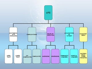
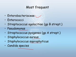
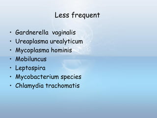
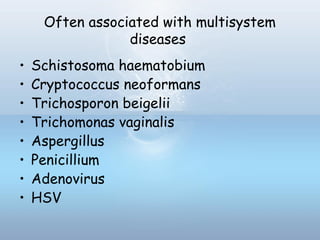
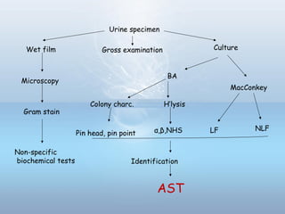
Urinary Tract Infections
Dr. Kanwal Deep Singh Lyall
1. Definition
2. Anatomical structure
3. Classification
4. Etiology
5. Pathogenesis
6. Sign & symptoms
7. Specimen collection & transport
8. Lab diagnosis & interpretation
9. AST
10. Treatment
11. References
• A spectrum of diseases caused by
microbial invasion of GU tract that
extends from renal cortex of the
kidney to the urethral meatus
• An extremely common medical problem
• Commonest sample received
Urinary tract
Resident Microflora of Urethra
• Coagulase-negative staphylococci (excluding S.
saprophyticus)
• Viridians & non-hemolytic streptococci
• Lactobacilli
• Diphtheroids (Corynebacterium spp.)
• Nonpathogenic Neisseria spp.
• Anaerobic cocci
• Propionibacterium spp.
• Anaerobic gram-negative bacilli
• Commensal Mycobacterium spp.
• Commensal Mycoplasma spp.
Classification of UTI
UTI
ANATOMICAL
SITE
INVOLOVED
COMPLICATIONS
ROUTE OF
INFECTION
LOCALIZATION
OF
PATIENT
UPPER
TRACT
LOWER
TRACT
UN-
COMPLICATED
COMPLICATED ASCENDING DESCENDING
COMMUNITY
ACQUIRED
HOSPITAL
ACQUIRED
CAUSATIVE
AGENT
BACTERIA
VIRUS
FUNGI
PARASITES
Upper urinary tract infections
Acute pyelonephritis Enterobacteriaceae
Staphylococcus aureus
Subclinical
pyelonephritis
CONS
Candida
Mycobacterium
Mycoplasma hominis
Lower urinary tract infections
Acute bacterial
cystitis
E coli
Klebsiella spp.
Proteus
Enterococci
CONS
Urethritis
Acute urethral
syndrome
Chlamydia trachomatis
Neisseria gonorrhoeae
Ureaplasma
urealyticum
Etiology
Most frequent
• Enterobacteriaceae
• Enterococci
• Streptococcus agalacitiae (gp B strept.)
• Pseudomonas
• Streptococcus pyogenes (gp A strept.)
• Staphylococcus aureus
• Staphylococcus saprophyticus
• Candida species
Less frequent
• Gardnerella vaginalis
• Ureaplasma urealyticum
• Mycoplasma hominis
• Mobiluncus
• Leptospira
• Mycobacterium species
• Chlamydia trachomatis
Often associated with multisystem
diseases
• Schistosoma haematobium
• Cryptococcus neoformans
• Trichosporon beigelii
• Trichomonas vaginalis
• Aspergillus
• Penicillium
• Adenovirus
• HSV
Pathogenesis
Risk factors for UTI
Females Males
All ages Previous UTI
Uroligical instrumentation or surgery
Urethral catheterization
Urinary tract obstruction
Neurogenic bladder
Renal transplantation
Lack of circumcision
Uroligical instrumentation or
surgery
Urethral catheterization
Urinary tract obstruction
Neurogenic bladder
Renal transplantation
HomosexualsAdults Sexual intercourse
Spermicidal contraceptive jellies
Diaphragm use
Pregnancy
Lower socioeconomic group
Diabetes
Sickle cell trait in pregnancy
HIV +
Diabetes
HIV+
Older age Estrogen deficiency
Loss of vaginal lactobacilli
Bladder prolapse
Functional & mental
impairment
Prostatic enlargement
Condom catheter drainage
Antibacterial host defenses in urinary tract
• Urine (osmolality, pH, organic acids)
• Urine flow & micturation
• Urinary tract mucosa (bactericidal activity, cytokines)
Urinary inhibitors of bacterial adherence
1. Tamm-Horsfall protein
2. Bladder mucopolysaccharide
3. Low-molecular-weight oligosaccharides
4. Secretory immunoglobulin A (SIgA)
5. lactoferrin
Inflammatory response
1. PMNs
2. Cytokines
Immune system
1. Humoral immunity
2. Cell-mediated immunity
• Prostatic secretions
Uropathogenic strains
• Not all E coli cause UTI
• Serogroups O1, O2, O4, O6, O7,O8, O75, O150 &
O18ab
• Certain O,K & H serogroups also correlate with
clinical severity
Recognized virulence factors include
• Adhesins →pili or fimbriae
• Nonfilamentous proteins in outer membrane
• P fimbriae
• K antigen (K1, K5, K12)
• α & β hemolysins
• Aerobactin
• Siderphores
• Endotoxins
• Sat protein
• Motility
• Proteus adhere produce→ →
urease hydrolyze urea increases pH of→ →
urine direct toxicity to kidney cells→
stimulates formation of kidney stones→
• Similar findings with Klebsiella spp.
• S. saprophyticus adheres better than
S.aureus or S. epidermidis
• invade superficial epithelial cells →
replicate large foci triggers host→ →
response exfoliation of superficial cells→
Signs & Symptoms
1. Urethritis
2. Cystitis
3. Pyelonephritis
4. Asymptomatic bacteriuria
5. Urethral syndrome
Types of UTI & their clinical manifestations
Specimen collection & transport
Specimen Patient preparation Special instructions
Clean voided mid stream
urine
Females: clean area with
soap & water; hold labia
apart & begin voiding in
commode; after passing
several ml, collect MSU
Males: clean glans with
soap & water, rinse with
water, retract the
foreskin, after passing
several ml, collect MSU
Specimen Patient preparation Special instructions
Suprapubic aspirate Disinfect skin Needle aspiration above
the symphysis pubis
through abdominal wall
into full bladder
Specimen Patient preparation Special instructions
Indwelling catheter
(Foley)
Disinfect catheter
Collection port
Aspirate 5-10ml of urine
with needle & syringe
Specimen Patient preparation Special instructions
Straight catheter (in &
out)
Clean urethral area (soap
& water) & rinse with
water
Insert catheter Into
bladder; allow 1st
15ml to
pass; collect remainder
• 1st
urine passed in morning → most concentrated
• Renal failure/young child → few milliliters of urine
• Label (date & time of collection, name & number)
• Explain to patient
• Deliver promptly
• Delay unavoidable → store in refrigerator at 4°C
• Or transport in refrigerated container
• Or collection & transport in container with boric acid at
a final bacteriostatic conc. 1.8%
• Samples not so treated and delayed more than 5hrs
should be discarded & doctor should be informed
Tuberculosis of urinary tract
• 1st
urine passed in the day (early morning
urine; EMU)
• 3 to 6 consecutive days
• 3 complete EMU should be sent to laboratory
• Individual specimens refrigerated pending
process
Urethritis
• Initial flow rather than the mid stream
collected
Prostatitis
• Prostatic secretions
• Urethral urine & MSU obtained before &
after massage
Laboratory Examination of Urine
Urine specimen
Wet film Culture
Microscopy
Gram stain
Non-specific
biochemical tests
Gross examination
BA
MacConkey
LF NLF
Colony charc. H’lysis
α,β,NHPin head, pin point
Identification
AST
Appearance Possible Cause
Cloudy
Urine has an unpleasant
smell & contains WBCs
•Bacterial urinary infection
Red & cloudy
Due to red cells
•Urinary schistosomiais
•Bacterial infection
Brown & cloudy → Due to
haemoglobin
•Blackwater fever
•Intravascular Haemolysis
Yellow-brown, green brown
→ Due to bilirubin
•Acute viral hepatitis
•Obstructive jaundice
Yellow-orange → Due to
urobilin, i.e. oxidized
urobilinogen
•Haemolysis
•Hepatocellular jaundice
Milky-white→ Due to chyle •Bancroftian filariasis
Microscopy
Examine wet preparation, 40X to detect
• Pus cells
• Red cells
• Casts
• Yeast cells
• Trichomonas vaginalis motile trophozoites
• Schistosoma haematobium eggs
• Bacteria (freshly collected urine)
WBC Epithelial cells RBC
Tyrosine crystals calcium oxalate "coffin-lid" triple phosphate
Uric acid crystals
Cholesterol crystals
Cystine crystals
Hyaline castGranular cast Cellular cast
RBC cast of glomerular
pathology
Spermatozoa WBC cast of pyelonephritis
S. haematobium
Budding yeast
bacteria and many white cells
Trichomonas vaginalis
Renal epithelial cells
Transitional Epi
cells
Examination of a gram stained smear
• Both uncentrifuged & centrifuged specimens
• 1 or more bacterial cells/OIF x at least 5
fields (uncentrifuged) ≥ 105
CFU/ml
• If negative → smear sediment → bacterial cells
indicate < 105
CFU/ml
Non-specific biochemical tests
1. Griess nitrite test
2. Leucocyte esterase test
3. Catalase test
4. Triphenyltetrazolium chloride (TTC)
5. Glucose test paper method
Urine specimen
Wet film
Culture
Microscopy
Gram stain
Non-specific
biochemical tests
Gross examination
Culture the
specimen
Quantitative
methods
Semi
quantitative
methods
Pour plate
method
Surface viable
count
By
spreading
method
Standard loop
method
Filter paper
method
Dip-slide
method
Miles & Misra
method
Standard loop method
No. of CFUs x 1000 (0.001 ml loop) or x 100 ( 0.01 ml loop) = no. of bacteria/ml
Dip slide method
Urine specimen
Wet film Culture
Microscopy
Gram stain
Non-specific
biochemical tests
Gross examination
BA
MacConkey
LF NLF
Colony charc. H’lysis
α,β,NHPin head, pin point
Identification
AST
MacConkey
LF
NLF
LF
•Enterobacter
(M,indole-, citrate+)
•E.coli (M,indole+, citrate-)
•Klebsiella
(NM,indole-,urease+,citrate+)
LLF
•Citrobacter (M)
•Serratia (M)
Contd.
NLF
Oxidase - Oxidase +
Motile MotileNon-Motile
Non-Motile
•Chrysobacterium
meningosepticum
•Empedobacter brevis
•Spingobacterium multivorum
•Spingobacterium spiritivorum
•Spingobacterium thalpophilium
•Shigella
•Acinetobacter
Contd.Contd.
NLF, Oxidase +, motile
• Pseudomonas aeruginosa
• Pseudomonas flurescens
• Pseudomonas mendocina
• Pseudomonas monteilli
• Pseudomonas putida
• Pseudomonas stutzeri
• Burkholderia cepacia
• Aeromonas
• Plesiomonas
NLF, Oxidase –ve, motile
• Edwardsiella tarda
• Edwardsiella tostinae
• Hafnia (variable motility)
• Morganella morganii
• Proteus mirabilis
• Proteus penneri
• Proteus vulgaris
• Providentia stuartii
• Providentia rettgerii
BA
Haemolysis Colony characters
βα NH
Pin head size Pin point size
Staphylococcus Streptococcus
Contd.
Haemolysis
β-haemolytic
Strept pyogenes
Strept agalactiae
Staphylococcus aureus
α-haemolytic
Strept viridans
Strept pneumniae
NHS
Enterococcus faecalis
Enterococcus faecium
Enterococcus durans
Enterococcus avium
Streptococcus mutans
Fungal culture
Wet mount/gram stain fungal cells or hyphae or fungal infection suspected→
Few drops of sediment
Fungal bottle containing Emmons modification of SDA & SDA with actidione
Incubate @ room t° upto 2wks
BHIA x room t°x 4wks
(Diamorphic fungi suspected)
Low colony count as significant as high
Mycobacterium
Urine
50ml x 3600g x 30mnts
2ml sediment
Decontamination x oxalic acid x 30mnts
Inoculate
BACTEC medium LJ medium
Incubate x 37°C ≥ 6wks
Growth +
Confirmation by AFB smear
Incubate x 37°C ≥ 6wks
Growth +
Confirmation by AFB smear
Interpretation
Significant bacteriuria
• MSU most easily & commonly collected sample
• Contaminants → very bacteria which cause UTI
• Isolation not proof of UTI
• Proof of UTI → demonstration of pathogens in
freshly voided urine in No. > those likely to result
from contamination
• Edward Kass suggested that this number, taken to
indicate significant bacteriuria, is about 105
/ml
• In properly collected sample contamination accounts
for less than 104
/ml & usually for less than 103
/ml
General interpretation guidelines for urine cultures
Result Specific specimen type/
associated clinical
condition, if known
Workup
≥104
CFU/ml of a single
potential pathogen or of
each of two potential
pathogens
MSU/ Pyelonephritis,
acute cystitis,
asymptomatic bacteriuria
or catheterized urines
Complete
≥103
CFU/ml of a single
potential pathogen
MSU/symptomatic males
or catheterized urines or
acute urethral syndrome
Complete
≥three organisms with no
predominating organism
MSU or catheterized
urines
None. Because of possible
contamination, ask for
another specimen
Either two or three
organisms with
predominant growth of one
organism type & <104
CFU/ml of other
organisms
MSU Complete workup for
predominant organism.
description of other
organisms
≥102
CFU/ml of any number
of organism
Suprapubic aspirates complete
Oxoid Chromogenic UTI Clarity Agar
• Chromogenic media → aid diagnosis
• Distinguish b/w colonies of
different species on a culture plate.
• Good growth of the main UTI
pathogens
• Prevents swarming of Proteus
• Coliforms dark blue/purple→
colonies
• Enterococci blue/turquoise→
colonies
• Improved recovery of S aureus
• Correct presumptive
identification of Citrobacter freundii
• Results 16-24 hours
• Ready poured plates
• Or dehydrated culture medium
Automated screening methods
Automated Principle
Bioluminescence
UTI screen
Detects bacterial ATP utilizing
enzymatic bioluminescent
reaction of ATP with luciferin &
lucifrase
Photometry Vitek If significant no. of organisms
present grow in medium to a→
detectable conc. Utilizing
photometry
Colorimetric particle filtration
Bac-T-Screen
Automated combination testing
for both bacteria & WBCs by
membrane filtration & detection
utilizing
Safranin O dye
Antimicrobial sensitivity testing
Urine specimen
Wet film Culture
Microscopy
Gram stain
Non-specific
biochemical tests
Gross examination
BA
MacConkey
LF NLF
Colony charc. H’lysis
α,β,NHSPin head, pin point
Identification
AST
• CAUTI
• HAI
• AST → Mueller-Hinton agar by Kirby-Bauer disc
diffusion method
• Antibiotics concentrated in urine → high-content test
discs
For OPD patients → oral drugs
• e.g. amoxycillin or ampicillin (25μg), cephalexin (30μg),
nalidixic acid (30μg), ciprofloxacin or norfloxacin
(5μg), nitrofurantion (50μg), trimethoprim (2.5μg), co-
amoxiclav (30μg)
For patients in hospital → parenteral drugs
• Sensitivity to cefuroxime (30μg), gentamycin (10μg),
amikacin, netlimycin, pipracillin & ceftazidime may be
tested
Enterobact
eriaceae
P aeruginosa
& non-
enterobact.
Staph. Enterococci Strept.
Carbenic.
Lomeflox.,
Norflox., or
Oflox.
Nitrofuran.
Trimethop.
Carbenic.
Ceftizoxime
Tetracyc.
Lomeflox.,
Norflox., or
Oflox.
Lomeflox.,
Norflox., or
Oflox.
Nitrofuran.
Trimethop.
Ciproflox.
Norflox.
Nitrofuran.
Tetracyc.
Norflox.
Nitrofuran.
Antomicrobials usually tested
Treatment
Drug Dose Duration
of course
Dose Duration
of course
dose Duration
of course
dose
Trimetho
prim
300mg
daily
3days 300mg
daily
7-14days 200mg
12hrly
4-6
weeks
100mg/ni
ght
Co-
amoxycl
av
250mg
8-hrly
3days 250-
500mg
8hrly
7-14days 250mg/ni
ght
Gentamy
cin
3-5mg/kg
i.v. daily
7-14days
Ciproflox
acin
250-
500mg
12hrly
3days 250mg
12hrly
oral or
750mg
6-8hrly
i.v.
7-14days
start
treatmen
t i.v. in
seriously
ill patient
250mg
12hrly
4-6
weeks
Cefalexin 7-14days 250mg/ni
ght
Erythrom
ycin
250mg
6hrly
4-6
weeks
Treatment of
presumed UTI
Treatment of
presumed
pyelonephritis
Treatment of acute
prostatitis
Prophyla
ctic or
suppressi
ve
threaphy
References
• Mackie & McCartney practical medical microbiology
• Bailey & Scott’s diagnostic microbiology
• Mahon Manusalis textbook of diagnostic microbiology
• Monica cheesbrough textbook of diagnostic
microbiology
• Mandel’s

Urinary tract infections

  • 1.
    Urinary Tract Infections Dr.Kanwal Deep Singh Lyall
  • 2.
    1. Definition 2. Anatomicalstructure 3. Classification 4. Etiology 5. Pathogenesis 6. Sign & symptoms 7. Specimen collection & transport 8. Lab diagnosis & interpretation 9. AST 10. Treatment 11. References
  • 3.
    • A spectrumof diseases caused by microbial invasion of GU tract that extends from renal cortex of the kidney to the urethral meatus • An extremely common medical problem • Commonest sample received
  • 4.
  • 5.
    Resident Microflora ofUrethra • Coagulase-negative staphylococci (excluding S. saprophyticus) • Viridians & non-hemolytic streptococci • Lactobacilli • Diphtheroids (Corynebacterium spp.) • Nonpathogenic Neisseria spp. • Anaerobic cocci • Propionibacterium spp. • Anaerobic gram-negative bacilli • Commensal Mycobacterium spp. • Commensal Mycoplasma spp.
  • 6.
  • 7.
  • 8.
    Upper urinary tractinfections Acute pyelonephritis Enterobacteriaceae Staphylococcus aureus Subclinical pyelonephritis CONS Candida Mycobacterium Mycoplasma hominis
  • 9.
    Lower urinary tractinfections Acute bacterial cystitis E coli Klebsiella spp. Proteus Enterococci CONS Urethritis Acute urethral syndrome Chlamydia trachomatis Neisseria gonorrhoeae Ureaplasma urealyticum
  • 10.
  • 11.
    Most frequent • Enterobacteriaceae •Enterococci • Streptococcus agalacitiae (gp B strept.) • Pseudomonas • Streptococcus pyogenes (gp A strept.) • Staphylococcus aureus • Staphylococcus saprophyticus • Candida species
  • 12.
    Less frequent • Gardnerellavaginalis • Ureaplasma urealyticum • Mycoplasma hominis • Mobiluncus • Leptospira • Mycobacterium species • Chlamydia trachomatis
  • 13.
    Often associated withmultisystem diseases • Schistosoma haematobium • Cryptococcus neoformans • Trichosporon beigelii • Trichomonas vaginalis • Aspergillus • Penicillium • Adenovirus • HSV
  • 14.
  • 15.
    Risk factors forUTI Females Males All ages Previous UTI Uroligical instrumentation or surgery Urethral catheterization Urinary tract obstruction Neurogenic bladder Renal transplantation Lack of circumcision Uroligical instrumentation or surgery Urethral catheterization Urinary tract obstruction Neurogenic bladder Renal transplantation HomosexualsAdults Sexual intercourse Spermicidal contraceptive jellies Diaphragm use Pregnancy Lower socioeconomic group Diabetes Sickle cell trait in pregnancy HIV + Diabetes HIV+ Older age Estrogen deficiency Loss of vaginal lactobacilli Bladder prolapse Functional & mental impairment Prostatic enlargement Condom catheter drainage
  • 16.
    Antibacterial host defensesin urinary tract • Urine (osmolality, pH, organic acids) • Urine flow & micturation • Urinary tract mucosa (bactericidal activity, cytokines) Urinary inhibitors of bacterial adherence 1. Tamm-Horsfall protein 2. Bladder mucopolysaccharide 3. Low-molecular-weight oligosaccharides 4. Secretory immunoglobulin A (SIgA) 5. lactoferrin Inflammatory response 1. PMNs 2. Cytokines Immune system 1. Humoral immunity 2. Cell-mediated immunity • Prostatic secretions
  • 17.
    Uropathogenic strains • Notall E coli cause UTI • Serogroups O1, O2, O4, O6, O7,O8, O75, O150 & O18ab • Certain O,K & H serogroups also correlate with clinical severity
  • 18.
    Recognized virulence factorsinclude • Adhesins →pili or fimbriae • Nonfilamentous proteins in outer membrane • P fimbriae • K antigen (K1, K5, K12) • α & β hemolysins • Aerobactin • Siderphores • Endotoxins • Sat protein • Motility
  • 19.
    • Proteus adhereproduce→ → urease hydrolyze urea increases pH of→ → urine direct toxicity to kidney cells→ stimulates formation of kidney stones→ • Similar findings with Klebsiella spp. • S. saprophyticus adheres better than S.aureus or S. epidermidis • invade superficial epithelial cells → replicate large foci triggers host→ → response exfoliation of superficial cells→
  • 20.
  • 21.
    1. Urethritis 2. Cystitis 3.Pyelonephritis 4. Asymptomatic bacteriuria 5. Urethral syndrome Types of UTI & their clinical manifestations
  • 22.
  • 23.
    Specimen Patient preparationSpecial instructions Clean voided mid stream urine Females: clean area with soap & water; hold labia apart & begin voiding in commode; after passing several ml, collect MSU Males: clean glans with soap & water, rinse with water, retract the foreskin, after passing several ml, collect MSU
  • 24.
    Specimen Patient preparationSpecial instructions Suprapubic aspirate Disinfect skin Needle aspiration above the symphysis pubis through abdominal wall into full bladder
  • 25.
    Specimen Patient preparationSpecial instructions Indwelling catheter (Foley) Disinfect catheter Collection port Aspirate 5-10ml of urine with needle & syringe
  • 26.
    Specimen Patient preparationSpecial instructions Straight catheter (in & out) Clean urethral area (soap & water) & rinse with water Insert catheter Into bladder; allow 1st 15ml to pass; collect remainder
  • 27.
    • 1st urine passedin morning → most concentrated • Renal failure/young child → few milliliters of urine • Label (date & time of collection, name & number) • Explain to patient • Deliver promptly • Delay unavoidable → store in refrigerator at 4°C • Or transport in refrigerated container • Or collection & transport in container with boric acid at a final bacteriostatic conc. 1.8% • Samples not so treated and delayed more than 5hrs should be discarded & doctor should be informed
  • 28.
    Tuberculosis of urinarytract • 1st urine passed in the day (early morning urine; EMU) • 3 to 6 consecutive days • 3 complete EMU should be sent to laboratory • Individual specimens refrigerated pending process
  • 29.
    Urethritis • Initial flowrather than the mid stream collected Prostatitis • Prostatic secretions • Urethral urine & MSU obtained before & after massage
  • 30.
  • 31.
    Urine specimen Wet filmCulture Microscopy Gram stain Non-specific biochemical tests Gross examination BA MacConkey LF NLF Colony charc. H’lysis α,β,NHPin head, pin point Identification AST
  • 32.
    Appearance Possible Cause Cloudy Urinehas an unpleasant smell & contains WBCs •Bacterial urinary infection Red & cloudy Due to red cells •Urinary schistosomiais •Bacterial infection Brown & cloudy → Due to haemoglobin •Blackwater fever •Intravascular Haemolysis Yellow-brown, green brown → Due to bilirubin •Acute viral hepatitis •Obstructive jaundice Yellow-orange → Due to urobilin, i.e. oxidized urobilinogen •Haemolysis •Hepatocellular jaundice Milky-white→ Due to chyle •Bancroftian filariasis
  • 33.
    Microscopy Examine wet preparation,40X to detect • Pus cells • Red cells • Casts • Yeast cells • Trichomonas vaginalis motile trophozoites • Schistosoma haematobium eggs • Bacteria (freshly collected urine)
  • 34.
    WBC Epithelial cellsRBC Tyrosine crystals calcium oxalate "coffin-lid" triple phosphate Uric acid crystals Cholesterol crystals Cystine crystals
  • 35.
    Hyaline castGranular castCellular cast RBC cast of glomerular pathology Spermatozoa WBC cast of pyelonephritis
  • 36.
    S. haematobium Budding yeast bacteriaand many white cells Trichomonas vaginalis Renal epithelial cells Transitional Epi cells
  • 37.
    Examination of agram stained smear • Both uncentrifuged & centrifuged specimens • 1 or more bacterial cells/OIF x at least 5 fields (uncentrifuged) ≥ 105 CFU/ml • If negative → smear sediment → bacterial cells indicate < 105 CFU/ml
  • 38.
    Non-specific biochemical tests 1.Griess nitrite test 2. Leucocyte esterase test 3. Catalase test 4. Triphenyltetrazolium chloride (TTC) 5. Glucose test paper method
  • 39.
    Urine specimen Wet film Culture Microscopy Gramstain Non-specific biochemical tests Gross examination
  • 40.
    Culture the specimen Quantitative methods Semi quantitative methods Pour plate method Surfaceviable count By spreading method Standard loop method Filter paper method Dip-slide method Miles & Misra method
  • 41.
    Standard loop method No.of CFUs x 1000 (0.001 ml loop) or x 100 ( 0.01 ml loop) = no. of bacteria/ml
  • 42.
  • 43.
    Urine specimen Wet filmCulture Microscopy Gram stain Non-specific biochemical tests Gross examination BA MacConkey LF NLF Colony charc. H’lysis α,β,NHPin head, pin point Identification AST
  • 44.
    MacConkey LF NLF LF •Enterobacter (M,indole-, citrate+) •E.coli (M,indole+,citrate-) •Klebsiella (NM,indole-,urease+,citrate+) LLF •Citrobacter (M) •Serratia (M) Contd.
  • 45.
    NLF Oxidase - Oxidase+ Motile MotileNon-Motile Non-Motile •Chrysobacterium meningosepticum •Empedobacter brevis •Spingobacterium multivorum •Spingobacterium spiritivorum •Spingobacterium thalpophilium •Shigella •Acinetobacter Contd.Contd.
  • 46.
    NLF, Oxidase +,motile • Pseudomonas aeruginosa • Pseudomonas flurescens • Pseudomonas mendocina • Pseudomonas monteilli • Pseudomonas putida • Pseudomonas stutzeri • Burkholderia cepacia • Aeromonas • Plesiomonas
  • 47.
    NLF, Oxidase –ve,motile • Edwardsiella tarda • Edwardsiella tostinae • Hafnia (variable motility) • Morganella morganii • Proteus mirabilis • Proteus penneri • Proteus vulgaris • Providentia stuartii • Providentia rettgerii
  • 48.
    BA Haemolysis Colony characters βαNH Pin head size Pin point size Staphylococcus Streptococcus Contd.
  • 49.
    Haemolysis β-haemolytic Strept pyogenes Strept agalactiae Staphylococcusaureus α-haemolytic Strept viridans Strept pneumniae NHS Enterococcus faecalis Enterococcus faecium Enterococcus durans Enterococcus avium Streptococcus mutans
  • 50.
  • 51.
    Wet mount/gram stainfungal cells or hyphae or fungal infection suspected→ Few drops of sediment Fungal bottle containing Emmons modification of SDA & SDA with actidione Incubate @ room t° upto 2wks BHIA x room t°x 4wks (Diamorphic fungi suspected) Low colony count as significant as high
  • 52.
  • 53.
    Urine 50ml x 3600gx 30mnts 2ml sediment Decontamination x oxalic acid x 30mnts Inoculate BACTEC medium LJ medium Incubate x 37°C ≥ 6wks Growth + Confirmation by AFB smear Incubate x 37°C ≥ 6wks Growth + Confirmation by AFB smear
  • 54.
  • 55.
    Significant bacteriuria • MSUmost easily & commonly collected sample • Contaminants → very bacteria which cause UTI • Isolation not proof of UTI • Proof of UTI → demonstration of pathogens in freshly voided urine in No. > those likely to result from contamination • Edward Kass suggested that this number, taken to indicate significant bacteriuria, is about 105 /ml • In properly collected sample contamination accounts for less than 104 /ml & usually for less than 103 /ml
  • 56.
    General interpretation guidelinesfor urine cultures Result Specific specimen type/ associated clinical condition, if known Workup ≥104 CFU/ml of a single potential pathogen or of each of two potential pathogens MSU/ Pyelonephritis, acute cystitis, asymptomatic bacteriuria or catheterized urines Complete ≥103 CFU/ml of a single potential pathogen MSU/symptomatic males or catheterized urines or acute urethral syndrome Complete ≥three organisms with no predominating organism MSU or catheterized urines None. Because of possible contamination, ask for another specimen Either two or three organisms with predominant growth of one organism type & <104 CFU/ml of other organisms MSU Complete workup for predominant organism. description of other organisms ≥102 CFU/ml of any number of organism Suprapubic aspirates complete
  • 57.
    Oxoid Chromogenic UTIClarity Agar • Chromogenic media → aid diagnosis • Distinguish b/w colonies of different species on a culture plate. • Good growth of the main UTI pathogens • Prevents swarming of Proteus • Coliforms dark blue/purple→ colonies • Enterococci blue/turquoise→ colonies • Improved recovery of S aureus • Correct presumptive identification of Citrobacter freundii • Results 16-24 hours • Ready poured plates • Or dehydrated culture medium
  • 58.
    Automated screening methods AutomatedPrinciple Bioluminescence UTI screen Detects bacterial ATP utilizing enzymatic bioluminescent reaction of ATP with luciferin & lucifrase Photometry Vitek If significant no. of organisms present grow in medium to a→ detectable conc. Utilizing photometry Colorimetric particle filtration Bac-T-Screen Automated combination testing for both bacteria & WBCs by membrane filtration & detection utilizing Safranin O dye
  • 59.
  • 60.
    Urine specimen Wet filmCulture Microscopy Gram stain Non-specific biochemical tests Gross examination BA MacConkey LF NLF Colony charc. H’lysis α,β,NHSPin head, pin point Identification AST
  • 61.
  • 62.
    • AST →Mueller-Hinton agar by Kirby-Bauer disc diffusion method • Antibiotics concentrated in urine → high-content test discs For OPD patients → oral drugs • e.g. amoxycillin or ampicillin (25μg), cephalexin (30μg), nalidixic acid (30μg), ciprofloxacin or norfloxacin (5μg), nitrofurantion (50μg), trimethoprim (2.5μg), co- amoxiclav (30μg) For patients in hospital → parenteral drugs • Sensitivity to cefuroxime (30μg), gentamycin (10μg), amikacin, netlimycin, pipracillin & ceftazidime may be tested
  • 63.
    Enterobact eriaceae P aeruginosa & non- enterobact. Staph.Enterococci Strept. Carbenic. Lomeflox., Norflox., or Oflox. Nitrofuran. Trimethop. Carbenic. Ceftizoxime Tetracyc. Lomeflox., Norflox., or Oflox. Lomeflox., Norflox., or Oflox. Nitrofuran. Trimethop. Ciproflox. Norflox. Nitrofuran. Tetracyc. Norflox. Nitrofuran. Antomicrobials usually tested
  • 64.
  • 65.
    Drug Dose Duration ofcourse Dose Duration of course dose Duration of course dose Trimetho prim 300mg daily 3days 300mg daily 7-14days 200mg 12hrly 4-6 weeks 100mg/ni ght Co- amoxycl av 250mg 8-hrly 3days 250- 500mg 8hrly 7-14days 250mg/ni ght Gentamy cin 3-5mg/kg i.v. daily 7-14days Ciproflox acin 250- 500mg 12hrly 3days 250mg 12hrly oral or 750mg 6-8hrly i.v. 7-14days start treatmen t i.v. in seriously ill patient 250mg 12hrly 4-6 weeks Cefalexin 7-14days 250mg/ni ght Erythrom ycin 250mg 6hrly 4-6 weeks Treatment of presumed UTI Treatment of presumed pyelonephritis Treatment of acute prostatitis Prophyla ctic or suppressi ve threaphy
  • 67.
  • 68.
    • Mackie &McCartney practical medical microbiology • Bailey & Scott’s diagnostic microbiology • Mahon Manusalis textbook of diagnostic microbiology • Monica cheesbrough textbook of diagnostic microbiology • Mandel’s