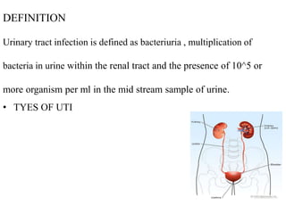
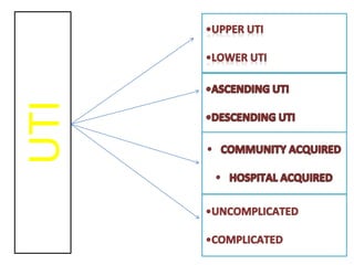
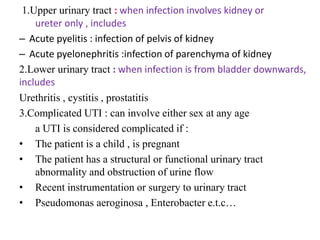
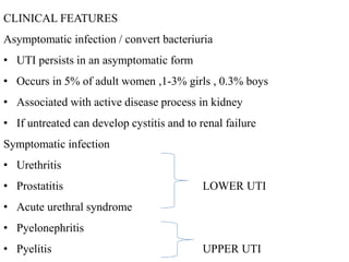
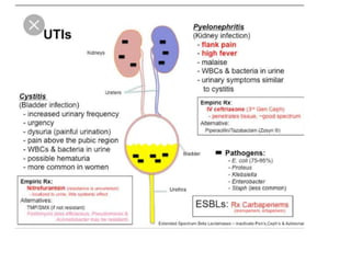
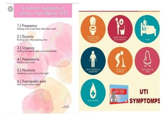
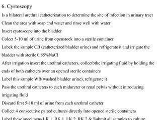
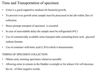
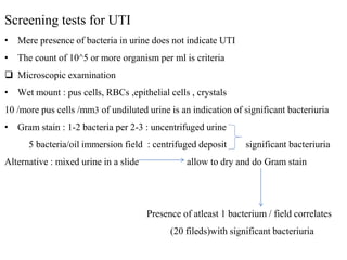
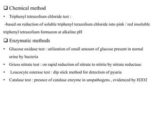
URINARY TRACT INFECTION
DEFINITION
Urinary tract infection is defined as bacteriuria , multiplication of
bacteria in urine within the renal tract and the presence of 10^5 or
more organism per ml in the mid stream sample of urine.
• TYES OF UTI
UTI
1.Upper urinary tract : when infection involves kidney or
ureter only , includes
– Acute pyelitis : infection of pelvis of kidney
– Acute pyelonephritis :infection of parenchyma of kidney
2.Lower urinary tract : when infection is from bladder downwards,
includes
Urethritis , cystitis , prostatitis
3.Complicated UTI : can involve either sex at any age
a UTI is considered complicated if :
• The patient is a child , is pregnant
• The patient has a structural or functional urinary tract
abnormality and obstruction of urine flow
• Recent instrumentation or surgery to urinary tract
• Pseudomonas aeroginosa , Enterobacter e.t.c…
4. Uncomplicated UTI : is usually considered to be cystitis or
pyelonephritis that occurs in premenopausal adult women with no
structural or functional anomalily of urinary tract and who are not
pregnant
• E.coli , Klebsiella , Proteus , Streptococcus faecalis e.t.c…
PREDISPOSING FACTORS
• Age : increases greatly with age
• Sex : sexually active female
• Pregnancy : dilation of uterus and renal pelvis, stasis in ureter
• Instrumentation and surgery : catheterization and surgical procedure
• Diabetes mellitus
• Immunosuppressive agents : steroids , cytotoxic drugs
• Structural and functional abnormalities of UTIs
obstruction : calculus formation , tumour ,pregnancy e.t.c
neurogenic bladder
genital prolapse
vesicourethral reflux
Urinary Tract Infection
More among females
•Short urethra
•Close proximity to
perirectal region
•Bacteria reach the bladder
more easily in females
ORGANISMS CAUSING UTI
Bacterial causes
More common
• E.Coli (80%)
• S.Saprophyticus
• Proteus mirabilis
• K.Species
• S.Epidermidis
• Enterococci
• P.Aeruginosa(common
in hospital acquired
UTI)
Less common
• Enterobacter species
• Citrobacter speecies
• Acinetobacter species
• Providencia species
• Morganella species
• Serratia marcescens
• Alcaligens faecalis
• streptococci A&B
• Mycobacterium
tuberculosis
Rare causes
• Streptococci other
than A &B
• Leptospira species
• Salmonella species
• Haemophilus
influenzae
• Mycoplasma hominis
• Ureapasalma
urealyticum
• Neisseria gonorrhoeae
• Gardenalla vaginalis
Fungal causes
• Candida albicans and other candida species
• Torulopsis galbrata
• Cryptococcus neoformans
• Histoplasma duboissi
Viral causes
• Viruses are rarely involved in UTI
• Adenovirus , CMV , Measles virus , Rubella virus , HIV e.t.c…
Parasitic causes
• Schistosoma haematobium
• Enterobius vermicularis occasional causes
• Trichomonas vaginalis
PATHOGENESIS
• Adhesion of organism is an imp: factor in pathogenesis of UTI
• Organism have the ability to adhere mucosal lining, are able to resist
elimination at voiding and are able to produce significant bacteriuria
• Adhesion is mediated by pili and coloniozation factor
• Once adhered , it spreads and produces pathogenic effects by
resisting bactericidal and bacteriostatic effect of local tissue
• Mainly 4 routes of bacterial entry to urinary tract
*ascending infection
*descending / haematogenous spread
*lymphatogenous spread
*direct extention from other organs
*Ascending route
Organism from faecal flora perineum and periurethral site
ascend up
kidneys urethra bladder
sometimesupto
*Descending route
Infection of any site blood bacteraemia
via lympatic system haematogenous spread
kidneys
sometimes
*Lymphatogenous spread
• Men through rectal and colonic lymphatic vessels to prostrate and
bladder
• Women through periuterine lymphatics to urinary tract
*Direct extension from other organs
• Pelvic inflammatory disease
• Genito urinary tract fistulas
o Most infections involving kidneys are acquired by ascending route
o Yeast ,Mycobacterium tuberculosis ,Salmonella species e.t.c..in urine
Indicates pyelonephritis acquired via descending route
CLINICAL FEATURES
Asymptomatic infection / convert bacteriuria
• UTI persists in an asymptomatic form
• Occurs in 5% of adult women ,1-3% girls , 0.3% boys
• Associated with active disease process in kidney
• If untreated can develop cystitis and to renal failure
Symptomatic infection
• Urethritis
• Prostatitis LOWER UTI
• Acute urethral syndrome
• Pyelonephritis
• Pyelitis UPPER UTI
Compilcations
• Septicaemia : particularly in elderly
• Renal / perirenal abscess
• Chronic renal failure : due to renal scarring because of persistent or
recurrent UTI in the young
• Preterm delivery and low birth weight because of asymptomatic
bacteriuria in pregnancy
LABORATORY DIAGNOSIS
 Specimen Collection
• Prevent contamination by normal vaginal, perineal and urethral
flora.
1. Clean Catch Mid Stream Urine
2. Straight Catheterized Urine
3. Suprapubic Bladder Aspiration
4. Indwelling Catheter
5. Illeal conduit
6. Cystoscopy
1.Clean-catch MSU
• Most convenient and most commonly used method
• Patient Education is a must
– Clean periurethral area with 2 separate washes of soap and water
– Well rinsed with sterile warm water to remove detergent
– Retract labial folds/glans penis
– Void first part of urine (contains commensals)
– Collect middle portion of urine (in sterile wide mouthed container)
– Void last portion
– Close the container and transport to lab within 2hrs
*For tuberculosis of urinary tract MSU is not useful because of intermittent
excretion of tubercle bacilli.. Early morning specimen on 3 consecutive days
/alternative is collection of 24 hrs sample of urine
2. Straight catheterized urine
• Slightly invasive
• Allows collection of bladder uncontaminated urine
• Performed by physician or trained professional
• There is a risk of introducing infection
• Might introduce urethral flora into bladder with catheter
3. Suprapubic Aspiration
• Contamination free!
• Bladder must be full
• Disinfect skin over bladder
• Puncture using a needle and syringe
– Usually in Infants when urine is difficult to obtain
– Small chlidren , pregnant women e.t.c..
4.Indwelling Catheter
• Aseptic techniques must be followed
• Wear gloves
• Catheter tubing is clamped off above the portion
• Port or wall of tube is cleaned thoroughly with 70% ethanol
• Urine is aspirated with a needle and syringe
• Specimen from collection bag -INAPPROPRIATE
5.Illeal conduit
• Remove external device
• Cleanse the stoma with 70% alcohol followed by iodine
• Remove the iodine with alcohol
• Insert a double catheter into the cleansed stoma , to a depth beyond the fascial level ,
collect the urine
6. Cystoscopy
Is a bilateral urethral catheterization to determine the site of infection in urinary tract
Clean the area with soap and water and rinse well with water
Insert cystoscope into the bladder
Colect 5-10 ml of urine from openstock into a sterile container
Labek the sample CB (catheterized bladder urine) and refrigerate it and irrigate the
bladder with sterile 0.85%NaCl
After irrigation insert the urethral catheters, collectbthe irrigating fluid by holding the
ends of both catheters over an opened sterile containers
Label this sample WB(washed bladder urine), refrigerate it
Pass the urethral catheters to each midureter or renal pelvis without introducing
irrigating fluid
Discard first 5-10 ml of urine from each urethral catheter
Collect 4 consecutive paired cultures directly into opened sterile containers
Time and Transportation of specimen
• Urine is a good supportive medium for bacterial growth.
• To prevent over growth urine sample must be processed in the lab within 2hrs of
collection.
• Hence prompt transport of specimen is essential
• In case of unavoidable delay the sample must be refrigerated (4oC)
• Use of commercially available urine transport tube containing boric acid , glycerol
sodium formate
• Use of container with boric acid (1.8%) which is bacteriostatic
TIMING OF SPECIMEN COLLECTION
• Obtain early morning specimens whenever possible
• Allowing urine to remain in the bladder overnight or for atleast 4 hr will decrease
the no : of false negative results
Screening tests for UTI
• Mere presence of bacteria in urine does not indicate UTI
• The count of 10^5 or more organism per ml is criteria
 Microscopic examination
• Wet mount : pus cells, RBCs ,epithelial cells , crystals
10 /more pus cells /mm3 of undiluted urine is an indication of significant bacteriuria
• Gram stain : 1-2 bacteria per 2-3 : uncentrifuged urine
5 bacteria/oil immersion field : centrifuged deposit significant bacteriuria
Alternative : mixed urine in a slide allow to dry and do Gram stain
Presence of atleast 1 bacterium / field correlates
(20 fileds)with significant bacteriuria
 Chemical method
• Triphenyl tetrazolium chloride test :
-based on reduction of soluble triphenyl terazolium chloride into pink / red insoluble
triphenyl tetrazolium formazon at alkaline pH
 Enzymatic methods
• Glucose oxidase test : utilization of small amount of glucose present in nornal
urine by bacteria
• Griess nitrate test : on rapid reduction of nitrate to nitrite by nitrate reductase
• Leucocyte esterase test : dip stick method for detection of pyuria
• Catalase test : presnce of catalase enzyme in uropathogens , evidenced by H2O2
 Culture
• Most accurate and acceptable methods
• Both quantitative and semiquantitative methods are used
 Quantitative method
• Pour plate method : 10 fold dilution of urine sample is done
1 ml of diluted urine added to melted and cooled BA(45°C) , mixed well and
pour to plate incubate at 37°C at 18 – 24 hrs
After incubation no: of colonies are counted and assuming each bacterium forms one
colony and total number of bacteria/ml calculated
• Pipette dilution method
• Simplified spread plate method
 Semiquantitative culture
• Calibrated loop method
o Most convienient method
o Size of loop carefully controlled
o A standard calibrated loop that can transfer fixed small volumes of urine ( 0.001
ml) is used
o Culture is set up on SBA & MacConkey Agar CLED
o Incubation at 35-37°C for 24 hours and count the colonies
o Total no: of bacteria per ml of urine = no: of colonies *1000
o More siutable to detect large number of urine samples
• Dip slide culture method
o Commercially available plastic slides coated with CLED agar on 1 side &MA on
other side
o Inoculate by immersing into freshly passed urine
o Slides are placed in sterile containers
o Incubated colonies are counted
Advantages :
avoid problem of transfer of urine specimen to lab
permit screening in clinic itself
Disadvantage :
expensive
• Filter paper strip technique
Filter paper strip is dipped into urine and transferred to conventional agar plate and
incubated and colonies are counted
 OTHER SCREENING METHODS
• Automated screening test
commercially available kits are used for rapid screening of urine by using
lighy scatter photometry in 4-5 hrs e.g.
 Pfizer’s autobac
 MS-2 Abbot
 Auto microbic system ( Vitek system)
• Gas liquid chromatography
INTERPRETATION OF COLONY COUNT
• Significant bacteriuria was introduced by Kass
• 10^5 or more bacteria/ml of urine : significant bacteriuria , sensitivity test is to be
done
• Count between 10^4 and 10^5 /ml : doubtful significance : repeat the culture
• Count less than 10^4/ml of urine : no significant bacteriuria and considered as
contamination
• Conditions where ≤105 CFU/ml is considered significant
– Suprapubic specimen/Nephrostomy specimen/Catheterized specimen
– Antibiotic therapy
– Gram positive bacterial infection
– Complicated UTI
– Acute urethral syndrome
IDENTIFICATION OF BACTERIA
• Cultures of screened positive urine samples are identified by using standard
biochemical and / serological tests
LABORATORY DIAGNOSIS OF TUBERCULOSIS OT URINART TRACT
• Demonstration of acid-alcohol fast bacilli : Z-N staining
• Isolation on LJ medium
OTHER INVESTIGATIONS
• Immunofluorescence test
• Haemtological investigations : Hb , TLC ,DLC e.t.c…
• Biochemical investigations : urea , creatinine
• X- ray of kidney, ureter , bladder
• Ultrasonography and cystography
TREATMENT OF UTI
• On basis of susceptibility reports as multiple resistance to drugs may occur in
uropathogens
• Sometimes combination drugs is required
• Quinolones,cotrimoxazole,nitrofurantoin can be given
UTI.pptx for educational purposes for students

UTI.pptx for educational purposes for students

  • 1.
  • 2.
    DEFINITION Urinary tract infectionis defined as bacteriuria , multiplication of bacteria in urine within the renal tract and the presence of 10^5 or more organism per ml in the mid stream sample of urine. • TYES OF UTI
  • 3.
  • 4.
    1.Upper urinary tract: when infection involves kidney or ureter only , includes – Acute pyelitis : infection of pelvis of kidney – Acute pyelonephritis :infection of parenchyma of kidney 2.Lower urinary tract : when infection is from bladder downwards, includes Urethritis , cystitis , prostatitis 3.Complicated UTI : can involve either sex at any age a UTI is considered complicated if : • The patient is a child , is pregnant • The patient has a structural or functional urinary tract abnormality and obstruction of urine flow • Recent instrumentation or surgery to urinary tract • Pseudomonas aeroginosa , Enterobacter e.t.c…
  • 5.
    4. Uncomplicated UTI: is usually considered to be cystitis or pyelonephritis that occurs in premenopausal adult women with no structural or functional anomalily of urinary tract and who are not pregnant • E.coli , Klebsiella , Proteus , Streptococcus faecalis e.t.c…
  • 6.
    PREDISPOSING FACTORS • Age: increases greatly with age • Sex : sexually active female • Pregnancy : dilation of uterus and renal pelvis, stasis in ureter • Instrumentation and surgery : catheterization and surgical procedure • Diabetes mellitus • Immunosuppressive agents : steroids , cytotoxic drugs • Structural and functional abnormalities of UTIs obstruction : calculus formation , tumour ,pregnancy e.t.c neurogenic bladder genital prolapse vesicourethral reflux
  • 7.
    Urinary Tract Infection Moreamong females •Short urethra •Close proximity to perirectal region •Bacteria reach the bladder more easily in females
  • 8.
    ORGANISMS CAUSING UTI Bacterialcauses More common • E.Coli (80%) • S.Saprophyticus • Proteus mirabilis • K.Species • S.Epidermidis • Enterococci • P.Aeruginosa(common in hospital acquired UTI) Less common • Enterobacter species • Citrobacter speecies • Acinetobacter species • Providencia species • Morganella species • Serratia marcescens • Alcaligens faecalis • streptococci A&B • Mycobacterium tuberculosis Rare causes • Streptococci other than A &B • Leptospira species • Salmonella species • Haemophilus influenzae • Mycoplasma hominis • Ureapasalma urealyticum • Neisseria gonorrhoeae • Gardenalla vaginalis
  • 9.
    Fungal causes • Candidaalbicans and other candida species • Torulopsis galbrata • Cryptococcus neoformans • Histoplasma duboissi Viral causes • Viruses are rarely involved in UTI • Adenovirus , CMV , Measles virus , Rubella virus , HIV e.t.c… Parasitic causes • Schistosoma haematobium • Enterobius vermicularis occasional causes • Trichomonas vaginalis
  • 10.
    PATHOGENESIS • Adhesion oforganism is an imp: factor in pathogenesis of UTI • Organism have the ability to adhere mucosal lining, are able to resist elimination at voiding and are able to produce significant bacteriuria • Adhesion is mediated by pili and coloniozation factor • Once adhered , it spreads and produces pathogenic effects by resisting bactericidal and bacteriostatic effect of local tissue • Mainly 4 routes of bacterial entry to urinary tract *ascending infection *descending / haematogenous spread *lymphatogenous spread *direct extention from other organs
  • 11.
    *Ascending route Organism fromfaecal flora perineum and periurethral site ascend up kidneys urethra bladder sometimesupto *Descending route Infection of any site blood bacteraemia via lympatic system haematogenous spread kidneys sometimes
  • 12.
    *Lymphatogenous spread • Menthrough rectal and colonic lymphatic vessels to prostrate and bladder • Women through periuterine lymphatics to urinary tract *Direct extension from other organs • Pelvic inflammatory disease • Genito urinary tract fistulas o Most infections involving kidneys are acquired by ascending route o Yeast ,Mycobacterium tuberculosis ,Salmonella species e.t.c..in urine Indicates pyelonephritis acquired via descending route
  • 13.
    CLINICAL FEATURES Asymptomatic infection/ convert bacteriuria • UTI persists in an asymptomatic form • Occurs in 5% of adult women ,1-3% girls , 0.3% boys • Associated with active disease process in kidney • If untreated can develop cystitis and to renal failure Symptomatic infection • Urethritis • Prostatitis LOWER UTI • Acute urethral syndrome • Pyelonephritis • Pyelitis UPPER UTI
  • 15.
    Compilcations • Septicaemia :particularly in elderly • Renal / perirenal abscess • Chronic renal failure : due to renal scarring because of persistent or recurrent UTI in the young • Preterm delivery and low birth weight because of asymptomatic bacteriuria in pregnancy
  • 17.
    LABORATORY DIAGNOSIS  SpecimenCollection • Prevent contamination by normal vaginal, perineal and urethral flora. 1. Clean Catch Mid Stream Urine 2. Straight Catheterized Urine 3. Suprapubic Bladder Aspiration 4. Indwelling Catheter 5. Illeal conduit 6. Cystoscopy
  • 18.
    1.Clean-catch MSU • Mostconvenient and most commonly used method • Patient Education is a must – Clean periurethral area with 2 separate washes of soap and water – Well rinsed with sterile warm water to remove detergent – Retract labial folds/glans penis – Void first part of urine (contains commensals) – Collect middle portion of urine (in sterile wide mouthed container) – Void last portion – Close the container and transport to lab within 2hrs *For tuberculosis of urinary tract MSU is not useful because of intermittent excretion of tubercle bacilli.. Early morning specimen on 3 consecutive days /alternative is collection of 24 hrs sample of urine
  • 19.
    2. Straight catheterizedurine • Slightly invasive • Allows collection of bladder uncontaminated urine • Performed by physician or trained professional • There is a risk of introducing infection • Might introduce urethral flora into bladder with catheter 3. Suprapubic Aspiration • Contamination free! • Bladder must be full • Disinfect skin over bladder • Puncture using a needle and syringe – Usually in Infants when urine is difficult to obtain – Small chlidren , pregnant women e.t.c..
  • 20.
    4.Indwelling Catheter • Aseptictechniques must be followed • Wear gloves • Catheter tubing is clamped off above the portion • Port or wall of tube is cleaned thoroughly with 70% ethanol • Urine is aspirated with a needle and syringe • Specimen from collection bag -INAPPROPRIATE 5.Illeal conduit • Remove external device • Cleanse the stoma with 70% alcohol followed by iodine • Remove the iodine with alcohol • Insert a double catheter into the cleansed stoma , to a depth beyond the fascial level , collect the urine
  • 21.
    6. Cystoscopy Is abilateral urethral catheterization to determine the site of infection in urinary tract Clean the area with soap and water and rinse well with water Insert cystoscope into the bladder Colect 5-10 ml of urine from openstock into a sterile container Labek the sample CB (catheterized bladder urine) and refrigerate it and irrigate the bladder with sterile 0.85%NaCl After irrigation insert the urethral catheters, collectbthe irrigating fluid by holding the ends of both catheters over an opened sterile containers Label this sample WB(washed bladder urine), refrigerate it Pass the urethral catheters to each midureter or renal pelvis without introducing irrigating fluid Discard first 5-10 ml of urine from each urethral catheter Collect 4 consecutive paired cultures directly into opened sterile containers
  • 22.
    Time and Transportationof specimen • Urine is a good supportive medium for bacterial growth. • To prevent over growth urine sample must be processed in the lab within 2hrs of collection. • Hence prompt transport of specimen is essential • In case of unavoidable delay the sample must be refrigerated (4oC) • Use of commercially available urine transport tube containing boric acid , glycerol sodium formate • Use of container with boric acid (1.8%) which is bacteriostatic TIMING OF SPECIMEN COLLECTION • Obtain early morning specimens whenever possible • Allowing urine to remain in the bladder overnight or for atleast 4 hr will decrease the no : of false negative results
  • 23.
    Screening tests forUTI • Mere presence of bacteria in urine does not indicate UTI • The count of 10^5 or more organism per ml is criteria  Microscopic examination • Wet mount : pus cells, RBCs ,epithelial cells , crystals 10 /more pus cells /mm3 of undiluted urine is an indication of significant bacteriuria • Gram stain : 1-2 bacteria per 2-3 : uncentrifuged urine 5 bacteria/oil immersion field : centrifuged deposit significant bacteriuria Alternative : mixed urine in a slide allow to dry and do Gram stain Presence of atleast 1 bacterium / field correlates (20 fileds)with significant bacteriuria
  • 24.
     Chemical method •Triphenyl tetrazolium chloride test : -based on reduction of soluble triphenyl terazolium chloride into pink / red insoluble triphenyl tetrazolium formazon at alkaline pH  Enzymatic methods • Glucose oxidase test : utilization of small amount of glucose present in nornal urine by bacteria • Griess nitrate test : on rapid reduction of nitrate to nitrite by nitrate reductase • Leucocyte esterase test : dip stick method for detection of pyuria • Catalase test : presnce of catalase enzyme in uropathogens , evidenced by H2O2
  • 26.
     Culture • Mostaccurate and acceptable methods • Both quantitative and semiquantitative methods are used  Quantitative method • Pour plate method : 10 fold dilution of urine sample is done 1 ml of diluted urine added to melted and cooled BA(45°C) , mixed well and pour to plate incubate at 37°C at 18 – 24 hrs After incubation no: of colonies are counted and assuming each bacterium forms one colony and total number of bacteria/ml calculated • Pipette dilution method • Simplified spread plate method
  • 27.
     Semiquantitative culture •Calibrated loop method o Most convienient method o Size of loop carefully controlled o A standard calibrated loop that can transfer fixed small volumes of urine ( 0.001 ml) is used o Culture is set up on SBA & MacConkey Agar CLED o Incubation at 35-37°C for 24 hours and count the colonies o Total no: of bacteria per ml of urine = no: of colonies *1000 o More siutable to detect large number of urine samples • Dip slide culture method o Commercially available plastic slides coated with CLED agar on 1 side &MA on other side
  • 28.
    o Inoculate byimmersing into freshly passed urine o Slides are placed in sterile containers o Incubated colonies are counted Advantages : avoid problem of transfer of urine specimen to lab permit screening in clinic itself Disadvantage : expensive • Filter paper strip technique Filter paper strip is dipped into urine and transferred to conventional agar plate and incubated and colonies are counted
  • 29.
     OTHER SCREENINGMETHODS • Automated screening test commercially available kits are used for rapid screening of urine by using lighy scatter photometry in 4-5 hrs e.g.  Pfizer’s autobac  MS-2 Abbot  Auto microbic system ( Vitek system) • Gas liquid chromatography INTERPRETATION OF COLONY COUNT • Significant bacteriuria was introduced by Kass • 10^5 or more bacteria/ml of urine : significant bacteriuria , sensitivity test is to be done • Count between 10^4 and 10^5 /ml : doubtful significance : repeat the culture
  • 30.
    • Count lessthan 10^4/ml of urine : no significant bacteriuria and considered as contamination • Conditions where ≤105 CFU/ml is considered significant – Suprapubic specimen/Nephrostomy specimen/Catheterized specimen – Antibiotic therapy – Gram positive bacterial infection – Complicated UTI – Acute urethral syndrome IDENTIFICATION OF BACTERIA • Cultures of screened positive urine samples are identified by using standard biochemical and / serological tests
  • 32.
    LABORATORY DIAGNOSIS OFTUBERCULOSIS OT URINART TRACT • Demonstration of acid-alcohol fast bacilli : Z-N staining • Isolation on LJ medium OTHER INVESTIGATIONS • Immunofluorescence test • Haemtological investigations : Hb , TLC ,DLC e.t.c… • Biochemical investigations : urea , creatinine • X- ray of kidney, ureter , bladder • Ultrasonography and cystography TREATMENT OF UTI • On basis of susceptibility reports as multiple resistance to drugs may occur in uropathogens • Sometimes combination drugs is required • Quinolones,cotrimoxazole,nitrofurantoin can be given

Editor's Notes

  • #5 Community acquired : e.coli , staph , kleb etc…associated with mdr organism such as esbl e.coli