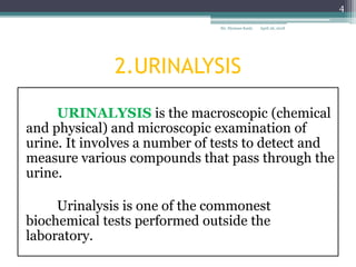
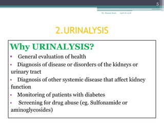
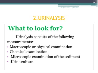
Sir. Stymass Kasty
SOKOINE UNIVERSITY OF AGRICULTURE(SUA)
MOROGORO-TANZANIA
RENAL FUNCTION TEST-1
URINALYSIS AND RENAL
PLASMA FLOW TEST
April 26, 2018Sir. Stymass Kasty
1
CONTENTS
1.Introduction to Renal
Function Tests (RFT).
2.Urinalysis.
3.Renal plasma flow test.
4.Conclusion.
5.References.
April 26, 2018Sir. Stymass Kasty
2
1. INTRODUCTION TO RFTs
• Renal function tests are common lab tests used
to evaluate how well the kidneys are working.
• Biochemical Tests of Renal Function classified based
on
Urinalysis
 Measurement of renal blood flow (RBF) or
effective renal plasma flow (ERPF)
Measurement of GFR
Clearance tests
Plasma creatinine
Tubular function tests
April 26, 2018Sir. Stymass Kasty
3
2.URINALYSIS
URINALYSIS is the macroscopic (chemical
and physical) and microscopic examination of
urine. It involves a number of tests to detect and
measure various compounds that pass through the
urine.
Urinalysis is one of the commonest
biochemical tests performed outside the
laboratory.
April 26, 2018Sir. Stymass Kasty
4
2.URINALYSIS
Why URINALYSIS?
 General evaluation of health
• Diagnosis of disease or disorders of the kidneys or
urinary tract
• Diagnosis of other systemic disease that affect kidney
function
• Monitoring of patients with diabetes
• Screening for drug abuse (eg. Sulfonamide or
aminoglycosides)
April 26, 2018Sir. Stymass Kasty
5
2.URINALYSIS
What to look for?
Urinalysis consists of the following
measurements: –
• Macroscopic or physical examination
• Chemical examination
• Microscopic examination of the sediment
• Urine culture
April 26, 2018Sir. Stymass Kasty
6
2.URINALYSIS
Physical Examination of urine
I) Colour:
Freshly excreted urine is colorless to straw colored
The normal color of urine is due to the presence of
pigment urochrome
Trances of other substances, such as- Uroerythrin,
urobilin, uroporphyrin and coproporphyrins also
contribute to the color of urine
April 26, 2018Sir. Stymass Kasty
7
2.URINALYSIS
Physiological Variations of Urine colour
Dark yellow _______Concentrated urine-
Mild dehydration
Vitamin B complex
therapy
Orange ___________Drug induced
Pinkish ___________ Excessive beet root
intake
April 26, 2018Sir. Stymass Kasty
8
2.URINALYSIS
II) Volume of urine:
 Normal volume-800-2,500 ml/day with an average
of 1500 ml/day.
 Approximately 500 ml/day is the minimum volume
of urine needed in normal health to remove waste
products
 The volume of urine is affected by-
• Fluid intake
• Fluid loss
• Type of diet
• Cardio-vascular status and
• Renal functions
April 26, 2018Sir. Stymass Kasty
9
2.URINALYSIS
Variations in volume of urine excreted
• A) Polyuria: Polyuria implies an increased volume of
urine excreted per day, generally volume of urine
exceeding 2,500 ml/day is termed as Polyuria.
• Conditions causing Polyuria
• Diabetes mellitus (Melichuria)
• Diabetes Insipidus
• Late stage of chronic glomerulonephritis
•Drug induced- Diuretics
• Alcohol
• Compulsive polydipsia
April 26, 2018Sir. Stymass Kasty
10
2.URINALYSIS
B) Oliguria- Volume of urine less than 500 ml/day
is termed oliguria.
Conditions causing oliguria
• Fever
• Diarrhea (loss of fluid from extra renal sites)
• Severe edema
• Acute nephritis
• Early stage of acute glomerulonephritis
• Cardiac failure and hypertension (reduced
circulatory volume)
April 26, 2018Sir. Stymass Kasty
11
2.URINALYSIS
C) Anuria- Complete cessation of urine or
volume of excreted urine less than 100 ml/day.
• Conditions causing Anuria
• Acute tubular necrosis
• Blood transfusion reaction
• Surgical shock
• Bilateral renal stones
• Sulphonamide therapy
April 26, 2018Sir. Stymass Kasty
12
2.URINALYSIS
III)Specific Gravity:
The specific gravity indicates the concentrating ability
of the kidneys.
IT IS CRUDE ESTIMATION OF URINARY
OSMOLALITY
• In normal health the urinary specific gravity ranges
between 1.016-1.025, the average being 1.020.
• The specific gravity is affected by-
• Volume of urine excreted and
• The amount of solids present in the urine
April 26, 2018Sir. Stymass Kasty
13
2.URINALYSIS
Variations in specific gravity of urine
A) Low specific gravity- 1.016 or less
Conditions
• Compulsive polydipsia
• Diabetes Insipidus
• Glomerulonephritis
• Pyelonephritis
April 26, 2018Sir. Stymass Kasty
14
2.URINALYSIS
B) High specific gravity of urine- 1.025 or more
Conditions causing high specific gravity of urine
• Severe dehydration
• Nephrotic syndrome (Due to proteinuria)
• Diabetes mellitus ( Due to glycosuria)
• Adrenal insufficiency(Excess of sodium in urine)
• Congestive heart failure
• Hepatic diseases
• Extra renal water losses (fever, vomiting and
diarrhea)
April 26, 2018Sir. Stymass Kasty
15
2.URINALYSIS
C)Fixed specific gravity (Isosthenuria)
Fixed specific gravity is seen in chronic renal
failure.
• Specific gravity of urine is based on tubular
function
• In the late stages of chronic renal failure, kidneys
fail to concentrate or dilute urine, which has a
constant specific gravity ranging between 1.008-
1.012(average 1.010) same as that of plasma.
April 26, 2018Sir. Stymass Kasty
16
2.URINALYSIS
Measurement of specific gravity
The specific gravity is measured by Refractometer
April 26, 2018Sir. Stymass Kasty
17
2.URINALYSIS
IV) Odor of urine
Normal urine has an aromatic odor
Variations
• Ammoniacal Odor- On keeping sample for a
long time
• Acetone like Odor- Ketonuria such as Diabetic
ketoacidosis or starvation
• Foul smell due to bacterial infections
April 26, 2018Sir. Stymass Kasty
18
2.URINALYSIS
V) pH
Normal urine is acidic, pH ranges between 4.5-8.0 with a
mean of 6.0 in 24 hours
Variations of urinary pH
A) Acidic urine Physiologically,
It is found after
• A protein rich diet
• Heavy exercise
• Pathologically , It is found in conditions of acidosis, such
as
• diabetic ketoacidosis, respiratory acidosis, and high fever
(break down of tissue proteins)
April 26, 2018Sir. Stymass Kasty
19
2.URINALYSIS
Variations of urinary pH
• B) Alkaline pH
Physiologically it is found after
• Heavy meals
• Diet rich in citrus fruits
• Excessive intake of milk and antacids
Pathologically, it is found in-
• Urinary tract infections
• Conditions of alkalosis
April 26, 2018Sir. Stymass Kasty
20
2.URINALYSIS
• Urinary pH is measured by-
 pH papers.
 Litmus papers.
Multitrix sticks
(disposable strips).
April 26, 2018Sir. Stymass Kasty
21
2.URINALYSIS
Chemical methods
USING DISPOSABLE STRIPS
• Urinalysis is commonly measure the following
indices in urine sample;
▫ Specific gravity and osmolality
▫ pH
▫ Glucose
▫ Protein
▫ Urinary sediments
April 26, 2018Sir. Stymass Kasty
22
2.URINALYSIS
•Biochemical testing of urine use commercially
available disposable strips. Urine Dipstick
Glucose
Bilirubin
Ketones
Specific Gravity
Blood
pH
Protein
Urobilinogen
Nitrite
Leukocyte Esterase
April 26, 2018Sir. Stymass Kasty
23
2.URINALYSIS
PRINCIPLE
When the strip is manually immersed in the
urine sample , the reagents react with a specific
component of urine in such a way that to form
color which is proportional to the conc. of the
component being tested for.
April 26, 2018Sir. Stymass Kasty
24
2.URINALYSIS
Procedure to test a urine sample :
fresh urine is collected into a clean dry
container
the sample is not centrifuged
 the disposable strip is briefly immersed in
the urine specimen;
The colour of the test areas are compared
with those provided on a colour chart ( next
slide )
April 26, 2018Sir. Stymass Kasty
25
2.URINALYSIS
multistix testing of urine sample
(a) strip immersed in urine, (b) excess urine removed
(c) Test strip is compared with colour chart on the bottle.
April 26, 2018Sir. Stymass Kasty
26
2.URINALYSIS
Microscopic examination of
the sediment.
URINE SEDIMENTS.
- Microscopic examination of sediment from freshly passed
urine involves looking for cells, casts, fat droplets .Example,
- Red Cell casts could indicate glomerular disease
- Leucocytes in the urine suggests acute inflammation and
the presence of a urinary tract infection.
April 26, 2018Sir. Stymass Kasty
27
2. URINALYSIS
 The urine
specimen is
centrifuged
and the liquid
portion is
poured off.
April 26, 2018Sir. Stymass Kasty
28
2.URINALYSIS
• The concentrated cellular sediment is then placed
on a microscope slide, covered with a coverslip and
viewed under a microscope.
• A variety of normal and abnormal cellular elements
may be seen in urine sediment such as:
▫ Red blood cells
▫ White blood cells
▫ Mucus
▫ Various epithelial cells
▫ Various crystals
▫ Bacteria
April 26, 2018Sir. Stymass Kasty
29
2.URINALYSIS
 Red blood cells
 presence of a few
is normal
 higher numbers
are indicator of
renal disease
 result of bleeding
at any point in
urinary system
40x objective
April 26, 2018Sir. Stymass Kasty
30
2.URINALYSIS
 White blood
cells
 a few are normal
 high numbers
indicate
inflammation or
infection
somewhere along
the urinary or
genital tract
40x objective
April 26, 2018Sir. Stymass Kasty
31
2.URINALYSIS
 Mucus
 look like long,
ribbon-like threads
 common finding in
urine sediment
 secreted by glands in
the lower urinary
tract
40x objective
April 26, 2018Sir. Stymass Kasty
32
2.URINALYSIS
Epithelial cells
cells are large and flat
 normal cells that
line the urinary
and genital tract
or renal tubules
40x objective
April 26, 2018Sir. Stymass Kasty
33
2.URINALYSIS
 A variety of
normal and
abnormal
crystals
may be
present in
the urine
sediment.
April 26, 2018Sir. Stymass Kasty
34
3.RENAL PLASMA FLOW TEST
• RENAL PLASMA FLOW
▫ Is the volume of plasma that reaches the kidneys
per unit time.
▫ Tests to Measure Renal Plasma Flow / or effective
renal plasma flow (ERPF) are:
I. Para-Amino hippurate (PAH) test
II. Filtration fraction
April 26, 2018Sir. Stymass Kasty
35
3.RENAL PLASMA FLOW TEST
A. MEASUREMENT OF RENAL PLASMA FLOW.
PARA-AMINO HIPPURATE (PAH)
Is filtered at the glomeruli and secreted by the
tubules. At low blood concentrations of plasma, PAH is
removed completely during a single circulation of the blood
through the kidneys. Tubular capacity for excreting PAH of
low blood levels is great. Thus, the amount of PAH in the
urine becomes a measure for the value of plasma cleared of
PAH in a unit time.
April 26, 2018Sir. Stymass Kasty
36
3.RENAL PLASMA FLOW TEST
PARA-AMINO HIPPURATE TEST OR
PAH CLEARANCE
• Since the Renal plasma flow is given by the Fick
principle
• Where,
- Pa arterial plasma
concentration of the substance,
- Pv is its venous plasma conc.,
-Ux is its urineconcentration,
and V is the urine flow rate
April 26, 2018Sir. Stymass Kasty
37
3.RENAL PLASMA FLOW TEST
• Values of Pv are difficult to obtain in patients. In
practice, PAH clearance is used instead.
Accordingly, the venous plasma concentration of
PAH is approximately zero (PAH at low doses, is
completely cleared from the blood during a
single pass through the kidney) and then__
• For PAH
which is the equation
for renal clearance
April 26, 2018Sir. Stymass Kasty
38
3.RENAL PLASMA FLOW TEST
IMPORTANT;
Since the venous plasma concentration of
PAH is not exactly zero (in fact, it is usually 10% of
the PAH arterial plasma concentration), eRPF
usually underestimates RPF by approximately
10%. This margin of error is generally acceptable
considering the ease with which PAH infusion
allows eRPF to be measured.
April 26, 2018Sir. Stymass Kasty
39
3.RENAL PLASMA FLOW TEST
B.FILTRATION FRACTION
The filtration fraction (FF) Is the fraction of plasma passing
through the kidney which is filtered at the glomerulus and is
obtained by dividing the inulin clearance by the PAH
clearance.
Example;
Assume GFR = 125 and RPF = 594, then the
Filtration Fraction =125/574 = 0.217 (21.7%)
Normal range: 0.16 to 0.21 in an adult
April 26, 2018Sir. Stymass Kasty
40
3.RENAL PLASMA FLOW TEST
INTERPRETATIONS
The FF tends to be normal in early essential
hypertension, but as the disease progresses, the decrease in
RPF is greater than the decrease in the GFR. This produces
an increase in FF.
i. In the malignant phase of hypertension: These
changes are much greater, consequently the FF rises
considerably.
ii. A rise in FF is also observed early in congestive
cardiac failure.
April 26, 2018Sir. Stymass Kasty
41
3.RENAL PLASMA FLOW TEST
• ALSO:
▫ Catecholamines (Norepinephrine and
Epinephrine) increase the filtration fraction by
vasoconstriction of afferent and efferent
arterioles, which is possibly activated by alpha 1
adrenergic receptors.
▫ Severe haemorrhage will also result in an
increased filtration fraction.
April 26, 2018Sir. Stymass Kasty
42
4. CONCLUSION
▫ Renal function tests should be ordered on patients
who are at risk of kidney disease.
• They are used to monitor renal function, stage
chronic kidney disease, classify acute renal
failure, and dose medications.
• Knowing the various tests available and the
idiocrancies of each test will provide patients
with a better health care plan and monitoring.
April 26, 2018Sir. Stymass Kasty
43
5.REFERENCES
1. Boron, Walter F., Boulpaep, Emile L.
(2005). Medical Physiology: A Cellular and
Molecular Approach. Philadelphia, PA:
Elsevier/Saunders. ISBN 1-4160-2328-3,
2. Eaton, Douglas C., Pooler, John P.
(2004). Vander's Renal Physiology (8th
edition ed.). Lange Medical Books/McGraw-
Hill. ISBN 0-07-135728-9.
3. MN Chatterjea, Rana Shinde.(2012, Text
book of Medical Biochemistry. 8th Edition.
April 26, 2018Sir. Stymass Kasty
44
Email.Stymass1@gmail.com_
Contact 0755527871
April 26, 2018Sir. Stymass Kasty
45

URINALYSIS AND RPFT

  • 1.
    Sir. Stymass Kasty SOKOINEUNIVERSITY OF AGRICULTURE(SUA) MOROGORO-TANZANIA RENAL FUNCTION TEST-1 URINALYSIS AND RENAL PLASMA FLOW TEST April 26, 2018Sir. Stymass Kasty 1
  • 2.
    CONTENTS 1.Introduction to Renal FunctionTests (RFT). 2.Urinalysis. 3.Renal plasma flow test. 4.Conclusion. 5.References. April 26, 2018Sir. Stymass Kasty 2
  • 3.
    1. INTRODUCTION TORFTs • Renal function tests are common lab tests used to evaluate how well the kidneys are working. • Biochemical Tests of Renal Function classified based on Urinalysis  Measurement of renal blood flow (RBF) or effective renal plasma flow (ERPF) Measurement of GFR Clearance tests Plasma creatinine Tubular function tests April 26, 2018Sir. Stymass Kasty 3
  • 4.
    2.URINALYSIS URINALYSIS is themacroscopic (chemical and physical) and microscopic examination of urine. It involves a number of tests to detect and measure various compounds that pass through the urine. Urinalysis is one of the commonest biochemical tests performed outside the laboratory. April 26, 2018Sir. Stymass Kasty 4
  • 5.
    2.URINALYSIS Why URINALYSIS?  Generalevaluation of health • Diagnosis of disease or disorders of the kidneys or urinary tract • Diagnosis of other systemic disease that affect kidney function • Monitoring of patients with diabetes • Screening for drug abuse (eg. Sulfonamide or aminoglycosides) April 26, 2018Sir. Stymass Kasty 5
  • 6.
    2.URINALYSIS What to lookfor? Urinalysis consists of the following measurements: – • Macroscopic or physical examination • Chemical examination • Microscopic examination of the sediment • Urine culture April 26, 2018Sir. Stymass Kasty 6
  • 7.
    2.URINALYSIS Physical Examination ofurine I) Colour: Freshly excreted urine is colorless to straw colored The normal color of urine is due to the presence of pigment urochrome Trances of other substances, such as- Uroerythrin, urobilin, uroporphyrin and coproporphyrins also contribute to the color of urine April 26, 2018Sir. Stymass Kasty 7
  • 8.
    2.URINALYSIS Physiological Variations ofUrine colour Dark yellow _______Concentrated urine- Mild dehydration Vitamin B complex therapy Orange ___________Drug induced Pinkish ___________ Excessive beet root intake April 26, 2018Sir. Stymass Kasty 8
  • 9.
    2.URINALYSIS II) Volume ofurine:  Normal volume-800-2,500 ml/day with an average of 1500 ml/day.  Approximately 500 ml/day is the minimum volume of urine needed in normal health to remove waste products  The volume of urine is affected by- • Fluid intake • Fluid loss • Type of diet • Cardio-vascular status and • Renal functions April 26, 2018Sir. Stymass Kasty 9
  • 10.
    2.URINALYSIS Variations in volumeof urine excreted • A) Polyuria: Polyuria implies an increased volume of urine excreted per day, generally volume of urine exceeding 2,500 ml/day is termed as Polyuria. • Conditions causing Polyuria • Diabetes mellitus (Melichuria) • Diabetes Insipidus • Late stage of chronic glomerulonephritis •Drug induced- Diuretics • Alcohol • Compulsive polydipsia April 26, 2018Sir. Stymass Kasty 10
  • 11.
    2.URINALYSIS B) Oliguria- Volumeof urine less than 500 ml/day is termed oliguria. Conditions causing oliguria • Fever • Diarrhea (loss of fluid from extra renal sites) • Severe edema • Acute nephritis • Early stage of acute glomerulonephritis • Cardiac failure and hypertension (reduced circulatory volume) April 26, 2018Sir. Stymass Kasty 11
  • 12.
    2.URINALYSIS C) Anuria- Completecessation of urine or volume of excreted urine less than 100 ml/day. • Conditions causing Anuria • Acute tubular necrosis • Blood transfusion reaction • Surgical shock • Bilateral renal stones • Sulphonamide therapy April 26, 2018Sir. Stymass Kasty 12
  • 13.
    2.URINALYSIS III)Specific Gravity: The specificgravity indicates the concentrating ability of the kidneys. IT IS CRUDE ESTIMATION OF URINARY OSMOLALITY • In normal health the urinary specific gravity ranges between 1.016-1.025, the average being 1.020. • The specific gravity is affected by- • Volume of urine excreted and • The amount of solids present in the urine April 26, 2018Sir. Stymass Kasty 13
  • 14.
    2.URINALYSIS Variations in specificgravity of urine A) Low specific gravity- 1.016 or less Conditions • Compulsive polydipsia • Diabetes Insipidus • Glomerulonephritis • Pyelonephritis April 26, 2018Sir. Stymass Kasty 14
  • 15.
    2.URINALYSIS B) High specificgravity of urine- 1.025 or more Conditions causing high specific gravity of urine • Severe dehydration • Nephrotic syndrome (Due to proteinuria) • Diabetes mellitus ( Due to glycosuria) • Adrenal insufficiency(Excess of sodium in urine) • Congestive heart failure • Hepatic diseases • Extra renal water losses (fever, vomiting and diarrhea) April 26, 2018Sir. Stymass Kasty 15
  • 16.
    2.URINALYSIS C)Fixed specific gravity(Isosthenuria) Fixed specific gravity is seen in chronic renal failure. • Specific gravity of urine is based on tubular function • In the late stages of chronic renal failure, kidneys fail to concentrate or dilute urine, which has a constant specific gravity ranging between 1.008- 1.012(average 1.010) same as that of plasma. April 26, 2018Sir. Stymass Kasty 16
  • 17.
    2.URINALYSIS Measurement of specificgravity The specific gravity is measured by Refractometer April 26, 2018Sir. Stymass Kasty 17
  • 18.
    2.URINALYSIS IV) Odor ofurine Normal urine has an aromatic odor Variations • Ammoniacal Odor- On keeping sample for a long time • Acetone like Odor- Ketonuria such as Diabetic ketoacidosis or starvation • Foul smell due to bacterial infections April 26, 2018Sir. Stymass Kasty 18
  • 19.
    2.URINALYSIS V) pH Normal urineis acidic, pH ranges between 4.5-8.0 with a mean of 6.0 in 24 hours Variations of urinary pH A) Acidic urine Physiologically, It is found after • A protein rich diet • Heavy exercise • Pathologically , It is found in conditions of acidosis, such as • diabetic ketoacidosis, respiratory acidosis, and high fever (break down of tissue proteins) April 26, 2018Sir. Stymass Kasty 19
  • 20.
    2.URINALYSIS Variations of urinarypH • B) Alkaline pH Physiologically it is found after • Heavy meals • Diet rich in citrus fruits • Excessive intake of milk and antacids Pathologically, it is found in- • Urinary tract infections • Conditions of alkalosis April 26, 2018Sir. Stymass Kasty 20
  • 21.
    2.URINALYSIS • Urinary pHis measured by-  pH papers.  Litmus papers. Multitrix sticks (disposable strips). April 26, 2018Sir. Stymass Kasty 21
  • 22.
    2.URINALYSIS Chemical methods USING DISPOSABLESTRIPS • Urinalysis is commonly measure the following indices in urine sample; ▫ Specific gravity and osmolality ▫ pH ▫ Glucose ▫ Protein ▫ Urinary sediments April 26, 2018Sir. Stymass Kasty 22
  • 23.
    2.URINALYSIS •Biochemical testing ofurine use commercially available disposable strips. Urine Dipstick Glucose Bilirubin Ketones Specific Gravity Blood pH Protein Urobilinogen Nitrite Leukocyte Esterase April 26, 2018Sir. Stymass Kasty 23
  • 24.
    2.URINALYSIS PRINCIPLE When the stripis manually immersed in the urine sample , the reagents react with a specific component of urine in such a way that to form color which is proportional to the conc. of the component being tested for. April 26, 2018Sir. Stymass Kasty 24
  • 25.
    2.URINALYSIS Procedure to testa urine sample : fresh urine is collected into a clean dry container the sample is not centrifuged  the disposable strip is briefly immersed in the urine specimen; The colour of the test areas are compared with those provided on a colour chart ( next slide ) April 26, 2018Sir. Stymass Kasty 25
  • 26.
    2.URINALYSIS multistix testing ofurine sample (a) strip immersed in urine, (b) excess urine removed (c) Test strip is compared with colour chart on the bottle. April 26, 2018Sir. Stymass Kasty 26
  • 27.
    2.URINALYSIS Microscopic examination of thesediment. URINE SEDIMENTS. - Microscopic examination of sediment from freshly passed urine involves looking for cells, casts, fat droplets .Example, - Red Cell casts could indicate glomerular disease - Leucocytes in the urine suggests acute inflammation and the presence of a urinary tract infection. April 26, 2018Sir. Stymass Kasty 27
  • 28.
    2. URINALYSIS  Theurine specimen is centrifuged and the liquid portion is poured off. April 26, 2018Sir. Stymass Kasty 28
  • 29.
    2.URINALYSIS • The concentratedcellular sediment is then placed on a microscope slide, covered with a coverslip and viewed under a microscope. • A variety of normal and abnormal cellular elements may be seen in urine sediment such as: ▫ Red blood cells ▫ White blood cells ▫ Mucus ▫ Various epithelial cells ▫ Various crystals ▫ Bacteria April 26, 2018Sir. Stymass Kasty 29
  • 30.
    2.URINALYSIS  Red bloodcells  presence of a few is normal  higher numbers are indicator of renal disease  result of bleeding at any point in urinary system 40x objective April 26, 2018Sir. Stymass Kasty 30
  • 31.
    2.URINALYSIS  White blood cells a few are normal  high numbers indicate inflammation or infection somewhere along the urinary or genital tract 40x objective April 26, 2018Sir. Stymass Kasty 31
  • 32.
    2.URINALYSIS  Mucus  looklike long, ribbon-like threads  common finding in urine sediment  secreted by glands in the lower urinary tract 40x objective April 26, 2018Sir. Stymass Kasty 32
  • 33.
    2.URINALYSIS Epithelial cells cells arelarge and flat  normal cells that line the urinary and genital tract or renal tubules 40x objective April 26, 2018Sir. Stymass Kasty 33
  • 34.
    2.URINALYSIS  A varietyof normal and abnormal crystals may be present in the urine sediment. April 26, 2018Sir. Stymass Kasty 34
  • 35.
    3.RENAL PLASMA FLOWTEST • RENAL PLASMA FLOW ▫ Is the volume of plasma that reaches the kidneys per unit time. ▫ Tests to Measure Renal Plasma Flow / or effective renal plasma flow (ERPF) are: I. Para-Amino hippurate (PAH) test II. Filtration fraction April 26, 2018Sir. Stymass Kasty 35
  • 36.
    3.RENAL PLASMA FLOWTEST A. MEASUREMENT OF RENAL PLASMA FLOW. PARA-AMINO HIPPURATE (PAH) Is filtered at the glomeruli and secreted by the tubules. At low blood concentrations of plasma, PAH is removed completely during a single circulation of the blood through the kidneys. Tubular capacity for excreting PAH of low blood levels is great. Thus, the amount of PAH in the urine becomes a measure for the value of plasma cleared of PAH in a unit time. April 26, 2018Sir. Stymass Kasty 36
  • 37.
    3.RENAL PLASMA FLOWTEST PARA-AMINO HIPPURATE TEST OR PAH CLEARANCE • Since the Renal plasma flow is given by the Fick principle • Where, - Pa arterial plasma concentration of the substance, - Pv is its venous plasma conc., -Ux is its urineconcentration, and V is the urine flow rate April 26, 2018Sir. Stymass Kasty 37
  • 38.
    3.RENAL PLASMA FLOWTEST • Values of Pv are difficult to obtain in patients. In practice, PAH clearance is used instead. Accordingly, the venous plasma concentration of PAH is approximately zero (PAH at low doses, is completely cleared from the blood during a single pass through the kidney) and then__ • For PAH which is the equation for renal clearance April 26, 2018Sir. Stymass Kasty 38
  • 39.
    3.RENAL PLASMA FLOWTEST IMPORTANT; Since the venous plasma concentration of PAH is not exactly zero (in fact, it is usually 10% of the PAH arterial plasma concentration), eRPF usually underestimates RPF by approximately 10%. This margin of error is generally acceptable considering the ease with which PAH infusion allows eRPF to be measured. April 26, 2018Sir. Stymass Kasty 39
  • 40.
    3.RENAL PLASMA FLOWTEST B.FILTRATION FRACTION The filtration fraction (FF) Is the fraction of plasma passing through the kidney which is filtered at the glomerulus and is obtained by dividing the inulin clearance by the PAH clearance. Example; Assume GFR = 125 and RPF = 594, then the Filtration Fraction =125/574 = 0.217 (21.7%) Normal range: 0.16 to 0.21 in an adult April 26, 2018Sir. Stymass Kasty 40
  • 41.
    3.RENAL PLASMA FLOWTEST INTERPRETATIONS The FF tends to be normal in early essential hypertension, but as the disease progresses, the decrease in RPF is greater than the decrease in the GFR. This produces an increase in FF. i. In the malignant phase of hypertension: These changes are much greater, consequently the FF rises considerably. ii. A rise in FF is also observed early in congestive cardiac failure. April 26, 2018Sir. Stymass Kasty 41
  • 42.
    3.RENAL PLASMA FLOWTEST • ALSO: ▫ Catecholamines (Norepinephrine and Epinephrine) increase the filtration fraction by vasoconstriction of afferent and efferent arterioles, which is possibly activated by alpha 1 adrenergic receptors. ▫ Severe haemorrhage will also result in an increased filtration fraction. April 26, 2018Sir. Stymass Kasty 42
  • 43.
    4. CONCLUSION ▫ Renalfunction tests should be ordered on patients who are at risk of kidney disease. • They are used to monitor renal function, stage chronic kidney disease, classify acute renal failure, and dose medications. • Knowing the various tests available and the idiocrancies of each test will provide patients with a better health care plan and monitoring. April 26, 2018Sir. Stymass Kasty 43
  • 44.
    5.REFERENCES 1. Boron, WalterF., Boulpaep, Emile L. (2005). Medical Physiology: A Cellular and Molecular Approach. Philadelphia, PA: Elsevier/Saunders. ISBN 1-4160-2328-3, 2. Eaton, Douglas C., Pooler, John P. (2004). Vander's Renal Physiology (8th edition ed.). Lange Medical Books/McGraw- Hill. ISBN 0-07-135728-9. 3. MN Chatterjea, Rana Shinde.(2012, Text book of Medical Biochemistry. 8th Edition. April 26, 2018Sir. Stymass Kasty 44
  • 45.