These were the Class notes for the students undertaking the Basic Technician Certificate in Animal Health and Production at Kilacha Agriculture and Livestock Training Institute, the Class of 2023
KILACHA AGRICULTURE ANDLIVESTOCK TRAINING INSTITUTE
MODULE 02
CODE: AHT 04102
NAME: BASIC MICROBIOLOGY
NTA LEVEL 4
Lesson 0
GENERAL OVERVIEW
Instructor: Mr. Kasti M Frederick
2.
MODULE DESCRIPTION
ModuleCode: AHT 04102
Module Name: Basic Microbiology
Number of credits: 08
Pre-requisite Modules: Nil
Learning context: Lecture, Group discussion,
Brainstorming, Exercise, Practical, visit to
VIC.
3.
MODULE DESCRIPTION
Learningmaterials: Chalkboard, Handouts,
Livestock production units, Text books,
video, OHP, internet, teaching manuals,
Charts, Posters and Preserved specimen.
Integrated Methods of Assessment:
Continuous Assessment 60 %
Semester Exams 40 %
4.
MODULE DESCRIPTION
At theend of this module, Students will be able to:
Classify common types of different microorganisms.
Describe morphological features of common
microorganisms.
Describe staining characteristics of common
microorganisms.
Outline common types of media used in bacteriology.
Apply stains to identify different common
microorganisms.
Use microscope to identify common microorganisms.
5.
MODULE DESCRIPTION
REFERENCES
• BradleyG. K. (2012). Cunningham's Textbook of
Veterinary Physiology. Saunder. 5th Edition.
• Brown, A. E (2008). Benson’s Microbiological
Applications. Laboratory Manual in General
Microbiology. McGraw-Hill Science/Engineering/Math.
11th Edition.
• Case, C. L. & T. R. Johnson (1984). Laboratory
Experiments in Microbiology. The Benjamin/Cummings
Publishing Company Ltd.
6.
MODULE DESCRIPTION
REFERENCES
• Hall,H.T.B (1977). Diseases and parasites of livestock in
the tropics. Longman – London.
• Hans P. R. and Burridge M. J. (1984). Impact of Diseases
on Livestock Production in the Tropics (Developments in
Animal & Veterinary Sciences). Elsevier.
• Shashi B. M & S. K. Dutta (1981).Veterinary Virology.
• Frederick M. E., Gibbs M. H., and M. Studdert. (1999).
Veterinary Virology. Elsevier. 3rd Edition.
• Harley Prescott. Laboratory Exercise in Microbiology. 5th
Edition.
7.
MODULE DESCRIPTION
REFERENCES
• Tortora,G. J; B. R. Funke & C. L. Case (2009). – An
Introduction to Microbiology. 10th Edition.
• Tortora, G. J. (2009). Study Guide for Microbiology.
Benjamin Cummings Publishing Company Ltd 10th
Edition.
• Walker, G. C. & D. Kaiser (1993). Frontiers of
Microbiology. American Society for Microbiology,
Washington, D.C.
• Subhash Chandra Parija. Textbook of Microbiology and
Immunology: 2nd Edition, Elsevier.
KILACHA AGRICULTURE ANDLIVESTOCK TRAINING INSTITUTE
MODULE 02
CODE: AHT 04102
NAME: BASIC MICROBIOLOGY
NTA LEVEL 4
Lesson 1
INTRODUCTION TO MICROBIOLOGY
Instructor: Mr. Kasti M Frederick
10.
INTRODUCTION
MICROBIOLOGY
Microbiology: Is theScientific study of Microorganisms.
Micro = Smallest, Measure in Micrometer (μm).
Bios = Life
Logos = Ology = Scientific study
OR:
Microbiology: Refers to the scientific study of living
organisms which can not be visualized by the human
naked eyes.
11.
INTRODUCTION
MICROBIOLOGY
Therefore:
Microorganisms: Are grosslydescribed as living
organisms which can not be visualized by the human
naked eyes. Example: Bacteria, Fungi and Viruses.
: It is generally believed that microorganisms are the
foundation for all life on earth.
: This means that higher plants and animals evolved
from microscopic organisms over billions of year.
: Microorganisms evolved in to diverse groups
varying in Size, Appearance and Biochemical processes.
12.
INTRODUCTION
BRANCHES OF MICROBIOLOGY
Microbiologyis divided in to two major branches:-
Pure Microbiology and
Applied microbiology.
……….Pure microbiology concern with the study of
single organism (in deep).
……….Applied microbiology means the use of
microbiology science in various fields.
13.
INTRODUCTION
BRANCHES OF MICROBIOLOGY
PUREMICROBIOLOGY:
Bacteriology: The study of Bacteria
Virology: The study of Virus
Mycology: The study of Fungi [Myco = Fungus] and
Immunology: The study of Immunity.
Protozoology: The study of protozoa.
Phycology [Algology]: The study of Algae.
Parasitology: The study of parasites.
14.
INTRODUCTION
BRANCHES OF MICROBIOLOGY
APPLIEDMICROBIOLOGY:
Industrial microbiology: Example: Fermentation and
Waste water treatment.
Food Microbiology
Microbial biotechnology
Pharmaceutical microbiology
Environmental microbiology
Medical Microbiology
Veterinary microbiology……. e.t.c
15.
INTRODUCTION
IMPORTANCES OF MICROORGANISM
Microorganismsare classified in to three groups
depending on their Pathogenicity.
1. Normal Micro flora or Useful microorganisms
2. Opportunistic Pathogens and
3. Pathogenic microorganisms.
All Pathogenic Microorganisms cause diseases in
Plants, Animals and human. Opportunistic Pathogens
cause disease only when the immunity of the animal’s
body is compromised. Normal micro flora do not cause
diseases in animals and man.
16.
INTRODUCTION
IMPORTANCES OF MICROORGANISM
1.Pathogenic microorganisms causes diseases in Animals,
Human and Plants.
Example
Microbial diseases or Infectious diseases
Food Poisoning and Toxicity
2. Beneficial microorganisms are used in different fields
for different purposes. Example: In Agriculture,
environment, medicine, biotechnology, pharmaceutical,
manufacturing and processing industries.
17.
INTRODUCTION
IMPORTANCES OF MICROORGANISM
Rolesof microorganism in Agriculture
o Some microorganisms fix atmospheric nitrogen.
Example: Azobacters, are bacteria which found on
legumes ‘Root nodule’, nitrogen is the essential mineral
in plants growth.
o Microorganisms are essential for the digestion process
in ruminant animals such as Cattle and Shoats. They
help to digest Cellulose, lignin, hemicellulose and other
polysaccharides.
18.
INTRODUCTION
IMPORTANCES OF MICROORGANISM
Rolesof microorganism in Agriculture
o Some microorganisms play important role in recycling of
important nutrients in plants nutrition including Carbon,
Nitrogen and sulphur to the form that can be readily
accessible to plants.
o Microorganisms in soil play an important roles in soil
fertility and soil structure to support the plants growth.
o Some Microorganisms causes plant diseases
contributing to major economic loss. Example: Rice
mottle virus cause disease in rice.
19.
INTRODUCTION
IMPORTANCES OF MICROORGANISM
Rolesof microorganism in industrial sector
o Some microorganisms cause food spoilage resulting in
wastage of vast amount of money every year. Example:
Mushrooms and black bread mold.
o Some microorganisms are used in the production of
economic value products such as Cheese, Yoghurt and
Butter milk.
o Some microorganisms are used in baking industries to
produce baked goods: Example: Yeast can make breads.
20.
INTRODUCTION
IMPORTANCES OF MICROORGANISM
Rolesof microorganism in industrial sector
o Some microorganism are used in beverage industries.
Example Yeast can make Beer.
o Some microorganisms are the source of foods. Example:
Mushrooms
Roles of microorganisms in Medicine and Pharmacy
o Microbes cause diseases. Example: Brucellosis
o Some Microbes produce antibiotics. Example: Penicillin
21.
INTRODUCTION
IMPORTANCES OF MICROORGANISM
Rolesof microorganisms in Medicine and Pharmacy
o Some microorganisms are used in the production of
Vaccines. Example: Influenza vaccines.
o Microbial proteins can be used to diagnose infectious
diseases. Example: Serological tests
o Some microbes can indirectly cause cancer. Example:
Leukemia, Cervical cancer and Hepatocellular
carcinoma.
o Microbes can cause several autoimmune diseases.
22.
INTRODUCTION
IMPORTANCES OF MICROORGANISM
Rolesof microorganisms in Biotechnology
o Scientists may use recombinant DNA technology to
produce various biological (Biomolecules) and chemical
products. Example: Insulin production, Vitamins and
Enzymes production [Streptokinase and Staphylokinase]
NB:
The technology employ the use of Genetic
engineering tools…. Example: Gene cloning.
INTRODUCTION
IMPORTANCES OF MICROORGANISM
Rolesof microorganisms in Environment
o Cleaning of environment: They are used in break down
of harmful substances. They decompose dead and
decaying organic matter from plants and animals to
simple substances readily used by plants.
o Some viruses that can infect bacteria are used in Sewage
treatment on environment. Example: Bacteriophage.
o They are the source of fuel. Example: Methane gas
production.
25.
INTRODUCTION
Microbial habitat [MicrobialDiversity]
Where can you find Microorganisms.?
o Soil
o Water
o Atmosphere or Air
o Other habitats [Extreme environments]
Examples of Extreme environments
High [Thermophiles] and low [Psychrophiles] temperature
Acidic [Acidophiles] and Basic [Basophiles] habitats:
High Water levels [Metalophiles] and Pressure [Barophiles]
KILACHA AGRICULTURE ANDLIVESTOCK TRAINING INSTITUTE
MODULE 02
CODE: AHT 04102
NAME: BASIC MICROBIOLOGY
NTA LEVEL 4
Lesson 2
CLASSIFICATION OF
MICROORGANISMS
Instructor: Mr. Kasti M Frederick
BACTERIA
Bacteria are Prokaryotes
GeneralCharacteristics of Prokaryotic cells
o They lack Cell Organelles: All the action takes
place in the cytosol or cytoplasmic membrane.
o Protein synthesis takes place in the Cytosol with
structurally different ribosome’s *70’S ribosomes+
o Most Bacteria possess cell wall with
Peptidoglycan containing Muramic acid.
o They don’t have Mitotic division.
BACTERIA
Bacteria Structure
- Bacteriaare small single-celled Organisms.
- They are found almost everywhere on Earth and
are vital to the planet’s ecosystem.
- Bacteria are Unicellular Prokaryotes, lacking cell
defined nuclei and membrane-bound organelles,
and with chromosomes composed of a single
closed DNA Circle.. ‘They have simple internal structures..’
- They have different shapes and sizes. Examples:
Spheres, Cylinder, Spiral, Rods and Filamentous.
BACTERIA
Bacteria Classification
Bacteria canbe classified in different ways as
follows:..:
1. Taxonomic classification: Example: Domain, Phyla,
Classes, Orders, Families, Genera and Species.
2. Motility.
3. Spore formation.
4. Cell shape and arrangement.
5. Classification on basis of Cell wall structure and
reaction to Gram staining.
37.
BACTERIA
Bacteria Classification
Taxonomic classification:Example: Domain, Phyla, Classes,
Orders, Families, Genera and Species.
………..Bacteria taxonomy consist of Classification,
Nomenclature and Identification of Bacteria.
………..All organisms on Earth originated from the Common
Ancestor, a Super-kingdom called Domain.
………..The three “DOMAIN OF LIFE” are Bacteria [Formally,
Eubacter], Eukarya and Archaea.
…………The “DOMAIN OF LIFE” is also known as
“PHYLOGENETIC TREE OF LIFE”
BACTERIA
Bacteria Classification
Taxonomic classification:Example: Domain, Phyla, Classes,
Orders, Families, Genera and Species.
Classification based on Biochemical processes
Example:
Methane Bacteria.
Nitrogen fixing Bacteria.
Cyanobecteria and
Nitrobacter.
41.
BACTERIA
Bacteria Classification
Classification basedon Bacteria Motility:
………..Most Bacteria move by locomotive structure called
FLAGELLUM (Plural: Flagella).
………...Bacteria Motility is the ability of Bacteria to move
independently using Metabolic energy.
………Swarming and Swimming movements are both
powered by rotating flagella.
.......There may be one or up to 20 flagella per cell.
…….Flagella help to place the cell in environments
favorable to growth and free from noxious influences.
42.
BACTERIA
Bacteria Classification
Classification basedon Bacteria Motility:
....... In some cases possession of flagella is thought to
contribute to the pathogenesis of disease
……….The word Motility, Movement and Locomotion are
used Synonymously.
……….The three major types of motility in Bacteria are:-
i. Flagella (Swarming and Swimming),
ii. Gliding, Sliding and
iii. Spirochaetal movement.
43.
BACTERIA
Bacteria Classification
Classification basedon Bacteria Motility:
.......Based on Flagella movement, Bacteria can be
classified as follow:
+ Atrichous
+ Monotrichous
+ Amphitrichous
+ Amphilophotrichous
+ Lophotrichous and
+ Peritrichous
44.
BACTERIA
Bacteria Classification
Classification basedon Bacteria Motility:
....... Note that: The Pili or Fimbriae [Have subunit called Pilin] is not
involved in movement but it is used in:
a. Cell Attachment and Protection
b. Sex Pili [Conjugation]
Types of Flagella
1. Monotrichous flagella
2. Amphitrichous flagella
3. Lophotrichous flagella
4. Peritrichous flagella
45.
BACTERIA
Bacteria Classification
Classification basedon Bacteria Motility:
The Arrangement of Flagella
Type of Flagella Number and arrangement Example
1 Atrichous No flagella -
2 Monotrichous Single flagella on one side Vibrio cholerae
3 Lophotrichous Tuft of flagella on one side
[Multiple polar flagella]
Bartonella bacillifornis
4 Amphitrichous Single or tuft on both sides Spirillum serpens
5 Peritrichous Surrounded by lateral flagella Escherichia coli
BACTERIA
Bacteria Classification
Classification basedon Spore formation:
SPORES
Some Bacteria have the ability to form highly
resistant resting stage called Spore.
Spore helps the Bacteria cell to overcome
adverse environmental conditions that are
unfavorable for vegetative growth of cell.
Each spore can give rise to only one Endospore
which play a role in heat resistance.
48.
BACTERIA
Bacteria Classification
Classification basedon Spore formation:
SPORES
Spores consists of three layers namely Core,
Cortex and Spore coat.
There are two types of Bacteria spores which are:
Endospore: Produced within the Bacteria cell.
Example: Bacillus, Clostridium and Sporosarcina.
Exospore: It is produced outside the cell.
Example: Methylosinus.
49.
BACTERIA
Bacteria Classification
Classification basedon Spore formation:
SPORES [Endospore Structure]
…….Bacterial spores are
resistant to ordinary
Boiling, Disinfectants, and
Heating.
…….Spores of all veterinary
important Bacteria are
destroyed by Autoclaving at
121oC for
15 minutes.
……The process of conversion of
a spore into Vegetative
Cell under suitable conditions is
known as Germination.
……The germination process
occurs in three stages:
Activation,
Initiation, and Outgrowth.
BACTERIA
Bacteria Classification
Classification basedon Spore formation:
SPORES [Endospore Structure]
…….Bacterial Spores shape,
location and arrangement
differ…….. Example:
o Central: Spindal shape
o Subterminal: Club shape
o Oval terminal: Tennis racket
shape
o Spherical terminal:
Drumstick appearance
53.
BACTERIA
SUMMARY
PHYSICAL CHARACTERISTICS OFBACTERIA:
Bacteria can be characterized according to their
physical appearance due to the presence of the following
common structures: [Explain the function of each]
A. Capsule [Slime layers and Glycocalyx]. (Attachment)
B. Pili {Fimbriae [Common pili] and Sex Pili}
C. Flagella. (For Movement)
D. Spores and (For protection in unfavorable environment)
E. Cell wall. {For cell protection and shape (Most Bacteria)}
KILACHA AGRICULTURE ANDLIVESTOCK TRAINING INSTITUTE
MODULE 02
CODE: AHT 04102
NAME: BASIC MICROBIOLOGY
NTA LEVEL 4
Lesson 3
CLASSIFICATION OF
MICROORGANISMS
Instructor: Mr. Kasti M Frederick
56.
BACTERIA
Bacteria Classification
Classification basedon Cell Shape and
Arrangement:
Bacteria vary in their shape and arrangement of cells.
Depending on their shape, Bacteria are classified into
Three main types.
1. SPHERES or ROUND: These are called Cocci.
2. ELONGATED or RODS: These are called Bacilli.
3. SPIRALS: These are Vibrios [Coma], Spirilla [Rigid
spiral] and Spirochaetes [Filamentous spiral].
57.
BACTERIA
Bacteria Classification
Classification basedon Cell Shape and
Arrangement:
4. ACTINOMYCETES: Are Branching filamentous Bacteria,
so called because of a fancied resemblance to the
radiating rays of the sun when seen in tissue lesions
*From ‘actis’ meaning ray and ‘mykes’ means fungus+.
5. MYCOPLASMA: Are Bacteria that are cell wall deficient
and hence do not possess a stable morphology [They
are Pleomorphic]. They occur as Round or Oval bodies
and as Interlacing filaments.
58.
BACTERIA
Bacteria Classification
Classification basedon Cell Shape and
Arrangement:
Bacteria Cell arrangement of different shapes can form
structures which looks like as follow:
1. Single. Example: Coccus, Bacillus and Coccobacillus [Oval]
2. Pair. Example: Diplococci and Diplobacilli.
3. Chain. Example: Streptococci and Streptobacillus.
4. Clusters. Example: Staphylococci.
5. Tetrad (Four)and Sarcina (Eight).
BACTERIA
Bacteria Classification
Classification basedon Cell wall structure and
reaction to Gram stain:
• Bacteria Cell wall is made up of Peptidoglycan [PG].
• Peptidoglycan is essential protective barrier for bacterial
cells that encapsulates the cytoplasmic membrane of
Bacteria.
• Peptidoglycan is rigid, highly conserved, complex
structure of polymeric carbohydrates and amino acids.
• Bacteria cell wall also helps maintain cell shape, which is
used for Growth, Locomotion and Reproduction
62.
BACTERIA
Bacteria Classification
Classification basedon Cell wall structure and
reaction to Gram stain:
• Bacteria cell wall shape is used to distinguish between
Gram Positive (G+) and Gram Negative (G-) Bacteria.
• The cell wall cannot be seen by direct light microscopy
and does not stain with simple stains.
• The presence and absence of some molecules such as
Teichoic acid [Antigen determinant and Ion transport]
and Lipids makes Gram staining possible to differentiate
between Gram negative and Gram Positive Bacteria.
KILACHA AGRICULTURE ANDLIVESTOCK TRAINING INSTITUTE
MODULE 02
CODE: AHT 04102
NAME: BASIC MICROBIOLOGY
NTA LEVEL 4
Lesson 4
CLASSIFICATION OF
MICROORGANISMS
Instructor: Mr. Kasti M Frederick
68.
BACTERIA
Bacteria Growth andMultiplication
Bacteria growth is explained by its ability to
divide and Increase in Number.
Bacteria can divide rapidly by BINARY FISSION.
Bacteria cell divides to form Two daughter cells.
Bacterial growth may be considered as two
levels, Increase in the size of individual cells and
Increase in number of cells.
Viable cell count gives the number of living cells
that can multiply in a population of Bacteria.
BACTERIA
Bacteria Growth andMultiplication
Note:
Bacteria can divide rapidly by Binary fission.
Binary fission: Is the most common asexual
reproduction mechanism shown by Bacteria for
Multiplication.
In addition, Bacteria use a sexual reproduction
method called Conjugation.
Conjugation: Is the process by which one
Bacterium transfer Genetic material to another
through Direct contact….*Donor and Recipient+
71.
BACTERIA
Bacteria Growth andMultiplication
Bacteria can be grown in an artificial medium
which contains essential nutrients for their
growth under optimum conditions.
The process of growing Bacteria in an artificial
media under optimal condition is known as
BACTERIA CULTURE.
When Bacteria are grown in a suitable medium
and incubated, its growth follow a definite
process.
72.
BACTERIA
Bacteria Growth andMultiplication
If bacterial counts are carried out at intervals
after Inoculation and plotted in relation to time,
a Growth Curve Is obtained.
The Curve describes four
major Phases
depending on the
Cell Viability.
73.
BACTERIA
Bacterial Growth Curve
The Bacterial Growth curve shows four major
Phases which are:
Lag Phase
Log Phase or Exponential Phase
Stationary Phase and
Decline or Death Phase
At different Phases, Bacteria are associated with
Morphological and Physiological changes.
BACTERIA
Bacterial Growth Curve
EXPLANATIONS
oInoculation: This refers to the introduction of
Bacteria in to the culture media
o LAG Phase: At this stage, there is No Increase in
number of living Bacterial cells.
Why.?
Bacterial cells engage in metabolic activity but
not cell division. During this stage, Bacteria
acclimate to the growth conditions.
77.
BACTERIA
Bacterial Growth Curve
EXPLANATIONS
oLOG Phase: At this stage, there is an Exponential
Increase in number of living Bacterial cells.
Why.?
Because, Rapid cell divisions occurs due to the
fact that, Bacterial have adopted the environment
and the Growth condition is favorable.
This stage is important for Beta-lactam antibiotics action such as Penicillin
because they require active dividing cells to interfere with cell wall formation.
78.
BACTERIA
Bacterial Growth Curve
EXPLANATIONS
oSTATIONARY Phase: At this stage, there is a
Plateau in number of living Bacterial cells. This
means that, the rate of cell division and cell
death is approximately equal.
Why.?
Because, Cell division stops due to depletion of
nutrient and accumulation of toxic products.
Nutrients are running Low and/or toxic levels are Elevated
79.
BACTERIA
Bacterial Growth Curve
EXPLANATIONS
oDECLINE Phase: At this stage, there is an
exponential decrease in number of living
Bacterial cells. However, some Bacteria may
remain viable during this stage.
Why.?
This is the phase when the population
decreased due to Cell death.
Involution [Swollen] forms are common in ageing cultures
80.
BACTERIA
Bacterial Growth Curve
EXPLANATIONS
oDECLINE Phase: At this stage, there is an
exponential decrease in number of living
Bacterial cells.
Why.?
The decline phase starts due to:
i. Accumulation of toxic products.
ii. Accumulation of Autolytic enzymes and
iii. Exhaustion of nutrients.
81.
BACTERIA
Bacterial Growth Curve
EXPLANATIONS
Summary:
Themaximum cell size is obtained towards the
end of the Lag Phase.
In the Log Phase, cells are smaller.
Sporulation [Spore formation] occurs at
Stationary Phase. Many Bacteria also produce
secondary metabolic products such as Exotoxins
and Antibiotics.
82.
BACTERIA
FACTORS AFFECTING GROWTHOF BACTERIA
Many Environmental factors affect the
generation time [Doubling] of the organism like
Nutrients, Temperature, Oxygen, Carbon dioxide,
Light, pH, Moisture and Salt [Osmotic pressure].
Meaning of Bacteria Generation time.
Bacterial generation time is the time taken
for a population to double in number during Log-
phase….. Generally, it refers to the time required
for a Bacteria cell to divide…. *Formula, G = t/n]
83.
BACTERIA
Example of BacteriaBinary Fission
…….Generation
time of Common
Bacteria…………
…….Many common
Bacteria: 20 ~ 60 Minutes
………most common
pathogens in animals
body: 05 ~ 10 Hours
84.
BACTERIA
FACTORS AFFECTING GROWTHOF BACTERIA
Nutrients
Nutrients requirements of Bacteria growth
o Water
o Carbon source
o Nitrogen source
o Minerals and
o Growth factors: They can’t be synthesized by the
Bacteria: Example: Amino acids and Vitamins.
85.
BACTERIA
FACTORS AFFECTING GROWTHOF BACTERIA
Nutrients
Bacteria on the basis of their source of
Carbon in nutrition, they can be classified in to:-
i. Autotrophs. Example: Photoautotrophs
[Cyanobacteria, Purple and Green Bacteria]
ii. Heterotrophs. Example: Photoheterotrophs
[Purple non-sulfur Bacteria] and
Chemoheterotrophs [Nitrifying Bacteria, Iron
oxidizing Bacteria].
86.
BACTERIA
FACTORS AFFECTING GROWTHOF BACTERIA
Light
Depending on the source of Energy Bacteria
make use of, they may be classified as:
• Phototrophs: Bacteria deriving energy from
sunlight and
• Chemotrophs: Bacteria deriving energy from
chemical sources.
87.
BACTERIA
FACTORS AFFECTING GROWTHOF BACTERIA
Oxygen
Bacteria on the basis of their Oxygen
requirements can be classified broadly into:-
Aerobic Bacteria and
Anaerobic Bacteria.
Aerobic Bacteria: They require oxygen for their
survival.
Anaerobic Bacteria: They grow only in the absence
of Oxygen.
88.
BACTERIA
FACTORS AFFECTING GROWTHOF BACTERIA
Oxygen
Based on Oxygen requirements, Bacteria can
be classified in to different groups as follow:
i. Obligate Aerobes
ii. Micro-aerophiles
iii. Obligate Anaerobes
iv. Facultative Aerobes and
v. Aerotolerant Anaerobes
89.
BACTERIA
FACTORS AFFECTING GROWTHOF BACTERIA
Oxygen
Based on Oxygen requirements, Bacteria can
be classified in to different groups as follow:
Obligate Aerobes
These are the group of Bacteria that can grow
only in the presence of Oxygen. Oxygen is required
for Aerobic respiration.
Example: Pseudomonas aeruginosa
90.
BACTERIA
FACTORS AFFECTING GROWTHOF BACTERIA
Oxygen
Based on Oxygen requirements, Bacteria can
be classified in to different groups as follow:
Microaerophilic Bacteria
These are the group of Bacteria that can grow
in the presence of low oxygen [Levels Below 0.2
atm]. No growth under anaerobic environment
Example: Campylobacter jejuni
91.
BACTERIA
FACTORS AFFECTING GROWTHOF BACTERIA
Oxygen
Based on Oxygen requirements, Bacteria can
be classified in to different groups as follow:
Obligate Anaerobes
These are group of Bacteria that can grow
only in the total absence of Oxygen.
Example: Clostridium botulinum and
Clostridium tetani
92.
BACTERIA
FACTORS AFFECTING GROWTHOF BACTERIA
Oxygen
Based on Oxygen requirements, Bacteria can
be classified in to different groups as follow:
Obligate Anaerobes
Why.?
Because these Bacteria lack Superoxide dismutase
and Catalase Enzymes therefore Oxygen is Lethal
[Oxygen is Toxic to them]
93.
BACTERIA
FACTORS AFFECTING GROWTHOF BACTERIA
Oxygen
Based on Oxygen requirements, Bacteria can
be classified in to different groups as follow:
Facultative Aerobes
These are the group of Bacteria that are
ordinary aerobes but can also grow without
oxygen. Oxygen is not required for growth but
utilized when available.
94.
BACTERIA
FACTORS AFFECTING GROWTHOF BACTERIA
Oxygen
Based on Oxygen requirements, Bacteria can
be classified in to different groups as follow:
Facultative Aerobes
Example: Escherichia coli
Note:
……..Most of the Pathogenic Bacteria are facultative
aerobes.
95.
BACTERIA
FACTORS AFFECTING GROWTHOF BACTERIA
Oxygen
Based on Oxygen requirements, Bacteria can
be classified in to different groups as follow:
Aerotolerant Anaerobes
These are the group of Bacteria that can grow
both at the presence or absence of Oxygen. For
them Oxygen is not required and when available
not utilized.
BACTERIA
FACTORS AFFECTING GROWTHOF BACTERIA
Carbon dioxide
The organisms that require higher amounts of
Carbon dioxide [CO2] for their growth are called
Capnophilic Bacteria.
……. They grow well in the presence of 5–10% CO2
and 15% O2. In candle jar, 3% CO2 can be achieved.
Example: Brucella abortus and Haemophilus
influenzae.
98.
BACTERIA
FACTORS AFFECTING GROWTHOF BACTERIA
Carbon dioxide
The organisms that require higher amounts of
Carbon dioxide [CO2] for their growth are called
Capnophilic Bacteria.
……. Note:
Sometimes, some Micro-aerophiles can
tolerate low concentration of CO2 at the amount of
below 4%.
99.
BACTERIA
FACTORS AFFECTING GROWTHOF BACTERIA
Temperature
The optimum temperature for most of the
Pathogenic Bacteria is 37oC.
Depending on the Temperature differences,
Bacteria can be grouped as follow:
i. Psychrophiles <…..> Psychrotroph
ii. Mesophiles and
iii. Thermophiles <…..> Hyperthermophiles
100.
BACTERIA
FACTORS AFFECTING GROWTHOF BACTERIA
Temperature
Psychrophiles
These Bacteria are Cold loving microbes that
grow within a temperature range of - < 0 – 20oC.
Most of soil and water Saprophytes belong to
this group.
Minimum temperature = Below 0, Maximum
temperature = Below 20: Average = 10 - 15
101.
BACTERIA
FACTORS AFFECTING GROWTHOF BACTERIA
Temperature
Psychrotrophs
These Bacteria can grow within a temperature
range of 0 – Above 25oC.
Able to grow at low temperature but prefer
moderate temperature.
Minimum temperature = 0, Maximum temperature
= Above 25: Average = 15 - 30
102.
BACTERIA
FACTORS AFFECTING GROWTHOF BACTERIA
Temperature
Mesophiles
These are moderate temperature loving
microbes that grow between 25oC and 40oC.
Most of pathogenic Bacteria belong to this
group. Esp: Warm-blooded animal Pathogens.
Minimum temperature = 10 - 15, Maximum
temperature = Below 45: Average = 30 - 40
103.
BACTERIA
FACTORS AFFECTING GROWTHOF BACTERIA
Temperature
Thermophiles
These are heat loving microbes.
They can grow at a high temperature range of 55
– 80oC
Minimum temperature = 45, Maximum
temperature = Above 100 [Water boiling]: Average
= 50 - 85
BACTERIA
FACTORS AFFECTING GROWTHOF BACTERIA
pH [Hydrogen Ion Concentration]
Most pathogenic bacteria grow between pH
7.2 and 7.6.
…….Very few Bacteria, such as Lactobacilli, can
grow at acidic pH below 4.0.
…….Many food items, such as Pickles and Cheese,
are prevented from spoilage by acids produced
during fermentation.
106.
BACTERIA
FACTORS AFFECTING GROWTHOF BACTERIA
Osmotic Pressure
Microbes obtain almost all their nutrients in
solution from surrounding water.
…….. Hence factors such as Osmotic pressure and
Salt concentration of the solution affect the growth
of Bacteria.
……... Organisms requiring high osmotic pressures
are called Osmophilic Bacteria.
107.
BACTERIA
FACTORS AFFECTING GROWTHOF BACTERIA
Osmotic Pressure
o Sudden exposure of bacteria to hypertonic
solution may cause osmotic withdrawal of water,
leading to osmotic shrinkage of the protoplasm
[Plasmolysis].
o On the other hand, sudden transfer of Bacteria
from concentrated solution to distilled water
may cause excessive imbibition of water leading
to swelling and bursting of cell [Plasmoptysis].
KILACHA AGRICULTURE ANDLIVESTOCK TRAINING INSTITUTE
MODULE 02
CODE: AHT 04102
NAME: BASIC MICROBIOLOGY
NTA LEVEL 4
Lesson 5
CLASSIFICATION OF
MICROORGANISMS
Instructor: Mr. Kasti M Frederick
113.
FUNGI
What is aFungus.?
A Fungus is an Eukaryotic, heterotrophic
organism devoid of chlorophyll that obtains its
nutrients by absorption, and reproduces by Spores.
………The Scientific study of fungus is known as
Mycology.
…….Fungi are very distinct from other Kingdoms.
They are classified in the kingdom called Protista.
Protista is a “Dumping ground” for organisms that
don’t fit into the other Eukaryotic Kingdoms.
114.
FUNGI
Features of Fungusin common
They have filamentous system of Cells with
Apical growth, Lateral branching and
heterotrophic nutrition.
They are characterized by a life cycle that begins
with Germination from Spores [Resting
structure], followed by a period of Growth and
the substrate is exploited to produce a Biomass.
Finally, there is a period of Sporulation
[Dissemination from the parent Mycelium]
115.
FUNGI
General Characteristics ofFungi
They are Eukaryotic, Non-vascular organisms.
They have an Alternation of generation.
Reproduce by means of Spores [Usually, Wind
dissemination].
Both Sexual [Meiosis] and Asexual [Mitosis]
Spores may be produced, depending on the
species and condition.
They are typically non motile, although a few
have a motile phase. Example: Chytrids.
116.
FUNGI
General Characteristics ofFungi
Most Fungi have very small nuclei, with little
repetitive DNA.
Fungal cell membrane have Sterol, Ergosterol,
which replaces cholesterol found in mammalian
cell membrane.
Fungi are Heterotrophic. They lack Chlorophyll.
….. Unlike animals, fungi produce Exoenzymes
which digest food before ingestion.
Most fungi store their food as Glycogen.
117.
FUNGI
Economic Importance ofFungi
Used in Fermentation industry [Anaerobic
process+…..Wine, Beer, Bread and Soy sauce.
Drug manufacturing….Antibiotics *Griseofulvin,
Penicillin and Cyclosporin]
Degradation of Complex organic materials in to
simple forms. Ecologically important process.
Fungi-Plant Symbiosis [Mycorrhizae]
They cause diseases. *Mycoses+… Systemic,
Subcutaneous, cutaneous and Opportunistic
118.
FUNGI
Classes of Fungi
oAlgal (Lower) Fungi: All are microscopic and
grow in water and damp soil. Example: Rhizopus.
o Sac Fungi: They grow in decaying citrus fruits, in
jellies and on leather. Examples: Yeasts [Candida
albacans] and Moulds [Blue and Green moulds]
o Imperfect Fungi: Fungi that grow in mildew walls
and spot clothes, as well as those that cause
plant diseases: Example: Mould [Fusarium]
o Club [Basidium] Fungi: Example: Mushrooms
FUNGI
Fungal Structure
FUNGI STRUCTUREMEANING/EXPLANATION
Molds and fleshy Fungi Hyphae Long filamentous
• Septate hyphae Cross wall
• Coenocytic or
aseptate hyphae
No cross wall, continuous mass
with many nuclei
Mycelium Hyphae Forms when environmental
conditions are right
Yeast Spherical or oval
- Facultative anaerobes
- Non-filamentous Unicellular
fungi
Dimorphic Fungi They can Grow as a
Mold or as a Yeast
- At 37oC = Yeast-like
- At 25oC = Mold-like
FUNGI
Reproduction in Fungi
•Fungi can use either/both Sexual and Asexual
modes of reproduction.
Types of Reproduction
o Asexual reproduction: It’s done by the
fragmentation of Hyphae.
o Sexual reproduction: It involve the union of
Compatible nuclei. The compatible gametes can
come from Same Mycelium or Different Mycelia.
FUNGI
Reproduction in Fungi
oAsexual reproduction: It’s done by the
fragmentation of Hyphae.
The group of Fungi with NO SEXUAL
Reproduction is called Deuteromycetes [Imperfect fungi]
……In Asexual reproduction, Progeny is identical to
Parent.
…….Example: Asexual Spores, Hyphae
fragmentation and Budding.
126.
FUNGI
Reproduction in Fungi
oAsexual reproduction: It’s done by the
fragmentation of Hyphae, Mitosis and
subsequent cell division.
Types of Asexual spores
…….…(i) Conidiospores ……… (ii) Blastospores
………(iii) Chlamydospores …..(iv) Sporangiospores
……….(v) Arthrospores …………
127.
FUNGI
Reproduction in Fungi
Typesof Asexual spores
Hypha can fragment to form cells that behave as
Spores. These cells are called Arthroconidiae or
Arthrospore.
If a cell is surrounded by a thick wall before
separation, they are called Chlamydospores.
If a Spore develop within a Sac [Sporangium] at a
hyphal tip, they are called Sporangiospore.
128.
FUNGI
Reproduction in Fungi
Typesof Asexual spores
If a Spores are not enclosed in a Sac but
produced at the tips or sides of the hypha, they
are called Conidiospores.
A Parent cell can divide in to two daughter cells
by central constriction and formation of new cell
wall. It forms Conidia [Macro and Micro-conidia]
Spores produced by vegetative mother cell by
Budding are called Blastospore.
FUNGI
Reproduction in Fungi
Typesof Asexual spores
Somatic vegetative cells may bud to produce new
Organisms. This is most common in Yeast.
Yeast Reproduction
i. Binary Fission: It’s called EVEN Reproduction:
Nucleus divides and form two identical Cells.
ii. Budding: It’s also called UNEVEN Reproduction:
Parent cell’s nucleus divides and migrates to
form a bud and then breaks away.
FUNGI
Reproduction in Fungi
Typesof Asexual spores
Asexual spore Appearance Example of Organism
Arthrospore Sliced bread pieces - Trichosporum spp
- Geotricum spp
Blastospore Buds on a twig Candida albicans
Chlamydospore Giant cell with oil Candida albicans
Conidiospores Fingers like - Aspergillus spp
- Penicillium spp
Sporangiospore Sac Rhizopus spp
133.
FUNGI
Reproduction in Fungi
oAsexual reproduction: It’s done by the
fragmentation of Hyphae, Mitosis and
subsequent cell division.
Importance of Asexual Spores
The Size, Shape, Color and Number are useful
in the identification of fungal species… *..You
shall know them by their fruits…+
They are used for fungal dissemination
134.
FUNGI
Reproduction in Fungi
oSexual reproduction: It involve the union of
compatible nuclei.
……. Some Fungi are self fertilizing and produce
sexually compatible gametes on the same
Mycelium. These Fungi are called Homothalic.
……… Other species require out crossing between
different but sexually compatible Mycelia. These
Fungi are called Heterothalic.
135.
FUNGI
Reproduction in Fungi
oSexual reproduction: It involve the union of
compatible nuclei.
Groups of True Fungi based on Sexual reproduction
Zygomycetes: Common Bread mold
[Phizopus+……. Zygospores
Basidiomycetes: Common mushrooms
…….Basidiospores
Ascomycetes: ………. Ascospores
FUNGI
Reproduction in Fungi
….Note
•Both Sexual and Asexual reproduction of Fungi
yields SPORES [Sexual spores and Asexual
Spores]
• Fungal Spores are different from Bacteria
Endospore. Endospore are not for Reproduction.
• Fungal Spores are for Reproduction: They do not
ensure resistance to environmental conditions.
KILACHA AGRICULTURE ANDLIVESTOCK TRAINING INSTITUTE
MODULE 02
CODE: AHT 04102
NAME: BASIC MICROBIOLOGY
NTA LEVEL 4
Lesson 6
CLASSIFICATION OF
MICROORGANISMS
Instructor: Mr. Kasti M Frederick
143.
VIRUS
Introduction
o Viruses aremicroscopic parasites that lack the
capacity to thrive and reproduce outside of a
host body… They contain no Organelles
o A virus cannot replicate on its own, therefore, It
must attach to and enter a host cell.
o It then uses the host cell’s energy to synthesize
protein, DNA, and RNA [Proteins and nucleic
acids+…. Viruses have RNA or DNA not both
o They can only be seen using Electron Microscope
VIRUS
Introduction
o Viruses areregarded as ‘Living’ and ‘Non-living’.
Why.?
As living: When they are in host cells they are living
organism because they use the host machinery to
synthesize proteins and Nucleic acids. They use the
host’s ribosomes to make their proteins.
As non-living: When they are outside the host cells
they can not replicate
Therefore: They are Obligate intracellular
146.
VIRUS
Introduction
o Viruses cannotreplicate outside the host cells.
o To replicate, Viruses have to enter into the host
cell.
o After successful entry, Viruses replicate within
the host cells.
o After successful replication, multiple progeny
Viruses are released from the host cell.
o Viruses hijack and utilize mechanisms present
within the host cell to be able to enter and
replicate within these cells.
147.
VIRUS
Introduction
Entry of virusesinto the host cells has
several stages including:
o Attachment to the host cell surface
o Internalization into the host cell [Entry: Direct
penetration, Membrane fusion and Endocytosis]
o Uncoating (Genetic materials are released out)
o Synthesis
o Assembly and Release
VIRUS
Virus Classification
Criteria forClassifying Viruses
i. Based on their Genome composition
ii. Baltimore classification of Viruses
iii. Based on Structure or Morphology
iv. Based on Symmetry of the Capsid [Capsid
protect Nucleic acids and aid in transfer to host]
v. Based on the host on host species [Holmes
classification]
150.
VIRUS
Virus Classification
Criteria forClassifying Viruses
Based on their Genome composition
dsDNA: Example: Adenoviridae, Poxviridae and
Parvoviridae
ssDNA: Example: Circoviridae and Anelloviridae
dsRNA: Example: Reoviridae
+ssRNA: Example: Coronaviridae and Togaviridae
-ssRNA: Example: Filoviridae and Rhabdoviridae
VIRUS
Virus Classification
Criteria forClassifying Viruses
Baltimore classification of Viruses
• Classifies genomes based on how these genomes
make their mRNA
VIRUS
Virus Classification
Criteria forClassifying Viruses
Baltimore classification of Viruses
Why do Viruses make viral proteins?
These proteins make up the capsid, virion enzymes and
glycoproteins for progeny viruses.
Viral proteins can assist the Virus to replicate.
Some viral proteins suppress the immune system of the
host and enable the Virus to escape detection and killing
by the host cell.
156.
VIRUS
Virus Classification
Criteria forClassifying Viruses
Based on Structure or Morphology
Enveloped: Genome has a nucleocapsid, which is
then surrounded by an envelope or a matrix and
an envelope: Example: Orthomyxoviridae,
Coronaviridae, Poxviridae and Rhabdoviridae.
Non-enveloped or naked Viruses: A genome
surrounded by a nucleocapsid: Example:
Parvoviridae, Adenoviridae and Circoviridae
VIRUS
Virus Classification
Criteria forClassifying Viruses
Based on Symmetry of the Capsid
Helical: Example: Filoviridae and Flaviviridae
Icosahedral: Example: Adenoviridae, Circoviridae
and Parvoviridae
160.
VIRUS
Virus Classification
Criteria forClassifying Viruses
Holmes Classification
o Phagina [Bacteria]: Example: Bacteriophages
o Phytophagina [Plants]: Geminiviridae, Tobaco
Mosaic Virus, Cassava Mosaic Virus.
o Zoophagina [Animals]: Retroviridae,
Orthomyxoviridae, Poxviridae, Human
Immunodeficiency Virus, Canine Parvovirus
161.
VIRUS
Virus Classification
Criteria forClassifying Viruses
The International Committee on Taxonomy of
Viruses [ICTV]
o Is a committee which is authorized and organizes
the taxonomic classification of Viruses.
VIRUS
Virus Nomenclature
Criteria fornaming Viruses
o Taxonomic nomenclature
o Based on size
Example: Picornaviridae is derived from Pico
meaning small
o Based on geographical location
o Based on the clinical picture of the disease
o Based on target organs or tissues
164.
VIRUS
Virus Nomenclature
Criteria fornaming Viruses
o Bases on the Shape of the virus
o Based on Structure or composition of nucleic
acids
o Based on their mode of replication
165.
VIRUS
Virus Nomenclature
Criteria fornaming Viruses
Based on Geographical location
• Rift valley fever: Because it is located in the rift valley
• West Nile Virus: Because they were found in the West of Nile
river
• Ebola Virus: Derived from the Ebola River in Zaire where the first
Ebola Virus disease outbreak occurred
• Marburg virus: Because it was first described in the city of
Marburg in German
• New Castle Disease Virus:
166.
VIRUS
Virus Nomenclature
Criteria fornaming Viruses
Based on Clinical Picture of the Disease
• HIV causes immunodeficiency in humans.
• Hapatitis Virus causes liver inflammation.
• Bovine Viral diarrhoea Virus, causes diarrhoea in cattle.
• Yellow fever Virus causes jaundice.
• Chikungunya Virus, in the Makonde language "that which
bends up”……..and Rabies Virus which causes ,ental confusion
• O'nyong'nyong, comes from the Nilotic language of Uganda
and Sudan and means “weakening of the joints“
• Foot and Mouth Disease Virus (FMD) in Animals
167.
VIRUS
Virus Nomenclature
Criteria fornaming Viruses
Based on target organ or tissue
• Pancreatic necrosis Virus ……….. Pancreas
• Encephalomyocarditis Virus ………… Brain and the Heart
Based on the Shape of the Virus
• Coronaviruses ………. Moon shaped
• Rhabdoviridae ………. Bullet-shaped
168.
VIRUS
Virus Nomenclature
Criteria fornaming Viruses
Based on composition of Nucleic Acids
• Circoviridae, circular ssDNA
• Hepadnaviridae ……... a hepatitis DNA Virus
• Picornaviridae ……. pico=small, small RNA Virus
Based on the Mode of replication
• Retroviridae ………….. reverse transcribing Virus
NB: Virion is a complete virus particle in its infectious form
169.
VIRUS
Common Livestock ViralDiseases
Foot and Mouth Disease (FMD)
Fowl Pox
New Castle Disease (NCD)
Rabies
African Swine Fever (ASF)
Peste des Petit Ruminants (PPR)
Lumpy Skin Disease (LSD)
Porcine Epidermic Diarrhea (PED) ……… e.t.c …..
KILACHA AGRICULTURE ANDLIVESTOCK TRAINING INSTITUTE
MODULE 02
CODE: AHT 04102
NAME: BASIC MICROBIOLOGY
NTA LEVEL 4
Lesson 7
CLASSIFICATION OF
MICROORGANISMS
Instructor: Mr. Kasti M Frederick
172.
RICKETTSIA
Introduction
The familyRickettsiae consist of Fastidious
Bacterial organisms which are Obligate
intracellular
Obligate intracellular means that, for their
survival they must spend part, or all of their lives
in living cells.
In that sense, they resemble Viruses.
173.
RICKETTSIA
Morphological features
• Theyare Pleomorphic occurring mostly in Single
cell measuring between 0.22 – 0.24 μm in
diameter.
• Other forms such as Coccoids, Rods, Filaments,
Pair and Short chains (0.8 – 1 μm) can be found
in tissue, or in culture deficient in essential
nutrients.
• They are non capsulated and non flagellated
except Rickettsia prowazekii
• They stain Gram Negative.
RICKETTSIA
General Properties
o Theyposses both RNA and DNA.
o They have Peptidoglycan, but not Teichoic acid in
their cell wall.
o They multiply by Transverse binary fission.
o They are Susceptible to Antibacterial agents.
o They require blood sucking vectors for part of
their natural life cycle. Example: Arthropods.
o They cause diseases, some of which can be fatal.
[Example: Heart water diseases and Q-Fever]
RICKETTSIA
General Properties
Why areRickettsia obligate intracellular.?
They have no own ATP [Energy]
They have a relatively permeable
membrane [They lack Teichoic acid]. This
means that, they need the dense cytoplasm
of the host cell for their osmotic stability.
KILACHA AGRICULTURE ANDLIVESTOCK TRAINING INSTITUTE
MODULE 02
CODE: AHT 04102
NAME: BASIC MICROBIOLOGY
NTA LEVEL 4
Lesson 8
CLASSIFICATION OF
MICROORGANISMS
Instructor: Mr. Kasti M Frederick
183.
CHLAMYDIA
Introduction
Chlamydiaceae is thefamily of Obligate
intracellular Gram –ve Bacteria Organisms, closely
similar to Rickettsia.
The two Genera of Pathogenic importance are:-
……Genus Chlamydia: Several species including
Chlamydia trachomatis which cause trachoma and
a Venereal condition in human called
Lymphogranuloma vanerum [LGV]
184.
CHLAMYDIA
Classification
Chlamydiaceae is thefamily of Obligate
intracellular Gram –ve Bacteria Organisms, closely
similar to Rickettsia. They depend on host cells for
Energy [ATP] Production
The two Genera of Pathogenic importance are:-
……Genus Chlamydophila: Several species
including Chlamydia psittaci, the causal agent of
zoonotic disease called Psittacosis [Synonym:
Ornithosis or Parrot fever]
CHLAMYDIA
Morphology
They arePleimorphic [Spherical to Coccoid
bodies]
Elementary bodies and Reticulate bodies [Exist in
two distinct form within the cytoplasm] like
Rickettsiae
They [Reticulate bodies] multiply by Binary fission
Transmission occur by Direct contact [In
Trachoma or Inclusion conjunctivitis] and
inhalation [Psittacosis]
KILACHA AGRICULTURE ANDLIVESTOCK TRAINING INSTITUTE
MODULE 02
CODE: AHT 04102
NAME: BASIC MICROBIOLOGY
NTA LEVEL 4
Lesson 9
CLASSIFICATION OF
MICROORGANISMS
Instructor: Mr. Kasti M Frederick
191.
MYCOPLASMA
General Characteristics
Smallest,self replicating extracellular Bacteria.
They Lack a cell wall: They are very flexible and
able to pass through filters which are 0.4
microns and which stop most bacteria.
Contain sterols in their cell membrane: They
cannot synthesize sterols so mycoplasmas
acquire sterols from hosts or special media.
They are Facultative anaerobes except for
Mycoplasma pneumoniae which is obligate
aerobe.
192.
MYCOPLASMA
General Characteristics
TheySpread by direct contact or fresh
respiratory droplets.
They bind to the exterior of cells and damage
epithelium from the outside.
Fastidious and slow to grow even on special
cholesterol-containing medium, so diagnosis is
usually clinical or serological with PCR becoming
more readily available.
They are consequently placed in a separate class
Mollicutes [mollis, soft; cutis, skin]
MYCOPLASMA
Diseases caused byMycoplasma
o Mycoplasma pneumoniae causes respiratory
tract infections. Examples: Contagious
Bovine/Caprine Pleuropneumonia [CBPP/CCPP]
o Mycoplasma hominis and Ureaplasma
urealyticum appear to be involved in
genitourinary tract infections.
MYCOPLASMA
Difference between Mycoplasmaand Typical
Bacteria
MYCOPLASMA TYPICAL BACTERIA
Define
Is a Bacterial genus which does not
contain a cell wall
Bacteria are Microscopic prokaryotic
organisms found everywhere on
earth
Shape
They are mostly spherical to
filamentous
They show different shapes such as
Coccus, Bacillus and Spirillum
Change in
Shape
Mycoplasma is highly Pleomorphic
[They do not posses a definite shape]
Bacterial cell possess a definite
shape due to the presence of a rigid
cell wall
Size of the
Genome
Mycoplasma is considered as the
smallest Bacteria with small Genome
Bacterial genome size varies
according to the species
198.
MYCOPLASMA
Difference between Mycoplasma,Bacteria,
Chlamydia/Rickettsia and Viruses
CHARACTER MYCOPLASMA BACTERIA CHLAMYDIA VIRUSES
Size 0.2-0.3µm 1-2µm 0.3µm 0.01-0.3µm
Cell wall - + + -
Presence of both DNA and RNA + + + -
Multiplication in cell-free medium + + - -
Multiplication dependent on host
nucleic acid
- - - +
Cholesterol requirement + - - -
Intrinsic energy metabolism + + + -
Narrow host range + - - +
Sensitivity to antibiotics inhibiting cell
wall synthesis
- + + -
Sensitivity to antibiotics inhibiting
protein synthesis
+ + + -
KILACHA AGRICULTURE ANDLIVESTOCK TRAINING INSTITUTE
MODULE 02
CODE: AHT 04102
NAME: BASIC MICROBIOLOGY
NTA LEVEL 4
Lesson 10
MICROBIAL CULTURE
Instructor: Mr. Kasti M Frederick
201.
MICROBIAL CULTURE
Introduction
o Microorganismsrequire a constant nutrient
supply for them to survive and grow.
o Naturally, they acquire nutrients from their
surroundings [free-living] or from a host
[parasites].
o Artificial media is used to grow Microorganisms
in a lab [in vitro]
o Artificial media provides the basic nutrients
required by the Microorganisms to grow.
202.
MICROBIAL CULTURE
Introduction
o AnIncubator, provides optimum temperature
for a Microorganisms to grow.
o For Capnophilic Bacteria and Microaerophiles,
Candle jar is used to provide CO2 and to reduce
the concentration of O2 respectively.
o Anaerobic jar is used only when culturing
Obligate anaerobic Bacteria.
o Both Candle jar and Anaerobic jar are put in an
Incubator to provide the required temperature
for different Bacteria.
203.
MICROBIAL CULTURE
Requirements forBacteria & Fungus Cultivation
Culture Media
Microbiological Incubator
Autoclave
Water bath
Refrigerator
Weighing balance
Conical flask and Measuring cylinder
Petri dishes [Plates] and Canister [For carying Petri dish]
Distilled water or Deionized water
204.
MICROBIAL CULTURE
Requirements forBacteria & Fungus Cultivation
Universal , Bijӧur and McCartney bottles.
Nickrom wire [Inoculating needle and inoculating loop]
Cool box with Ice packs
Washing bottle
Anaerobic jar
Candle jar
pH meter
Magnetic steer, glass rod and hot plate
Spatula, Test tubes and alluminium foil
205.
MICROBIAL CULTURE
Requirements forBacteria Cultivation
….…….Functions or Uses……..
Culture Media
Is a mixture of various nutrients that is
suitable for the growth of Microorganisms.
Weighing balance [Electronic top-pan Balance]
Used for weighing large quantities of media
and other chemicals when precise weighing is not
much important.
206.
MICROBIAL CULTURE
Requirements forBacteria Cultivation
….…….Functions or Uses……..
Microbiological Incubator
To provide and regulate optimum
temperature and humidity for microbial growth.
…….It has a Thermostat which is used to maintain
constant temperature, set according to
requirements.
……..Temperature can be monitored by Thermostat
or Thermometer fixed on the incubator.
207.
MICROBIAL CULTURE
Requirements forBacteria Cultivation
….…….Functions or Uses……..
Autoclave
For sterilization of liquid substances such as
Culture media, saline solutions and Glasswares at
sterilizing condition of 121oC, 15 Minutes and 15
lbs pressure.
……… Autoclave is the nucleus of Microbiology
laboratory.
………. It’s like Pressure cooker (Steam sterilization)
208.
MICROBIAL CULTURE
Requirements forBacteria Cultivation
….…….Functions or Uses……..
Water bath
Is used for Heating and Melting of Media,
solutions and samples at a temperature below
100oC.
Petri dishes
Used in preparation of solid media and
culturing microorganism.
209.
MICROBIAL CULTURE
Requirements forBacteria Cultivation
….…….Functions or Uses……..
Refrigerator
Is used for storing prepared media, samples
and some reagents
Distilled water/Deionized water
Used for dissolving media during media
preparation and to provide water for
microorganisms during culture.
210.
MICROBIAL CULTURE
Requirements forBacteria Cultivation
….…….Functions or Uses……..
Measuring Cylinder
Is used for measuring distilled water and
other liquid substances.
Conical flasks or Boiling bottles
Used for putting the mixture of agar and
distilled water for autoclaving [Sterilization]
211.
MICROBIAL CULTURE
Requirements forBacteria Cultivation
….…….Functions or Uses……..
Universal, Bijӧur and McCartney bottles
They are used for collecting and preserving
samples, storage of biochemical sugars and other
biochemical reagents.
Cool box
Used for carrying samples from sampling site
to the laboratory (Sample transportation).
212.
MICROBIAL CULTURE
Requirements forBacteria Cultivation
….…….Functions or Uses……..
Wire loop and inoculating needles
They are used for inoculation of
microorganisms in to the media and in preparing
slide smear. It’s made up of Nickel-chromium wire.
Washing bottles
Used for storing distilled water for washing
and measuring purposes
213.
MICROBIAL CULTURE
Requirements forBacteria Cultivation
….…….Functions or Uses……..
Candle jar
For cultivation of Microaerophilic
microorganisms and some capnophilic microbes
Anaerobic jar
For cultivation of strictly anaerobic
microorganisms
214.
MICROBIAL CULTURE
Requirements forBacteria Cultivation
….…….Functions or Uses……..
pH Meter
For measuring pH of the media
Hot plate and magnetic steer
For heating and mixing media [To dissolve]
Spatula and alluminium foil
For transferring and holding media in a balance
MICROBIAL CULTURE
CULTURE MEDIA
Commoningredients in Culture media
Peptone: Acid or enzymatic digest of meat, milk
or soya bean meal: Provides nitrogen for
growing microorganisms.
Meat extracts: Provides organisms with a
supply of amino acids, and also, with essential
growth factors, vitamins and minerals salts.
Carbohydrates: Simple or complex sugars are
added to many culture media to provide
Bacteria with sources of carbon and energy
220.
MICROBIAL CULTURE
CULTURE MEDIA
Commoningredients in Culture media
Mineral salts: Sulphates and Phosphates: traces
of magnesium, potassium, iron, calcium and
other elements, which are required for Bacterial
enzyme activity. Sodium chloride is also an
essential ingredient of most culture media
Agar: They are used to solidify culture media. It
gels below 45oC temperature and remain in
molten form above the temperature of 45oC.
221.
MICROBIAL CULTURE
CULTURE MEDIA
Commoningredients in Culture media
Water: Is essential for growth of all
Micoorganisms: Deionised or Distilled water
must be used in the preparation of culture
media.
222.
MICROBIAL CULTURE
CULTURE MEDIA
Typesof Culture media used in Bacteriology
Culture media can be classified depending on two
major criteria
i. Based on Consistence [Physical state] and
ii. Based on Chemical composition and use
Routine laboratory media
Synthetic media: These are chemically
defined media prepared from pure
chemical substances, commonly used in
research work.
223.
MICROBIAL CULTURE
CULTURE MEDIA
Typesof Culture media used in Bacteriology
Based on Consistence [Physical state]
Solid medium
It contain 1.5 – 2.5 % Agar. It is used for
isolation and identification of Bacteria.
Advantages
Bacteria can be identified by studying the
colony character.
Mixed bacteria can be separated.
224.
MICROBIAL CULTURE
CULTURE MEDIA
Typesof Culture media used in Bacteriology
Based on Consistence [Physical state]
Solid medium
It contain 1.5 – 2.5 % Agar.
Isolation of Pure Culture and
Identification of Microorganism
225.
MICROBIAL CULTURE
CULTURE MEDIA
Typesof Culture media used in Bacteriology
Based on Consistence [Physical state]
Agar:
'Agar' is most commonly used to prepare
solid media. Agar is polysaccharide extract
obtained from seaweed. Agar is an ideal
solidifying agent as it is : Agar is Bacteriologically
inert [no influence on Bacterial growth]. It remains
solid at 37°C, and it is transparent.
226.
MICROBIAL CULTURE
CULTURE MEDIA
Typesof Culture media used in Bacteriology
Based on Consistence [Physical state]
Semi - Solid medium
It contain 0.3 – 0.5 % Agar.
Advantages
It is used for observation of Bacteria motility.
It is also used for the preservation of Bacteria.
227.
MICROBIAL CULTURE
CULTURE MEDIA
Typesof Culture media used in Bacteriology
Based on Consistence [Physical state]
Semi - Solid medium
It contain 0.3 – 0.5 % Agar.
Observation of Bacteria motility
228.
MICROBIAL CULTURE
CULTURE MEDIA
Typesof Culture media used in Bacteriology
Based on Consistence [Physical state]
Liquid medium
It does not contain Agar. It is used for profuse
growth [Proliferation of Bacteria].
Disadvantage
Mixed organisms cannot be separated.
229.
MICROBIAL CULTURE
CULTURE MEDIA
Typesof Culture media used in Bacteriology
Based on Consistence [Physical state]
Liquid medium
Observations
• Pellicle: When a mass of organisms is floating on top
of the broth.
• Turbidity: When the organisms appear as a general
cloudiness throughout the broth.
• Sediment: When a mass of organisms appears as a
deposit at the bottom of the tube.
MICROBIAL CULTURE
CULTURE MEDIA
Typesof Culture media used in Bacteriology
Based on Chemical composition and use
The composition of a medium is depended on
the nutritional requirement of the microorganism
to be cultivated.
Copiotrophic Microorganisms: They require
medium rich in Nutrients: [Complete Medium], Most
Pathogenic Bacteria prefer this condition.
Oligotrophic microorganisms: They require low
concentration of Nutrients.
232.
MICROBIAL CULTURE
CULTURE MEDIA
Typesof Culture media used in Bacteriology
Based on Chemical composition and use
Routine laboratory media
- Basal media
- Enriched media
- Selective media
- Indicator or Differential media
- Transport media
- Storage or Maintenance media and
- Assay media and Complex media
233.
MICROBIAL CULTURE
CULTURE MEDIA
Typesof Culture media used in Bacteriology
Based on Chemical composition and use
Routine laboratory media
- Basal media
Basal media are those that may be used for growth
[culture] of Microorganisms that do not need enrichment
of the media.
Examples: Nutrient broth, Nutrient agar and
Peptone water [ Staphylococcus and Enterobacteriaceae
grow in these media] and Saboraud Dextrose Agar [Fungus]
234.
MICROBIAL CULTURE
CULTURE MEDIA
Typesof Culture media used in Bacteriology
Based on Chemical composition and use
Routine laboratory media
- Basal media
235.
MICROBIAL CULTURE
CULTURE MEDIA
Typesof Culture media used in Bacteriology
Based on Chemical composition and use
Routine laboratory media
- Basal media
236.
MICROBIAL CULTURE
CULTURE MEDIA
Typesof Culture media used in Bacteriology
Based on Chemical composition and use
Routine laboratory media
- Enriched media
The media are enriched usually by adding Blood,
Serum or Eggs. For growing fastidious microorganisms.
Examples: Blood agar, Selenite F Broth, Chocolate agar
and Lowenstein-Jensen Media Slant. Streptococci grow in
Blood agar media, Salmonella grow in Selenite media and
Mycobacterium grow in Lowenstein-Jensen media.
237.
MICROBIAL CULTURE
CULTURE MEDIA
Typesof Culture media used in Bacteriology
Based on Chemical composition and use
Routine laboratory media
- Enriched media
238.
MICROBIAL CULTURE
CULTURE MEDIA
Typesof Culture media used in Bacteriology
Based on Chemical composition and use
Routine laboratory media
- Selective media
These media favor the growth of a particular
Bacterium by inhibiting the growth of undesired Bacteria
and allowing growth of desirable Bacteria. Antibiotics may
be added to a medium for inhibition.
Examples: MacConkey agar, Lowenstein-Jensen media, Tellurite
Media, Brilliant Green Agar, Xylose lysine Deoxycholate [XLD] Agar
and PALCAM Agar (For Listeria Monocytogens).
239.
MICROBIAL CULTURE
CULTURE MEDIA
Typesof Culture media used in Bacteriology
Based on Chemical composition and use
Routine laboratory media
- Selective media
XLD Agar
240.
MICROBIAL CULTURE
CULTURE MEDIA
Typesof Culture media used in Bacteriology
Based on Chemical composition and use
Routine laboratory media
- Selective media
Polymixin Acriflavin Lithium Chloride Ceftazidine Aesculin Mannihot Agar [PALCAM]
241.
MICROBIAL CULTURE
CULTURE MEDIA
Typesof Culture media used in Bacteriology
Based on Chemical composition and use
Routine laboratory media
- Differential or Indicator media
An indicator is included in the medium. Example of
indicators are Blood, neutral red and tellurite. It
differentiates between different organisms growing on the
same plate on the basis of colonial morphology.
Examples: Blood agar, Salmonella-Shigella [SS] agar, Eosin
Methylene Blue [EMB] agar and MacConkey agar.
242.
MICROBIAL CULTURE
CULTURE MEDIA
Typesof Culture media used in Bacteriology
Based on Chemical composition and use
Routine laboratory media
- Differential or Indicator media
243.
MICROBIAL CULTURE
CULTURE MEDIA
Typesof Culture media used in Bacteriology
Based on Chemical composition and use
Routine laboratory media
- Differential or Indicator media
244.
MICROBIAL CULTURE
CULTURE MEDIA
Typesof Culture media used in Bacteriology
Based on Chemical composition and use
Routine laboratory media
- Differential or Indicator media
245.
MICROBIAL CULTURE
CULTURE MEDIA
Typesof Culture media used in Bacteriology
Based on Chemical composition and use
Routine laboratory media
- Differential or Indicator media
246.
MICROBIAL CULTURE
CULTURE MEDIA
Typesof Culture media used in Bacteriology
Based on Chemical composition and use
Routine laboratory media
- Transport media
These media are used when specimen cannot be
cultured soon after collection. Examples: Cary-Blair
medium, Amies medium and Stuart medium.
- Storage or Maintenance media
Media used for storing the bacteria for a long period
of time. Examples: Egg Saline Medium, Chalk Cooked Meat Broth
and Storage media on Sterile Swabs.
247.
MICROBIAL CULTURE
CULTURE MEDIA
Typesof Culture media used in Bacteriology
Based on Chemical composition and use
Routine laboratory media
- Assay media
Media of prescribed composition used for the
assay of vitamins, ammo acids or antibiotics by
microorganisms.
- Complex media
Media used for growth of most chemoheterotrophic
organisms. Also known as chemically undefined media.
248.
MICROBIAL CULTURE
CULTURE MEDIA
Typesof Culture media used in Bacteriology
Based on Chemical composition and use
Routine laboratory media
- Live medium
This is the media with living cells for the cultivation
of strictly intracellular microorganisms. Example: Liver
cells and Embryonated eggs for Virus and Obligate
intracellular Bacteria.
249.
MICROBIAL CULTURE
CULTURE MEDIAPREPARATION
PROCEDURES
o Calculations and Measurements: Instrument used:
Weighing balance and Measuring cylinder. Calculate the
mass of Agar to be used and the volume of distilled or
deionized water to suspend the Agar powder.
o Mixing of the Agar powder with distilled or deionized
water. Instruments used: Heating bottles/Conical flask,
Alluminium foil, Heat source (Bunsern burner, Water
bath or Hot plate), heat resistant gloves, steer.
250.
MICROBIAL CULTURE
CULTURE MEDIAPREPARATION
PROCEDURES
o Sterilization: Instrument: Autoclave and heat resistant
gloves
o Cooling and pouring to the Agar plates or petri dish.
o Storage: Instrument: Refrigerator
MICROBIAL CULTURE
CULTURE MEDIAPREPARATION
CALCULATIONS
Culture media from different manufactures
(Especially Solid media) comes with directions for use.
Most of the time, the manufacturer recommend a certain
amount of Agar powder to be mixed with one litre [1 Litre]
of Distilled Water.
………Because it’s not necessary to use the entire
recommended amount, we can prepare the culture media
to be used depends on the need by using simple
calculations to get the correct weight of Agar and D. Water
253.
MICROBIAL CULTURE
CULTURE MEDIAPREPARATION
CALCULATIONS
Example 1
You are required to prepare 12g of culture media for
the cultivation of a certain Bacteria. The stock media
directions are 24g to be dissolved in 1L of Distilled water.
What is the amount of distilled water will you need?
Solution: 24g in 1000ml 12g x 1000ml = 24g x ? ml
12g in ____ml? 24g 24g
Therefore: 500 mls [0.5 L] of Distilled water will be needed
254.
MICROBIAL CULTURE
CULTURE MEDIAPREPARATION
CALCULATIONS
Example 2
Suppose you want to prepare Bacteria culture media
in 20 Petri dishes of which each Petri dish has a capacity of
carrying 30 mls of prepared media for Bacteria Cultivation.
The Agar media directions for use are indicated as 30g to
be dissolved in 1 Litre of distilled water. Calculate the
amount of Agar media required to fill all your 20 petri
dishes with 30 mls each.
“Answer: 18 g of Agar media in 600 mls of DH2O”
KILACHA AGRICULTURE ANDLIVESTOCK TRAINING INSTITUTE
MODULE 02
CODE: AHT 04102
NAME: BASIC MICROBIOLOGY
NTA LEVEL 4
Lesson 11
CULTURE METHODS
Instructor: Mr. Kasti M Frederick
258.
CULTURE METHODS
Definition ofterms
Culture: A growth of microorganisms, viruses, or
tissue cells in a specially prepared Culture medium
under supervised conditions.
Inoculation: In Microbiology, inoculation refers to
the act of introducing micro-organisms or suspensions
of microorganisms into a culture medium.
Aseptic technique: Means using practices and
procedures to prevent contamination from pathogens.
All culture procedures must be done under aseptic
environments.
259.
CULTURE METHODS
Definition ofterms
Colony: This refers to a cluster of microorganisms
growing on a solid medium. It is directly visible and
arises from a single cell.
Inoculum: In Microbiology, inoculum is the
suspension of microorganisms to be cultured. It can
be in a suspected sample or from culture in transfer
technique [Pure culture isolation]
260.
CULTURE METHODS
Definition ofterms
Sample: This refers to the small portion of a material
taken from the specimen.
Example of samples that can be used as a source of
Microorganisms [Bacteria or Fungus]
- Urine
- Fecal materials
- Swabs
- Milk
- Blood, lymph
- Saliva, Mucus, Sputum
- Piece of meat and Organs *Liver, Lungs, …+
261.
CULTURE METHODS
Culture techniques
Streak plate Method or Surface plating.
o Quadrat streak
o Radiant streak
o Continuous streak
Pour plate method.
o Quantitative method [Serial dilution]
o Qualitative method
Spread plate Method
Liquid culture method
Anaerobic culture method
KILACHA AGRICULTURE ANDLIVESTOCK TRAINING INSTITUTE
MODULE 02
CODE: AHT 04102
NAME: BASIC MICROBIOLOGY
NTA LEVEL 4
Lesson 12
STAINING TECHNIQUES
Instructor: Mr. Kasti M Frederick
273.
STAINING TECHNIQUES
Introduction
Although livingmicroorganisms can be
directly examined with light microscopy. They often
must be fixed and stained:
Reasons for Staining Specimens
To increase visibility,
Accentuate specific Morphological features and
To Preserve microorganisms for future study.
274.
STAINING TECHNIQUES
Staining
Staining meansto exert color to the uncolored cells
for their easy identification.
• A stain is a substance that adheres to a cell, giving the
cell color. Dyes can be either Acidic or Basic
• They are used to stain Pre-prepared specimens on slides
Types of Microbiological stains
Simple stains
Differential stains and
Special stains
275.
STAINING TECHNIQUES
Staining
Types ofMicrobiological stains
Simple stains
Differential stains and
Special stains
Is a stain using only a single dye that does not
differentiate between different types of organisms.
It stains everything in the same color.
Example: Crystal violet, Malachite Green,
Methylene blue, Giemsa, Carbol-fuchsin & Auramine
276.
STAINING TECHNIQUES
Staining
Types ofMicrobiological stains
Simple stains
Differential stains and
Special stains
A differential stain uses more than one dye and
stains different kinds of organisms with different colors.
The first reagent is called as Primary Stain. The final
reagent or dye is called Counter Stain which is added after
the Decolorizing agent. The decolonizing agent is usually
an Alcohol. Example: Gram stains and Ziehl-Neelsen stains
277.
STAINING TECHNIQUES
Staining
Types ofMicrobiological stains
Simple stains
Differential stains and
Special stains
These are stains that Use a variety of dyes and
techniques to stain particular tissues, structures or
pathogens [such as Bacteria] or to assist pathologists with
tissue-based diagnosis. Example: Malachite green stains
Examples: Flagella staining, Spore staining, Capsule
[Negative] staining: ...... Dyes are Acid fuchsin, Congo red..
278.
STAINING TECHNIQUES
GRAM’S STAINING
Gram’sstaining method is used to divide Bacterial
cells in to two Major groups, Gram-Negative and Gram-
Positive. This depends on the thickness and composition
of the cell wall of a Bacteria.
Reagents Used:
i. Crystal violet: A primary dye which stain all cells Purple
ii. Gram’s Iodine: A Mordant which forms Crystal violet-
Iodine Complex, Cells appear Purple-black
iii. Ethyl alcohol: For decolorizing the complex, ……..
iv. Counter stain dye: Example Safranin: Secondary dye.
279.
STAINING TECHNIQUES
GRAM’S STAINING
Gram’sstaining Procedures
1. Prepare a Bacteria film or Smear and Fix it under heat
by passing it 1-2 times over the flame.
2. Flood the slide with Crystal Violet for 30-60 Seconds.
3. Drain and wash the slide with gentle stream of water
for approximately 2 seconds.
4. Rinse and flood the slide with Gram’s Iodine then leave
it for 60 seconds.
5. Drain and wash the slide with gentle stream of water
for about 2 seconds.
280.
STAINING TECHNIQUES
GRAM’S STAINING
Gram’sstaining Procedures
5. While holding the slide at an angle, run Alcohol [Ethyl
alcohol] over the surface until no more color emerges.
It takes about 2 seconds … Be careful at this stage,
avoid Over-decolorization.
6. Wash immediately with water, drain off excess.
7. Flood the slide with Counter stain for 30-45 seconds.
Secondary dye can be Safranin, Carbol fuchsin or
Neutral red.
8. Rinse the slide with a gentle stream of water then blot.
281.
STAINING TECHNIQUES
GRAM’S STAINING
Gram’sstaining Results
Gram Positive Bacteria and Yeast appear Blue or
Purple in color.
Gram Negative Organisms, Tissue, Cells and
Protein appear Red or Pink in color.
STAINING TECHNIQUES
SIMPLE STAINING
…..This is done by using a single basic dye such as
Methylene Blue, Methyl Violet and Basic Fuchsin. They are
used to demonstrate cell size, shape and arrangement.
Methylene Blue staining Procedures
Prepare a Bacteria film. Spreading and Drying
Flood the slide with Methylene blue. Leave for 1 minute.
Wash the slide in a gentle stream of water. Blot dry and
examine under light microscope.
>>>>>> Stained cells will appear Light Blue in Color.
KILACHA AGRICULTURE ANDLIVESTOCK TRAINING INSTITUTE
MODULE 02
CODE: AHT 04102
NAME: BASIC MICROBIOLOGY
NTA LEVEL 4
Lesson 13
MICROSCOPE
Instructor: Mr. Kasti M Frederick
289.
MICROSCOPE
Definition
A Microscope isan instrument for viewing
very small objects that can otherwise not be seen
by the eyes alone.
OR
A Microscope is an instrument designed to
produce magnified visual or photographic images
of objects too small to be seen with the naked
eyes.
290.
MICROSCOPE
Functions of aMicroscope
To produce a magnified image of the specimen.
To separate the details in the image
Render the details visible to the human eyes or
camera
The first microscope was discovered by a
genus Scientist “Antonie van Leeuwenhoek (1632-
1723)” a Dutch linen merchant and self-made
Microbiologist
291.
MICROSCOPE
Functions of aMicroscope
The first microscope was discovered by a genus
Scientist “Antonie van Leeuwenhoek (1632-1723)”
Antonie van Leeuwenhoek and his simple Microscope
292.
MICROSCOPE
Types of Microscope
Light Microscope: It is also known as Optical
Microscope. This is the type of Microscope that
commonly uses visible light and a system of
lenses to generate a magnified Images of small
Objects. They use Glass lenses.
Electron Microscope: This is the type of
Microscope that uses a beam of accelerated
Electrons as a source of illumination. They use
Electrostatic and Electromagnetic lenses.
MICROSCOPE
Types of Microscope
LightMicroscope
On the basis of how they utilize Light Image
• Bright field light Microscope
• Dark field light Microscope
• Fluorescent light Microscope
• Phase Contrast light Microscope
• Interference light Microscope and
• Polarized light Microscope
295.
MICROSCOPE
Types of Microscope
LightMicroscope
On the basis of Objectives Orientation
• Upright light Microscope
• Inverted light Microscope
On the Basis of the Number of Lenses used
• Simple light Microscope [Single lenses system]
• Compound light Microscope [Multiple lenses]
296.
MICROSCOPE
Types of Microscope
LightMicroscope
On the Basis of the Number of Lenses used
• Simple light Microscope : It consist of a biconvex
lens which may be hand held or placed in a
simple frame. It is sometimes referred to as a
Magnifying glass.
MICROSCOPE
Types of Microscope
LightMicroscope
On the Basis of the Number of Lenses used
• Compound light Microscope : It composed of
two convex lenses of short focal length placed in
a tube.
299.
MICROSCOPE
Types of Microscope
LightMicroscope
On the basis of Dimension
• Compound light Microscope
• Stereo or Dissecting or Stereo Zoom light
Microscope
Resolution: Is the shortest distance between two separate
points in a Microscope field of view that can still be
distinguished as distinct entities
MICROSCOPE
Types of Microscope
ElectronMicroscope
• Transmission Electron Microscope [TEM]
• Scanning Electron Microscope [SEM] and
• Reflection Electron microscope [REM]
The Main difference between SEM and TEM is that, SEM
Creates an Image by detecting reflected or knocked-off Electrons,
WHILE, TEM, uses transmitted Electrons [Electrons that passing
through the sample] to Create an Image.
MICROSCOPE
ANATOMY OF ALIGHT MICROSCOPE
[PARTS OF MICROSCOPE]
The Light Microscope consists of Mechanical and
Optical Components.
The Optical parts comprises of THREE lens
systems
The Condenser lens
Objectives lens and
Ocular lens
310.
MICROSCOPE
ANATOMY OF ALIGHT MICROSCOPE
[PARTS OF MICROSCOPE]
The Light Microscope consists of Mechanical and
Optical Components.
The Optical parts comprises of THREE lens
systems
The Condenser: Produce a cone of light that
illuminates the Object being Examined
311.
MICROSCOPE
ANATOMY OF ALIGHT MICROSCOPE
[PARTS OF MICROSCOPE]
The Light Microscope consists of Mechanical and
Optical Components.
The Optical parts comprises of THREE lens
systems
The Objective: Magnifies the Object and project
the image towards the Ocular.
312.
MICROSCOPE
ANATOMY OF ALIGHT MICROSCOPE
[PARTS OF MICROSCOPE]
The Light Microscope consists of Mechanical and
Optical Components.
The Optical parts comprises of THREE lens
systems
The Ocular: Enlarges the Image further and
Projects it in to the retina of the Observer.
313.
MICROSCOPE
ANATOMY OF ALIGHT MICROSCOPE
[PARTS OF MICROSCOPE]
Magnification:
Total (mt) = Eyepiece Power X Objective Power
Magnification of Lens (m) = I = V (Image Distance)
NOTE.! O = U (Object Distance)
If m > 1 - Image is magnified
If m = 1 - Image is same size as object
If m < 1 – Image is diminished
314.
MICROSCOPE
ANATOMY OF ALIGHT MICROSCOPE
[PARTS OF MICROSCOPE]
Magnification:
Total (mt) = Eyepiece Power X Objective Power
Example:
When you are using 10X ocular lens and 60X
objective lens, the total magnification of the image formed
will be (10 x 60)X = 600X.
315.
MICROSCOPE
ANATOMY OF ALIGHT MICROSCOPE
[PARTS OF MICROSCOPE]
Magnification:
The Simple Microscope magnifies an Object
by increasing the angle subtended at the eye by
the Object. The magnification of these Microscopes
depends also on the focal length of the lens. The
smaller the focal length, the greater is the
Magnification.
MICROSCOPE
ANATOMY OF ALIGHT MICROSCOPE
[PARTS OF MICROSCOPE]
Magnification:
For Compound Microscope, the magnified
image formed from Objective lens act as the Object
for the eye piece lens. The two lenses [Objective
and Eyepiece] are separated by a certain fixed
distance.
……The lens near the Object is called the Objective
lens while that near the eye is called Eyepiece lens
MICROSCOPE
ANATOMY OF ALIGHT MICROSCOPE
[PARTS OF MICROSCOPE]
Microscope Objective lenses color code
COLOR RING MAGNIFICATION
RED 4X
YELLOW 10X
GREEN 20X
BLUE 40X or 60X
WHITE 100X (Oil Immersion)
MICROSCOPE
PART FUNCTION
Eye piecelens To view and enlarge the Specimen
Revolving nose piece To hold objective lenses
Objectives lenses To magnify the Specimen
Stage To hold and support Microscopic slide
Clips To keep microscopic slides in a position
Diaphragm To control the amount of light entering the Objectives
Condenser To focus light on the specimen
Arm To support upper parts of a microscope and for carrying it
Course Adjustment knob Used to focus when using the lower Objective
Fine Adjustment knob Used to focus when using higher Objective
Base To support the Microscope
Illuminator To provide light
Mirror To reflect light into the Objectives
324.
MICROSCOPE
The Oil ImmersionObjective
Why Oil Immersion is used in 100X
Objective.?
Immersion Oil with Refractive Index equal to
that of a Glass slide in the space filled with Air,
More light is directed through the Objective and a
Clearer Image is Observed.
Note:
R.I of Air = 1.0 and R.I of Glass is 1.5
325.
MICROSCOPE
The Oil ImmersionObjective
Immersion oil, having the same refractive index as glass, prevents
light loss due to diffraction.
326.
MICROSCOPE
The Oil ImmersionObjective
Immersion oil, having the same refractive index as glass, prevents
light loss due to diffraction.
327.
MICROSCOPE
Care and Handlingof Microscope
Carry the Microscope with two hands. One
hand under base and the other on the arm.
Place the Microscope on the table with the
arm facing toward you.
Place the Microscope at least 10 cm from
the edge of the table.
Always wipe the lens with lens paper before
and after use
328.
MICROSCOPE
Care and Handlingof Microscope
Never touch lenses with your fingers. Oil
produced from your body smudges the
glass.
Be Careful..!
• Do not use Paper towel as it will scratch the
lenses. Use a soft cloth to clean other parts of
the Microscope.
• Do not Swing the Microscope
MICROSCOPE
How to useMicroscope
o Check that the Microscope is connected to the power
source [Turn it ON) or is placed where there is a light.
o Switch on the light source by means of the ON-OFF
button on Microscope.
o Place an object glass slide on the stage and adjust the
light intensity, then adjust the slide to the position of
light from the diaphragm.
o Adjust the inter-pupillary distance by moving the
eyepiece [For Binocular] in or out until a single circular
image can be seen [This is to get clear Resolution]
331.
MICROSCOPE
How to useMicroscope
o Use the lower magnification objective lens [4X or 10X]
to focus and bring the object on the slide in to focus.
o Once the image is sharply focused, swing the next
Objective to get magnification of interest.
o Once you start using higher Objective magnification
lenses [From 40X, 60X and 100X] use only fine focusing
adjustment knob.
o When looking a stained preparation of Microorganisms ,
use 100X with oil Immersion.
“NEVER adjust the COURSE FOCUS for high power Objectives”
KILACHA AGRICULTURE ANDLIVESTOCK TRAINING INSTITUTE
MODULE 02
CODE: AHT 04102
NAME: BASIC MICROBIOLOGY
NTA LEVEL 4
Lesson 14
IDENTIFICATION OF
MICROORGANISMS
Instructor: Mr. Kasti M Frederick
336.
MICROBES IDENTIFICATION
Introduction
Microorganisms canbe identified based on
their Physical characteristics, Biochemical reactions
they Catalyze and by using their Genetic
information. It involve the following:-
o Macroscopic Examination
o Microscopic Examination
o Biochemical Examination
o Serological test and
o Molecular test
337.
MICROBES IDENTIFICATION
Macroscopic Examination
Microorganismscan be partially identified
depending on the COLONY Morphology [For Solid
Media]. It can be influenced by their reaction to
different Media.
>>>> During Examination, Plate or Petri dish should be
tilted in various directions under bright direct illuminations
so that is reflected from various angles.
>>>>> Blood Agar Plates should be examined when
translluminated by bright light from behind the plate to
detect haemolysis reactions in the Agar.
338.
MICROBES IDENTIFICATION
Macroscopic Examination
Macro-morphologicalfeatures to be Observed
and Recorded
• Appearance: Example: Dry chalky, shiny
• Colony Size: Example :
• Colony Forms
• Colony Elevation
• Colony Margin : The edge of the Colony
• Color: Example: White, Yellow, Green, Pink or Black
• Consistence: Example: Friable[Dry], Mucoid, Butyrous [Butter like]
• Smell: Example: Fruity, Pungent, Urine, Fishy and faecal
MICROBES IDENTIFICATION
Macroscopic Examination
Macro-morphologicalfeatures to be Observed
and Recorded
• Optical Properties: Example: Opaque, Translucent or
Transparent
• Colony Surface: Example : Glistening, wrinkled,
powdery or Dull
• Texture: Example: Rough or Smooth
MICROBES IDENTIFICATION
RESULTS REPORT
Inreporting the results, the following four things are
important.
1. Technique used: Example: Gram staining, Methylene
Blue staining or ZN Staining.
2. Reaction: Example: Positive or Negative
3. Cell Shape: Example: Round [Cocci], Rod [Bacilli] or
Curved [Comma]
4. Cells arrangement: Example: Single, Chain, Pair or
Clusters


![INTRODUCTION
BRANCHES OF MICROBIOLOGY
PURE MICROBIOLOGY:
Bacteriology: The study of Bacteria
Virology: The study of Virus
Mycology: The study of Fungi [Myco = Fungus] and
Immunology: The study of Immunity.
Protozoology: The study of protozoa.
Phycology [Algology]: The study of Algae.
Parasitology: The study of parasites.](https://image.slidesharecdn.com/aht04102basicmicrobiology2022-23-250507060159-1ce2252f/85/Basic-Microbiology-Notes-for-Diploma-Students-13-320.jpg)


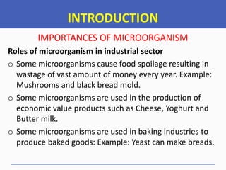


![INTRODUCTION
IMPORTANCES OF MICROORGANISM
Roles of microorganisms in Biotechnology
o Scientists may use recombinant DNA technology to
produce various biological (Biomolecules) and chemical
products. Example: Insulin production, Vitamins and
Enzymes production [Streptokinase and Staphylokinase]
NB:
The technology employ the use of Genetic
engineering tools…. Example: Gene cloning.](https://image.slidesharecdn.com/aht04102basicmicrobiology2022-23-250507060159-1ce2252f/85/Basic-Microbiology-Notes-for-Diploma-Students-22-320.jpg)


![INTRODUCTION
Microbial habitat [Microbial Diversity]
Where can you find Microorganisms.?
o Soil
o Water
o Atmosphere or Air
o Other habitats [Extreme environments]
Examples of Extreme environments
High [Thermophiles] and low [Psychrophiles] temperature
Acidic [Acidophiles] and Basic [Basophiles] habitats:
High Water levels [Metalophiles] and Pressure [Barophiles]](https://image.slidesharecdn.com/aht04102basicmicrobiology2022-23-250507060159-1ce2252f/85/Basic-Microbiology-Notes-for-Diploma-Students-25-320.jpg)


![Microorganisms
Examples
• Bacteria [Measures: 0.2 – 1.5 μm diameter, and
3-5 μm length]
• Fungi
• Virus
• Mycoplasma
• Rickettsiae
• Chlamydia and
• Protozoa](https://image.slidesharecdn.com/aht04102basicmicrobiology2022-23-250507060159-1ce2252f/85/Basic-Microbiology-Notes-for-Diploma-Students-28-320.jpg)


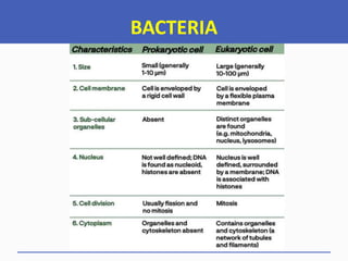


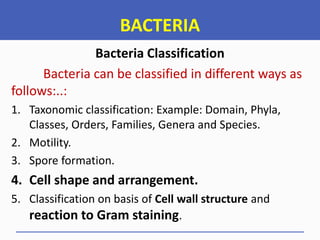
![BACTERIA
Bacteria Classification
Taxonomic classification: Example: Domain, Phyla, Classes,
Orders, Families, Genera and Species.
………..Bacteria taxonomy consist of Classification,
Nomenclature and Identification of Bacteria.
………..All organisms on Earth originated from the Common
Ancestor, a Super-kingdom called Domain.
………..The three “DOMAIN OF LIFE” are Bacteria [Formally,
Eubacter], Eukarya and Archaea.
…………The “DOMAIN OF LIFE” is also known as
“PHYLOGENETIC TREE OF LIFE”](https://image.slidesharecdn.com/aht04102basicmicrobiology2022-23-250507060159-1ce2252f/85/Basic-Microbiology-Notes-for-Diploma-Students-37-320.jpg)


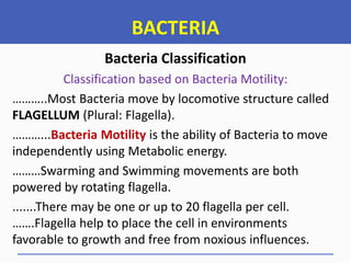


![BACTERIA
Bacteria Classification
Classification based on Bacteria Motility:
....... Note that: The Pili or Fimbriae [Have subunit called Pilin] is not
involved in movement but it is used in:
a. Cell Attachment and Protection
b. Sex Pili [Conjugation]
Types of Flagella
1. Monotrichous flagella
2. Amphitrichous flagella
3. Lophotrichous flagella
4. Peritrichous flagella](https://image.slidesharecdn.com/aht04102basicmicrobiology2022-23-250507060159-1ce2252f/85/Basic-Microbiology-Notes-for-Diploma-Students-44-320.jpg)
![BACTERIA
Bacteria Classification
Classification based on Bacteria Motility:
The Arrangement of Flagella
Type of Flagella Number and arrangement Example
1 Atrichous No flagella -
2 Monotrichous Single flagella on one side Vibrio cholerae
3 Lophotrichous Tuft of flagella on one side
[Multiple polar flagella]
Bartonella bacillifornis
4 Amphitrichous Single or tuft on both sides Spirillum serpens
5 Peritrichous Surrounded by lateral flagella Escherichia coli](https://image.slidesharecdn.com/aht04102basicmicrobiology2022-23-250507060159-1ce2252f/85/Basic-Microbiology-Notes-for-Diploma-Students-45-320.jpg)


![BACTERIA
Bacteria Classification
Classification based on Spore formation:
SPORES [Endospore Structure]
…….Bacterial spores are
resistant to ordinary
Boiling, Disinfectants, and
Heating.
…….Spores of all veterinary
important Bacteria are
destroyed by Autoclaving at
121oC for
15 minutes.
……The process of conversion of
a spore into Vegetative
Cell under suitable conditions is
known as Germination.
……The germination process
occurs in three stages:
Activation,
Initiation, and Outgrowth.](https://image.slidesharecdn.com/aht04102basicmicrobiology2022-23-250507060159-1ce2252f/85/Basic-Microbiology-Notes-for-Diploma-Students-49-320.jpg)
![BACTERIA
Bacteria Classification
Classification based on Spore formation:
SPORES [Endospore Structure]](https://image.slidesharecdn.com/aht04102basicmicrobiology2022-23-250507060159-1ce2252f/85/Basic-Microbiology-Notes-for-Diploma-Students-50-320.jpg)
![BACTERIA
Bacteria Classification
Classification based on Spore formation:
SPORES [Endospore Structure]](https://image.slidesharecdn.com/aht04102basicmicrobiology2022-23-250507060159-1ce2252f/85/Basic-Microbiology-Notes-for-Diploma-Students-51-320.jpg)
![BACTERIA
Bacteria Classification
Classification based on Spore formation:
SPORES [Endospore Structure]
…….Bacterial Spores shape,
location and arrangement
differ…….. Example:
o Central: Spindal shape
o Subterminal: Club shape
o Oval terminal: Tennis racket
shape
o Spherical terminal:
Drumstick appearance](https://image.slidesharecdn.com/aht04102basicmicrobiology2022-23-250507060159-1ce2252f/85/Basic-Microbiology-Notes-for-Diploma-Students-52-320.jpg)
![BACTERIA
SUMMARY
PHYSICAL CHARACTERISTICS OF BACTERIA:
Bacteria can be characterized according to their
physical appearance due to the presence of the following
common structures: [Explain the function of each]
A. Capsule [Slime layers and Glycocalyx]. (Attachment)
B. Pili {Fimbriae [Common pili] and Sex Pili}
C. Flagella. (For Movement)
D. Spores and (For protection in unfavorable environment)
E. Cell wall. {For cell protection and shape (Most Bacteria)}](https://image.slidesharecdn.com/aht04102basicmicrobiology2022-23-250507060159-1ce2252f/85/Basic-Microbiology-Notes-for-Diploma-Students-53-320.jpg)

![BACTERIA
Bacteria Classification
Classification based on Cell Shape and
Arrangement:
Bacteria vary in their shape and arrangement of cells.
Depending on their shape, Bacteria are classified into
Three main types.
1. SPHERES or ROUND: These are called Cocci.
2. ELONGATED or RODS: These are called Bacilli.
3. SPIRALS: These are Vibrios [Coma], Spirilla [Rigid
spiral] and Spirochaetes [Filamentous spiral].](https://image.slidesharecdn.com/aht04102basicmicrobiology2022-23-250507060159-1ce2252f/85/Basic-Microbiology-Notes-for-Diploma-Students-56-320.jpg)
![BACTERIA
Bacteria Classification
Classification based on Cell Shape and
Arrangement:
4. ACTINOMYCETES: Are Branching filamentous Bacteria,
so called because of a fancied resemblance to the
radiating rays of the sun when seen in tissue lesions
*From ‘actis’ meaning ray and ‘mykes’ means fungus+.
5. MYCOPLASMA: Are Bacteria that are cell wall deficient
and hence do not possess a stable morphology [They
are Pleomorphic]. They occur as Round or Oval bodies
and as Interlacing filaments.](https://image.slidesharecdn.com/aht04102basicmicrobiology2022-23-250507060159-1ce2252f/85/Basic-Microbiology-Notes-for-Diploma-Students-57-320.jpg)
![BACTERIA
Bacteria Classification
Classification based on Cell Shape and
Arrangement:
Bacteria Cell arrangement of different shapes can form
structures which looks like as follow:
1. Single. Example: Coccus, Bacillus and Coccobacillus [Oval]
2. Pair. Example: Diplococci and Diplobacilli.
3. Chain. Example: Streptococci and Streptobacillus.
4. Clusters. Example: Staphylococci.
5. Tetrad (Four)and Sarcina (Eight).](https://image.slidesharecdn.com/aht04102basicmicrobiology2022-23-250507060159-1ce2252f/85/Basic-Microbiology-Notes-for-Diploma-Students-58-320.jpg)
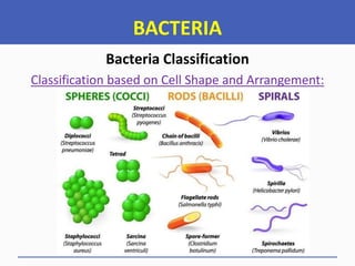

![BACTERIA
Bacteria Classification
Classification based on Cell wall structure and
reaction to Gram stain:
• Bacteria Cell wall is made up of Peptidoglycan [PG].
• Peptidoglycan is essential protective barrier for bacterial
cells that encapsulates the cytoplasmic membrane of
Bacteria.
• Peptidoglycan is rigid, highly conserved, complex
structure of polymeric carbohydrates and amino acids.
• Bacteria cell wall also helps maintain cell shape, which is
used for Growth, Locomotion and Reproduction](https://image.slidesharecdn.com/aht04102basicmicrobiology2022-23-250507060159-1ce2252f/85/Basic-Microbiology-Notes-for-Diploma-Students-61-320.jpg)
![BACTERIA
Bacteria Classification
Classification based on Cell wall structure and
reaction to Gram stain:
• Bacteria cell wall shape is used to distinguish between
Gram Positive (G+) and Gram Negative (G-) Bacteria.
• The cell wall cannot be seen by direct light microscopy
and does not stain with simple stains.
• The presence and absence of some molecules such as
Teichoic acid [Antigen determinant and Ion transport]
and Lipids makes Gram staining possible to differentiate
between Gram negative and Gram Positive Bacteria.](https://image.slidesharecdn.com/aht04102basicmicrobiology2022-23-250507060159-1ce2252f/85/Basic-Microbiology-Notes-for-Diploma-Students-62-320.jpg)


![BACTERIA
Bacterial Growth Curve
Well Sketched Bacterial Growth Curve [Population Dynamics]](https://image.slidesharecdn.com/aht04102basicmicrobiology2022-23-250507060159-1ce2252f/85/Basic-Microbiology-Notes-for-Diploma-Students-75-320.jpg)


![BACTERIA
Bacterial Growth Curve
EXPLANATIONS
o DECLINE Phase: At this stage, there is an
exponential decrease in number of living
Bacterial cells. However, some Bacteria may
remain viable during this stage.
Why.?
This is the phase when the population
decreased due to Cell death.
Involution [Swollen] forms are common in ageing cultures](https://image.slidesharecdn.com/aht04102basicmicrobiology2022-23-250507060159-1ce2252f/85/Basic-Microbiology-Notes-for-Diploma-Students-79-320.jpg)

![BACTERIA
Bacterial Growth Curve
EXPLANATIONS
Summary:
The maximum cell size is obtained towards the
end of the Lag Phase.
In the Log Phase, cells are smaller.
Sporulation [Spore formation] occurs at
Stationary Phase. Many Bacteria also produce
secondary metabolic products such as Exotoxins
and Antibiotics.](https://image.slidesharecdn.com/aht04102basicmicrobiology2022-23-250507060159-1ce2252f/85/Basic-Microbiology-Notes-for-Diploma-Students-81-320.jpg)
![BACTERIA
FACTORS AFFECTING GROWTH OF BACTERIA
Many Environmental factors affect the
generation time [Doubling] of the organism like
Nutrients, Temperature, Oxygen, Carbon dioxide,
Light, pH, Moisture and Salt [Osmotic pressure].
Meaning of Bacteria Generation time.
Bacterial generation time is the time taken
for a population to double in number during Log-
phase….. Generally, it refers to the time required
for a Bacteria cell to divide…. *Formula, G = t/n]](https://image.slidesharecdn.com/aht04102basicmicrobiology2022-23-250507060159-1ce2252f/85/Basic-Microbiology-Notes-for-Diploma-Students-82-320.jpg)


![BACTERIA
FACTORS AFFECTING GROWTH OF BACTERIA
Nutrients
Bacteria on the basis of their source of
Carbon in nutrition, they can be classified in to:-
i. Autotrophs. Example: Photoautotrophs
[Cyanobacteria, Purple and Green Bacteria]
ii. Heterotrophs. Example: Photoheterotrophs
[Purple non-sulfur Bacteria] and
Chemoheterotrophs [Nitrifying Bacteria, Iron
oxidizing Bacteria].](https://image.slidesharecdn.com/aht04102basicmicrobiology2022-23-250507060159-1ce2252f/85/Basic-Microbiology-Notes-for-Diploma-Students-85-320.jpg)


![BACTERIA
FACTORS AFFECTING GROWTH OF BACTERIA
Oxygen
Based on Oxygen requirements, Bacteria can
be classified in to different groups as follow:
Microaerophilic Bacteria
These are the group of Bacteria that can grow
in the presence of low oxygen [Levels Below 0.2
atm]. No growth under anaerobic environment
Example: Campylobacter jejuni](https://image.slidesharecdn.com/aht04102basicmicrobiology2022-23-250507060159-1ce2252f/85/Basic-Microbiology-Notes-for-Diploma-Students-90-320.jpg)

![BACTERIA
FACTORS AFFECTING GROWTH OF BACTERIA
Oxygen
Based on Oxygen requirements, Bacteria can
be classified in to different groups as follow:
Obligate Anaerobes
Why.?
Because these Bacteria lack Superoxide dismutase
and Catalase Enzymes therefore Oxygen is Lethal
[Oxygen is Toxic to them]](https://image.slidesharecdn.com/aht04102basicmicrobiology2022-23-250507060159-1ce2252f/85/Basic-Microbiology-Notes-for-Diploma-Students-92-320.jpg)


![BACTERIA
FACTORS AFFECTING GROWTH OF BACTERIA
Carbon dioxide
The organisms that require higher amounts of
Carbon dioxide [CO2] for their growth are called
Capnophilic Bacteria.
……. They grow well in the presence of 5–10% CO2
and 15% O2. In candle jar, 3% CO2 can be achieved.
Example: Brucella abortus and Haemophilus
influenzae.](https://image.slidesharecdn.com/aht04102basicmicrobiology2022-23-250507060159-1ce2252f/85/Basic-Microbiology-Notes-for-Diploma-Students-97-320.jpg)
![BACTERIA
FACTORS AFFECTING GROWTH OF BACTERIA
Carbon dioxide
The organisms that require higher amounts of
Carbon dioxide [CO2] for their growth are called
Capnophilic Bacteria.
……. Note:
Sometimes, some Micro-aerophiles can
tolerate low concentration of CO2 at the amount of
below 4%.](https://image.slidesharecdn.com/aht04102basicmicrobiology2022-23-250507060159-1ce2252f/85/Basic-Microbiology-Notes-for-Diploma-Students-98-320.jpg)


![BACTERIA
FACTORS AFFECTING GROWTH OF BACTERIA
Temperature
Thermophiles
These are heat loving microbes.
They can grow at a high temperature range of 55
– 80oC
Minimum temperature = 45, Maximum
temperature = Above 100 [Water boiling]: Average
= 50 - 85](https://image.slidesharecdn.com/aht04102basicmicrobiology2022-23-250507060159-1ce2252f/85/Basic-Microbiology-Notes-for-Diploma-Students-103-320.jpg)

![BACTERIA
FACTORS AFFECTING GROWTH OF BACTERIA
pH [Hydrogen Ion Concentration]
Most pathogenic bacteria grow between pH
7.2 and 7.6.
…….Very few Bacteria, such as Lactobacilli, can
grow at acidic pH below 4.0.
…….Many food items, such as Pickles and Cheese,
are prevented from spoilage by acids produced
during fermentation.](https://image.slidesharecdn.com/aht04102basicmicrobiology2022-23-250507060159-1ce2252f/85/Basic-Microbiology-Notes-for-Diploma-Students-105-320.jpg)

![BACTERIA
FACTORS AFFECTING GROWTH OF BACTERIA
Osmotic Pressure
o Sudden exposure of bacteria to hypertonic
solution may cause osmotic withdrawal of water,
leading to osmotic shrinkage of the protoplasm
[Plasmolysis].
o On the other hand, sudden transfer of Bacteria
from concentrated solution to distilled water
may cause excessive imbibition of water leading
to swelling and bursting of cell [Plasmoptysis].](https://image.slidesharecdn.com/aht04102basicmicrobiology2022-23-250507060159-1ce2252f/85/Basic-Microbiology-Notes-for-Diploma-Students-107-320.jpg)
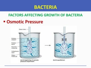


![FUNGI
Features of Fungus in common
They have filamentous system of Cells with
Apical growth, Lateral branching and
heterotrophic nutrition.
They are characterized by a life cycle that begins
with Germination from Spores [Resting
structure], followed by a period of Growth and
the substrate is exploited to produce a Biomass.
Finally, there is a period of Sporulation
[Dissemination from the parent Mycelium]](https://image.slidesharecdn.com/aht04102basicmicrobiology2022-23-250507060159-1ce2252f/85/Basic-Microbiology-Notes-for-Diploma-Students-114-320.jpg)
![FUNGI
General Characteristics of Fungi
They are Eukaryotic, Non-vascular organisms.
They have an Alternation of generation.
Reproduce by means of Spores [Usually, Wind
dissemination].
Both Sexual [Meiosis] and Asexual [Mitosis]
Spores may be produced, depending on the
species and condition.
They are typically non motile, although a few
have a motile phase. Example: Chytrids.](https://image.slidesharecdn.com/aht04102basicmicrobiology2022-23-250507060159-1ce2252f/85/Basic-Microbiology-Notes-for-Diploma-Students-115-320.jpg)

![FUNGI
Economic Importance of Fungi
Used in Fermentation industry [Anaerobic
process+…..Wine, Beer, Bread and Soy sauce.
Drug manufacturing….Antibiotics *Griseofulvin,
Penicillin and Cyclosporin]
Degradation of Complex organic materials in to
simple forms. Ecologically important process.
Fungi-Plant Symbiosis [Mycorrhizae]
They cause diseases. *Mycoses+… Systemic,
Subcutaneous, cutaneous and Opportunistic](https://image.slidesharecdn.com/aht04102basicmicrobiology2022-23-250507060159-1ce2252f/85/Basic-Microbiology-Notes-for-Diploma-Students-117-320.jpg)
![FUNGI
Classes of Fungi
o Algal (Lower) Fungi: All are microscopic and
grow in water and damp soil. Example: Rhizopus.
o Sac Fungi: They grow in decaying citrus fruits, in
jellies and on leather. Examples: Yeasts [Candida
albacans] and Moulds [Blue and Green moulds]
o Imperfect Fungi: Fungi that grow in mildew walls
and spot clothes, as well as those that cause
plant diseases: Example: Mould [Fusarium]
o Club [Basidium] Fungi: Example: Mushrooms](https://image.slidesharecdn.com/aht04102basicmicrobiology2022-23-250507060159-1ce2252f/85/Basic-Microbiology-Notes-for-Diploma-Students-118-320.jpg)


![FUNGI
Reproduction in Fungi
o Asexual reproduction: It’s done by the
fragmentation of Hyphae.
The group of Fungi with NO SEXUAL
Reproduction is called Deuteromycetes [Imperfect fungi]
……In Asexual reproduction, Progeny is identical to
Parent.
…….Example: Asexual Spores, Hyphae
fragmentation and Budding.](https://image.slidesharecdn.com/aht04102basicmicrobiology2022-23-250507060159-1ce2252f/85/Basic-Microbiology-Notes-for-Diploma-Students-125-320.jpg)

![FUNGI
Reproduction in Fungi
Types of Asexual spores
Hypha can fragment to form cells that behave as
Spores. These cells are called Arthroconidiae or
Arthrospore.
If a cell is surrounded by a thick wall before
separation, they are called Chlamydospores.
If a Spore develop within a Sac [Sporangium] at a
hyphal tip, they are called Sporangiospore.](https://image.slidesharecdn.com/aht04102basicmicrobiology2022-23-250507060159-1ce2252f/85/Basic-Microbiology-Notes-for-Diploma-Students-127-320.jpg)
![FUNGI
Reproduction in Fungi
Types of Asexual spores
If a Spores are not enclosed in a Sac but
produced at the tips or sides of the hypha, they
are called Conidiospores.
A Parent cell can divide in to two daughter cells
by central constriction and formation of new cell
wall. It forms Conidia [Macro and Micro-conidia]
Spores produced by vegetative mother cell by
Budding are called Blastospore.](https://image.slidesharecdn.com/aht04102basicmicrobiology2022-23-250507060159-1ce2252f/85/Basic-Microbiology-Notes-for-Diploma-Students-128-320.jpg)


![FUNGI
Reproduction in Fungi
….Note
• Both Sexual and Asexual reproduction of Fungi
yields SPORES [Sexual spores and Asexual
Spores]
• Fungal Spores are different from Bacteria
Endospore. Endospore are not for Reproduction.
• Fungal Spores are for Reproduction: They do not
ensure resistance to environmental conditions.](https://image.slidesharecdn.com/aht04102basicmicrobiology2022-23-250507060159-1ce2252f/85/Basic-Microbiology-Notes-for-Diploma-Students-137-320.jpg)


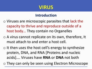


![VIRUS
Introduction
Entry of viruses into the host cells has
several stages including:
o Attachment to the host cell surface
o Internalization into the host cell [Entry: Direct
penetration, Membrane fusion and Endocytosis]
o Uncoating (Genetic materials are released out)
o Synthesis
o Assembly and Release](https://image.slidesharecdn.com/aht04102basicmicrobiology2022-23-250507060159-1ce2252f/85/Basic-Microbiology-Notes-for-Diploma-Students-147-320.jpg)

![VIRUS
Virus Classification
Criteria for Classifying Viruses
i. Based on their Genome composition
ii. Baltimore classification of Viruses
iii. Based on Structure or Morphology
iv. Based on Symmetry of the Capsid [Capsid
protect Nucleic acids and aid in transfer to host]
v. Based on the host on host species [Holmes
classification]](https://image.slidesharecdn.com/aht04102basicmicrobiology2022-23-250507060159-1ce2252f/85/Basic-Microbiology-Notes-for-Diploma-Students-149-320.jpg)


![VIRUS
Virus Classification
Criteria for Classifying Viruses
Holmes Classification
o Phagina [Bacteria]: Example: Bacteriophages
o Phytophagina [Plants]: Geminiviridae, Tobaco
Mosaic Virus, Cassava Mosaic Virus.
o Zoophagina [Animals]: Retroviridae,
Orthomyxoviridae, Poxviridae, Human
Immunodeficiency Virus, Canine Parvovirus](https://image.slidesharecdn.com/aht04102basicmicrobiology2022-23-250507060159-1ce2252f/85/Basic-Microbiology-Notes-for-Diploma-Students-160-320.jpg)
![VIRUS
Virus Classification
Criteria for Classifying Viruses
The International Committee on Taxonomy of
Viruses [ICTV]
o Is a committee which is authorized and organizes
the taxonomic classification of Viruses.](https://image.slidesharecdn.com/aht04102basicmicrobiology2022-23-250507060159-1ce2252f/85/Basic-Microbiology-Notes-for-Diploma-Students-161-320.jpg)
![VIRUS
The International Committee on Taxonomy of
Viruses [ICTV]](https://image.slidesharecdn.com/aht04102basicmicrobiology2022-23-250507060159-1ce2252f/85/Basic-Microbiology-Notes-for-Diploma-Students-162-320.jpg)


![RICKETTSIA
General Properties
o They posses both RNA and DNA.
o They have Peptidoglycan, but not Teichoic acid in
their cell wall.
o They multiply by Transverse binary fission.
o They are Susceptible to Antibacterial agents.
o They require blood sucking vectors for part of
their natural life cycle. Example: Arthropods.
o They cause diseases, some of which can be fatal.
[Example: Heart water diseases and Q-Fever]](https://image.slidesharecdn.com/aht04102basicmicrobiology2022-23-250507060159-1ce2252f/85/Basic-Microbiology-Notes-for-Diploma-Students-175-320.jpg)


![RICKETTSIA
General Properties
Why are Rickettsia obligate intracellular.?
They have no own ATP [Energy]
They have a relatively permeable
membrane [They lack Teichoic acid]. This
means that, they need the dense cytoplasm
of the host cell for their osmotic stability.](https://image.slidesharecdn.com/aht04102basicmicrobiology2022-23-250507060159-1ce2252f/85/Basic-Microbiology-Notes-for-Diploma-Students-179-320.jpg)


![CHLAMYDIA
Introduction
Chlamydiaceae is the family of Obligate
intracellular Gram –ve Bacteria Organisms, closely
similar to Rickettsia.
The two Genera of Pathogenic importance are:-
……Genus Chlamydia: Several species including
Chlamydia trachomatis which cause trachoma and
a Venereal condition in human called
Lymphogranuloma vanerum [LGV]](https://image.slidesharecdn.com/aht04102basicmicrobiology2022-23-250507060159-1ce2252f/85/Basic-Microbiology-Notes-for-Diploma-Students-183-320.jpg)
![CHLAMYDIA
Classification
Chlamydiaceae is the family of Obligate
intracellular Gram –ve Bacteria Organisms, closely
similar to Rickettsia. They depend on host cells for
Energy [ATP] Production
The two Genera of Pathogenic importance are:-
……Genus Chlamydophila: Several species
including Chlamydia psittaci, the causal agent of
zoonotic disease called Psittacosis [Synonym:
Ornithosis or Parrot fever]](https://image.slidesharecdn.com/aht04102basicmicrobiology2022-23-250507060159-1ce2252f/85/Basic-Microbiology-Notes-for-Diploma-Students-184-320.jpg)

![CHLAMYDIA
Morphology
They are Pleimorphic [Spherical to Coccoid
bodies]
Elementary bodies and Reticulate bodies [Exist in
two distinct form within the cytoplasm] like
Rickettsiae
They [Reticulate bodies] multiply by Binary fission
Transmission occur by Direct contact [In
Trachoma or Inclusion conjunctivitis] and
inhalation [Psittacosis]](https://image.slidesharecdn.com/aht04102basicmicrobiology2022-23-250507060159-1ce2252f/85/Basic-Microbiology-Notes-for-Diploma-Students-186-320.jpg)

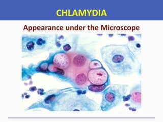


![MYCOPLASMA
General Characteristics
They Spread by direct contact or fresh
respiratory droplets.
They bind to the exterior of cells and damage
epithelium from the outside.
Fastidious and slow to grow even on special
cholesterol-containing medium, so diagnosis is
usually clinical or serological with PCR becoming
more readily available.
They are consequently placed in a separate class
Mollicutes [mollis, soft; cutis, skin]](https://image.slidesharecdn.com/aht04102basicmicrobiology2022-23-250507060159-1ce2252f/85/Basic-Microbiology-Notes-for-Diploma-Students-192-320.jpg)


![MYCOPLASMA
Diseases caused by Mycoplasma
o Mycoplasma pneumoniae causes respiratory
tract infections. Examples: Contagious
Bovine/Caprine Pleuropneumonia [CBPP/CCPP]
o Mycoplasma hominis and Ureaplasma
urealyticum appear to be involved in
genitourinary tract infections.](https://image.slidesharecdn.com/aht04102basicmicrobiology2022-23-250507060159-1ce2252f/85/Basic-Microbiology-Notes-for-Diploma-Students-195-320.jpg)
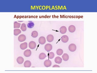
![MYCOPLASMA
Difference between Mycoplasma and Typical
Bacteria
MYCOPLASMA TYPICAL BACTERIA
Define
Is a Bacterial genus which does not
contain a cell wall
Bacteria are Microscopic prokaryotic
organisms found everywhere on
earth
Shape
They are mostly spherical to
filamentous
They show different shapes such as
Coccus, Bacillus and Spirillum
Change in
Shape
Mycoplasma is highly Pleomorphic
[They do not posses a definite shape]
Bacterial cell possess a definite
shape due to the presence of a rigid
cell wall
Size of the
Genome
Mycoplasma is considered as the
smallest Bacteria with small Genome
Bacterial genome size varies
according to the species](https://image.slidesharecdn.com/aht04102basicmicrobiology2022-23-250507060159-1ce2252f/85/Basic-Microbiology-Notes-for-Diploma-Students-197-320.jpg)


![MICROBIAL CULTURE
Introduction
o Microorganisms require a constant nutrient
supply for them to survive and grow.
o Naturally, they acquire nutrients from their
surroundings [free-living] or from a host
[parasites].
o Artificial media is used to grow Microorganisms
in a lab [in vitro]
o Artificial media provides the basic nutrients
required by the Microorganisms to grow.](https://image.slidesharecdn.com/aht04102basicmicrobiology2022-23-250507060159-1ce2252f/85/Basic-Microbiology-Notes-for-Diploma-Students-201-320.jpg)

![MICROBIAL CULTURE
Requirements for Bacteria & Fungus Cultivation
Culture Media
Microbiological Incubator
Autoclave
Water bath
Refrigerator
Weighing balance
Conical flask and Measuring cylinder
Petri dishes [Plates] and Canister [For carying Petri dish]
Distilled water or Deionized water](https://image.slidesharecdn.com/aht04102basicmicrobiology2022-23-250507060159-1ce2252f/85/Basic-Microbiology-Notes-for-Diploma-Students-203-320.jpg)
![MICROBIAL CULTURE
Requirements for Bacteria & Fungus Cultivation
Universal , Bijӧur and McCartney bottles.
Nickrom wire [Inoculating needle and inoculating loop]
Cool box with Ice packs
Washing bottle
Anaerobic jar
Candle jar
pH meter
Magnetic steer, glass rod and hot plate
Spatula, Test tubes and alluminium foil](https://image.slidesharecdn.com/aht04102basicmicrobiology2022-23-250507060159-1ce2252f/85/Basic-Microbiology-Notes-for-Diploma-Students-204-320.jpg)
![MICROBIAL CULTURE
Requirements for Bacteria Cultivation
….…….Functions or Uses……..
Culture Media
Is a mixture of various nutrients that is
suitable for the growth of Microorganisms.
Weighing balance [Electronic top-pan Balance]
Used for weighing large quantities of media
and other chemicals when precise weighing is not
much important.](https://image.slidesharecdn.com/aht04102basicmicrobiology2022-23-250507060159-1ce2252f/85/Basic-Microbiology-Notes-for-Diploma-Students-205-320.jpg)


![MICROBIAL CULTURE
Requirements for Bacteria Cultivation
….…….Functions or Uses……..
Measuring Cylinder
Is used for measuring distilled water and
other liquid substances.
Conical flasks or Boiling bottles
Used for putting the mixture of agar and
distilled water for autoclaving [Sterilization]](https://image.slidesharecdn.com/aht04102basicmicrobiology2022-23-250507060159-1ce2252f/85/Basic-Microbiology-Notes-for-Diploma-Students-210-320.jpg)


![MICROBIAL CULTURE
Requirements for Bacteria Cultivation
….…….Functions or Uses……..
pH Meter
For measuring pH of the media
Hot plate and magnetic steer
For heating and mixing media [To dissolve]
Spatula and alluminium foil
For transferring and holding media in a balance](https://image.slidesharecdn.com/aht04102basicmicrobiology2022-23-250507060159-1ce2252f/85/Basic-Microbiology-Notes-for-Diploma-Students-214-320.jpg)


![MICROBIAL CULTURE
Requirements for Bacteria Cultivation
Anaerobic
Jar
[GasPak
System]](https://image.slidesharecdn.com/aht04102basicmicrobiology2022-23-250507060159-1ce2252f/85/Basic-Microbiology-Notes-for-Diploma-Students-218-320.jpg)


![MICROBIAL CULTURE
CULTURE MEDIA
Types of Culture media used in Bacteriology
Culture media can be classified depending on two
major criteria
i. Based on Consistence [Physical state] and
ii. Based on Chemical composition and use
Routine laboratory media
Synthetic media: These are chemically
defined media prepared from pure
chemical substances, commonly used in
research work.](https://image.slidesharecdn.com/aht04102basicmicrobiology2022-23-250507060159-1ce2252f/85/Basic-Microbiology-Notes-for-Diploma-Students-222-320.jpg)
![MICROBIAL CULTURE
CULTURE MEDIA
Types of Culture media used in Bacteriology
Based on Consistence [Physical state]
Solid medium
It contain 1.5 – 2.5 % Agar. It is used for
isolation and identification of Bacteria.
Advantages
Bacteria can be identified by studying the
colony character.
Mixed bacteria can be separated.](https://image.slidesharecdn.com/aht04102basicmicrobiology2022-23-250507060159-1ce2252f/85/Basic-Microbiology-Notes-for-Diploma-Students-223-320.jpg)
![MICROBIAL CULTURE
CULTURE MEDIA
Types of Culture media used in Bacteriology
Based on Consistence [Physical state]
Solid medium
It contain 1.5 – 2.5 % Agar.
Isolation of Pure Culture and
Identification of Microorganism](https://image.slidesharecdn.com/aht04102basicmicrobiology2022-23-250507060159-1ce2252f/85/Basic-Microbiology-Notes-for-Diploma-Students-224-320.jpg)
![MICROBIAL CULTURE
CULTURE MEDIA
Types of Culture media used in Bacteriology
Based on Consistence [Physical state]
Agar:
'Agar' is most commonly used to prepare
solid media. Agar is polysaccharide extract
obtained from seaweed. Agar is an ideal
solidifying agent as it is : Agar is Bacteriologically
inert [no influence on Bacterial growth]. It remains
solid at 37°C, and it is transparent.](https://image.slidesharecdn.com/aht04102basicmicrobiology2022-23-250507060159-1ce2252f/85/Basic-Microbiology-Notes-for-Diploma-Students-225-320.jpg)
![MICROBIAL CULTURE
CULTURE MEDIA
Types of Culture media used in Bacteriology
Based on Consistence [Physical state]
Semi - Solid medium
It contain 0.3 – 0.5 % Agar.
Advantages
It is used for observation of Bacteria motility.
It is also used for the preservation of Bacteria.](https://image.slidesharecdn.com/aht04102basicmicrobiology2022-23-250507060159-1ce2252f/85/Basic-Microbiology-Notes-for-Diploma-Students-226-320.jpg)
![MICROBIAL CULTURE
CULTURE MEDIA
Types of Culture media used in Bacteriology
Based on Consistence [Physical state]
Semi - Solid medium
It contain 0.3 – 0.5 % Agar.
Observation of Bacteria motility](https://image.slidesharecdn.com/aht04102basicmicrobiology2022-23-250507060159-1ce2252f/85/Basic-Microbiology-Notes-for-Diploma-Students-227-320.jpg)
![MICROBIAL CULTURE
CULTURE MEDIA
Types of Culture media used in Bacteriology
Based on Consistence [Physical state]
Liquid medium
It does not contain Agar. It is used for profuse
growth [Proliferation of Bacteria].
Disadvantage
Mixed organisms cannot be separated.](https://image.slidesharecdn.com/aht04102basicmicrobiology2022-23-250507060159-1ce2252f/85/Basic-Microbiology-Notes-for-Diploma-Students-228-320.jpg)
![MICROBIAL CULTURE
CULTURE MEDIA
Types of Culture media used in Bacteriology
Based on Consistence [Physical state]
Liquid medium
Observations
• Pellicle: When a mass of organisms is floating on top
of the broth.
• Turbidity: When the organisms appear as a general
cloudiness throughout the broth.
• Sediment: When a mass of organisms appears as a
deposit at the bottom of the tube.](https://image.slidesharecdn.com/aht04102basicmicrobiology2022-23-250507060159-1ce2252f/85/Basic-Microbiology-Notes-for-Diploma-Students-229-320.jpg)
![MICROBIAL CULTURE
CULTURE MEDIA
Types of Culture media used in Bacteriology
Based on Consistence [Physical state]
Liquid medium
Observations](https://image.slidesharecdn.com/aht04102basicmicrobiology2022-23-250507060159-1ce2252f/85/Basic-Microbiology-Notes-for-Diploma-Students-230-320.jpg)
![MICROBIAL CULTURE
CULTURE MEDIA
Types of Culture media used in Bacteriology
Based on Chemical composition and use
The composition of a medium is depended on
the nutritional requirement of the microorganism
to be cultivated.
Copiotrophic Microorganisms: They require
medium rich in Nutrients: [Complete Medium], Most
Pathogenic Bacteria prefer this condition.
Oligotrophic microorganisms: They require low
concentration of Nutrients.](https://image.slidesharecdn.com/aht04102basicmicrobiology2022-23-250507060159-1ce2252f/85/Basic-Microbiology-Notes-for-Diploma-Students-231-320.jpg)

![MICROBIAL CULTURE
CULTURE MEDIA
Types of Culture media used in Bacteriology
Based on Chemical composition and use
Routine laboratory media
- Basal media
Basal media are those that may be used for growth
[culture] of Microorganisms that do not need enrichment
of the media.
Examples: Nutrient broth, Nutrient agar and
Peptone water [ Staphylococcus and Enterobacteriaceae
grow in these media] and Saboraud Dextrose Agar [Fungus]](https://image.slidesharecdn.com/aht04102basicmicrobiology2022-23-250507060159-1ce2252f/85/Basic-Microbiology-Notes-for-Diploma-Students-233-320.jpg)


![MICROBIAL CULTURE
CULTURE MEDIA
Types of Culture media used in Bacteriology
Based on Chemical composition and use
Routine laboratory media
- Selective media
These media favor the growth of a particular
Bacterium by inhibiting the growth of undesired Bacteria
and allowing growth of desirable Bacteria. Antibiotics may
be added to a medium for inhibition.
Examples: MacConkey agar, Lowenstein-Jensen media, Tellurite
Media, Brilliant Green Agar, Xylose lysine Deoxycholate [XLD] Agar
and PALCAM Agar (For Listeria Monocytogens).](https://image.slidesharecdn.com/aht04102basicmicrobiology2022-23-250507060159-1ce2252f/85/Basic-Microbiology-Notes-for-Diploma-Students-238-320.jpg)

![MICROBIAL CULTURE
CULTURE MEDIA
Types of Culture media used in Bacteriology
Based on Chemical composition and use
Routine laboratory media
- Selective media
Polymixin Acriflavin Lithium Chloride Ceftazidine Aesculin Mannihot Agar [PALCAM]](https://image.slidesharecdn.com/aht04102basicmicrobiology2022-23-250507060159-1ce2252f/85/Basic-Microbiology-Notes-for-Diploma-Students-240-320.jpg)
![MICROBIAL CULTURE
CULTURE MEDIA
Types of Culture media used in Bacteriology
Based on Chemical composition and use
Routine laboratory media
- Differential or Indicator media
An indicator is included in the medium. Example of
indicators are Blood, neutral red and tellurite. It
differentiates between different organisms growing on the
same plate on the basis of colonial morphology.
Examples: Blood agar, Salmonella-Shigella [SS] agar, Eosin
Methylene Blue [EMB] agar and MacConkey agar.](https://image.slidesharecdn.com/aht04102basicmicrobiology2022-23-250507060159-1ce2252f/85/Basic-Microbiology-Notes-for-Diploma-Students-241-320.jpg)


![MICROBIAL CULTURE
CULTURE MEDIA PREPARATION
CALCULATIONS
Culture media from different manufactures
(Especially Solid media) comes with directions for use.
Most of the time, the manufacturer recommend a certain
amount of Agar powder to be mixed with one litre [1 Litre]
of Distilled Water.
………Because it’s not necessary to use the entire
recommended amount, we can prepare the culture media
to be used depends on the need by using simple
calculations to get the correct weight of Agar and D. Water](https://image.slidesharecdn.com/aht04102basicmicrobiology2022-23-250507060159-1ce2252f/85/Basic-Microbiology-Notes-for-Diploma-Students-252-320.jpg)
![MICROBIAL CULTURE
CULTURE MEDIA PREPARATION
CALCULATIONS
Example 1
You are required to prepare 12g of culture media for
the cultivation of a certain Bacteria. The stock media
directions are 24g to be dissolved in 1L of Distilled water.
What is the amount of distilled water will you need?
Solution: 24g in 1000ml 12g x 1000ml = 24g x ? ml
12g in ____ml? 24g 24g
Therefore: 500 mls [0.5 L] of Distilled water will be needed](https://image.slidesharecdn.com/aht04102basicmicrobiology2022-23-250507060159-1ce2252f/85/Basic-Microbiology-Notes-for-Diploma-Students-253-320.jpg)


![CULTURE METHODS
Definition of terms
Colony: This refers to a cluster of microorganisms
growing on a solid medium. It is directly visible and
arises from a single cell.
Inoculum: In Microbiology, inoculum is the
suspension of microorganisms to be cultured. It can
be in a suspected sample or from culture in transfer
technique [Pure culture isolation]](https://image.slidesharecdn.com/aht04102basicmicrobiology2022-23-250507060159-1ce2252f/85/Basic-Microbiology-Notes-for-Diploma-Students-259-320.jpg)
![CULTURE METHODS
Definition of terms
Sample: This refers to the small portion of a material
taken from the specimen.
Example of samples that can be used as a source of
Microorganisms [Bacteria or Fungus]
- Urine
- Fecal materials
- Swabs
- Milk
- Blood, lymph
- Saliva, Mucus, Sputum
- Piece of meat and Organs *Liver, Lungs, …+](https://image.slidesharecdn.com/aht04102basicmicrobiology2022-23-250507060159-1ce2252f/85/Basic-Microbiology-Notes-for-Diploma-Students-260-320.jpg)
![CULTURE METHODS
Culture techniques
Streak plate Method or Surface plating.
o Quadrat streak
o Radiant streak
o Continuous streak
Pour plate method.
o Quantitative method [Serial dilution]
o Qualitative method
Spread plate Method
Liquid culture method
Anaerobic culture method](https://image.slidesharecdn.com/aht04102basicmicrobiology2022-23-250507060159-1ce2252f/85/Basic-Microbiology-Notes-for-Diploma-Students-261-320.jpg)
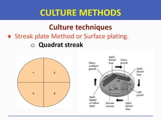


![CULTURE METHODS
Culture techniques
Pour plate method.
o Quantitative method [Serial dilution]
o Qualitative method](https://image.slidesharecdn.com/aht04102basicmicrobiology2022-23-250507060159-1ce2252f/85/Basic-Microbiology-Notes-for-Diploma-Students-267-320.jpg)


![STAINING TECHNIQUES
Staining
Types of Microbiological stains
Simple stains
Differential stains and
Special stains
These are stains that Use a variety of dyes and
techniques to stain particular tissues, structures or
pathogens [such as Bacteria] or to assist pathologists with
tissue-based diagnosis. Example: Malachite green stains
Examples: Flagella staining, Spore staining, Capsule
[Negative] staining: ...... Dyes are Acid fuchsin, Congo red..](https://image.slidesharecdn.com/aht04102basicmicrobiology2022-23-250507060159-1ce2252f/85/Basic-Microbiology-Notes-for-Diploma-Students-277-320.jpg)


![STAINING TECHNIQUES
GRAM’S STAINING
Gram’s staining Procedures
5. While holding the slide at an angle, run Alcohol [Ethyl
alcohol] over the surface until no more color emerges.
It takes about 2 seconds … Be careful at this stage,
avoid Over-decolorization.
6. Wash immediately with water, drain off excess.
7. Flood the slide with Counter stain for 30-45 seconds.
Secondary dye can be Safranin, Carbol fuchsin or
Neutral red.
8. Rinse the slide with a gentle stream of water then blot.](https://image.slidesharecdn.com/aht04102basicmicrobiology2022-23-250507060159-1ce2252f/85/Basic-Microbiology-Notes-for-Diploma-Students-280-320.jpg)


![STAINING TECHNIQUES
ZIEHL-NEELSEN STAINING
[> 30 Sec., >> 15-20 Sec., >>> 30 Sec.]](https://image.slidesharecdn.com/aht04102basicmicrobiology2022-23-250507060159-1ce2252f/85/Basic-Microbiology-Notes-for-Diploma-Students-283-320.jpg)


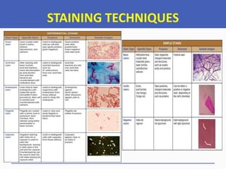


![MICROSCOPE
Types of Microscope
Light Microscope
On the basis of Objectives Orientation
• Upright light Microscope
• Inverted light Microscope
On the Basis of the Number of Lenses used
• Simple light Microscope [Single lenses system]
• Compound light Microscope [Multiple lenses]](https://image.slidesharecdn.com/aht04102basicmicrobiology2022-23-250507060159-1ce2252f/85/Basic-Microbiology-Notes-for-Diploma-Students-295-320.jpg)


![MICROSCOPE
Types of Microscope
Electron Microscope
• Transmission Electron Microscope [TEM]
• Scanning Electron Microscope [SEM] and
• Reflection Electron microscope [REM]
The Main difference between SEM and TEM is that, SEM
Creates an Image by detecting reflected or knocked-off Electrons,
WHILE, TEM, uses transmitted Electrons [Electrons that passing
through the sample] to Create an Image.](https://image.slidesharecdn.com/aht04102basicmicrobiology2022-23-250507060159-1ce2252f/85/Basic-Microbiology-Notes-for-Diploma-Students-303-320.jpg)
![MICROSCOPE
Types of Microscope
Electron Microscope
Transmission Electron
Microscope
[TEM]](https://image.slidesharecdn.com/aht04102basicmicrobiology2022-23-250507060159-1ce2252f/85/Basic-Microbiology-Notes-for-Diploma-Students-304-320.jpg)
![MICROSCOPE
Types of Microscope
Electron Microscope
Scanning Electron
Microscope
[SEM]](https://image.slidesharecdn.com/aht04102basicmicrobiology2022-23-250507060159-1ce2252f/85/Basic-Microbiology-Notes-for-Diploma-Students-305-320.jpg)
![MICROSCOPE
Types of Microscope
Electron Microscope
Reflection Electron
Microscope
[REM]](https://image.slidesharecdn.com/aht04102basicmicrobiology2022-23-250507060159-1ce2252f/85/Basic-Microbiology-Notes-for-Diploma-Students-306-320.jpg)


![MICROSCOPE
ANATOMY OF A LIGHT MICROSCOPE
[PARTS OF MICROSCOPE]
The Light Microscope consists of Mechanical and
Optical Components.
The Optical parts comprises of THREE lens
systems
The Condenser lens
Objectives lens and
Ocular lens](https://image.slidesharecdn.com/aht04102basicmicrobiology2022-23-250507060159-1ce2252f/85/Basic-Microbiology-Notes-for-Diploma-Students-309-320.jpg)
![MICROSCOPE
ANATOMY OF A LIGHT MICROSCOPE
[PARTS OF MICROSCOPE]
The Light Microscope consists of Mechanical and
Optical Components.
The Optical parts comprises of THREE lens
systems
The Condenser: Produce a cone of light that
illuminates the Object being Examined](https://image.slidesharecdn.com/aht04102basicmicrobiology2022-23-250507060159-1ce2252f/85/Basic-Microbiology-Notes-for-Diploma-Students-310-320.jpg)
![MICROSCOPE
ANATOMY OF A LIGHT MICROSCOPE
[PARTS OF MICROSCOPE]
The Light Microscope consists of Mechanical and
Optical Components.
The Optical parts comprises of THREE lens
systems
The Objective: Magnifies the Object and project
the image towards the Ocular.](https://image.slidesharecdn.com/aht04102basicmicrobiology2022-23-250507060159-1ce2252f/85/Basic-Microbiology-Notes-for-Diploma-Students-311-320.jpg)
![MICROSCOPE
ANATOMY OF A LIGHT MICROSCOPE
[PARTS OF MICROSCOPE]
The Light Microscope consists of Mechanical and
Optical Components.
The Optical parts comprises of THREE lens
systems
The Ocular: Enlarges the Image further and
Projects it in to the retina of the Observer.](https://image.slidesharecdn.com/aht04102basicmicrobiology2022-23-250507060159-1ce2252f/85/Basic-Microbiology-Notes-for-Diploma-Students-312-320.jpg)
![MICROSCOPE
ANATOMY OF A LIGHT MICROSCOPE
[PARTS OF MICROSCOPE]
Magnification:
Total (mt) = Eyepiece Power X Objective Power
Magnification of Lens (m) = I = V (Image Distance)
NOTE.! O = U (Object Distance)
If m > 1 - Image is magnified
If m = 1 - Image is same size as object
If m < 1 – Image is diminished](https://image.slidesharecdn.com/aht04102basicmicrobiology2022-23-250507060159-1ce2252f/85/Basic-Microbiology-Notes-for-Diploma-Students-313-320.jpg)
![MICROSCOPE
ANATOMY OF A LIGHT MICROSCOPE
[PARTS OF MICROSCOPE]
Magnification:
Total (mt) = Eyepiece Power X Objective Power
Example:
When you are using 10X ocular lens and 60X
objective lens, the total magnification of the image formed
will be (10 x 60)X = 600X.](https://image.slidesharecdn.com/aht04102basicmicrobiology2022-23-250507060159-1ce2252f/85/Basic-Microbiology-Notes-for-Diploma-Students-314-320.jpg)
![MICROSCOPE
ANATOMY OF A LIGHT MICROSCOPE
[PARTS OF MICROSCOPE]
Magnification:
The Simple Microscope magnifies an Object
by increasing the angle subtended at the eye by
the Object. The magnification of these Microscopes
depends also on the focal length of the lens. The
smaller the focal length, the greater is the
Magnification.](https://image.slidesharecdn.com/aht04102basicmicrobiology2022-23-250507060159-1ce2252f/85/Basic-Microbiology-Notes-for-Diploma-Students-315-320.jpg)
![MICROSCOPE
ANATOMY OF A LIGHT MICROSCOPE
[PARTS OF MICROSCOPE]](https://image.slidesharecdn.com/aht04102basicmicrobiology2022-23-250507060159-1ce2252f/85/Basic-Microbiology-Notes-for-Diploma-Students-316-320.jpg)
![MICROSCOPE
ANATOMY OF A LIGHT MICROSCOPE
[PARTS OF MICROSCOPE]
Magnification:
For Compound Microscope, the magnified
image formed from Objective lens act as the Object
for the eye piece lens. The two lenses [Objective
and Eyepiece] are separated by a certain fixed
distance.
……The lens near the Object is called the Objective
lens while that near the eye is called Eyepiece lens](https://image.slidesharecdn.com/aht04102basicmicrobiology2022-23-250507060159-1ce2252f/85/Basic-Microbiology-Notes-for-Diploma-Students-317-320.jpg)
![MICROSCOPE
ANATOMY OF A LIGHT MICROSCOPE
[PARTS OF MICROSCOPE]
Magnification:](https://image.slidesharecdn.com/aht04102basicmicrobiology2022-23-250507060159-1ce2252f/85/Basic-Microbiology-Notes-for-Diploma-Students-318-320.jpg)
![MICROSCOPE
ANATOMY OF A LIGHT MICROSCOPE
[PARTS OF MICROSCOPE]
Microscope Objective lenses color code
COLOR RING MAGNIFICATION
RED 4X
YELLOW 10X
GREEN 20X
BLUE 40X or 60X
WHITE 100X (Oil Immersion)](https://image.slidesharecdn.com/aht04102basicmicrobiology2022-23-250507060159-1ce2252f/85/Basic-Microbiology-Notes-for-Diploma-Students-319-320.jpg)


![MICROSCOPE
How to use Microscope
o Check that the Microscope is connected to the power
source [Turn it ON) or is placed where there is a light.
o Switch on the light source by means of the ON-OFF
button on Microscope.
o Place an object glass slide on the stage and adjust the
light intensity, then adjust the slide to the position of
light from the diaphragm.
o Adjust the inter-pupillary distance by moving the
eyepiece [For Binocular] in or out until a single circular
image can be seen [This is to get clear Resolution]](https://image.slidesharecdn.com/aht04102basicmicrobiology2022-23-250507060159-1ce2252f/85/Basic-Microbiology-Notes-for-Diploma-Students-330-320.jpg)
![MICROSCOPE
How to use Microscope
o Use the lower magnification objective lens [4X or 10X]
to focus and bring the object on the slide in to focus.
o Once the image is sharply focused, swing the next
Objective to get magnification of interest.
o Once you start using higher Objective magnification
lenses [From 40X, 60X and 100X] use only fine focusing
adjustment knob.
o When looking a stained preparation of Microorganisms ,
use 100X with oil Immersion.
“NEVER adjust the COURSE FOCUS for high power Objectives”](https://image.slidesharecdn.com/aht04102basicmicrobiology2022-23-250507060159-1ce2252f/85/Basic-Microbiology-Notes-for-Diploma-Students-331-320.jpg)


![MICROBES IDENTIFICATION
Macroscopic Examination
Microorganisms can be partially identified
depending on the COLONY Morphology [For Solid
Media]. It can be influenced by their reaction to
different Media.
>>>> During Examination, Plate or Petri dish should be
tilted in various directions under bright direct illuminations
so that is reflected from various angles.
>>>>> Blood Agar Plates should be examined when
translluminated by bright light from behind the plate to
detect haemolysis reactions in the Agar.](https://image.slidesharecdn.com/aht04102basicmicrobiology2022-23-250507060159-1ce2252f/85/Basic-Microbiology-Notes-for-Diploma-Students-337-320.jpg)
![MICROBES IDENTIFICATION
Macroscopic Examination
Macro-morphological features to be Observed
and Recorded
• Appearance: Example: Dry chalky, shiny
• Colony Size: Example :
• Colony Forms
• Colony Elevation
• Colony Margin : The edge of the Colony
• Color: Example: White, Yellow, Green, Pink or Black
• Consistence: Example: Friable[Dry], Mucoid, Butyrous [Butter like]
• Smell: Example: Fruity, Pungent, Urine, Fishy and faecal](https://image.slidesharecdn.com/aht04102basicmicrobiology2022-23-250507060159-1ce2252f/85/Basic-Microbiology-Notes-for-Diploma-Students-338-320.jpg)


![MICROBES IDENTIFICATION
Macroscopic Examination
Macro-morphological features to be Observed
and Recorded
MacConkey Agar Media
Pinkish Colonies [LF] and
Yellowish Colonies [NLF]](https://image.slidesharecdn.com/aht04102basicmicrobiology2022-23-250507060159-1ce2252f/85/Basic-Microbiology-Notes-for-Diploma-Students-341-320.jpg)
![MICROBES IDENTIFICATION
Macroscopic Examination
Macro-morphological features to be Observed
and Recorded
MacConkey Agar Media
Pinkish Colonies
Lactose fermenter [LF]](https://image.slidesharecdn.com/aht04102basicmicrobiology2022-23-250507060159-1ce2252f/85/Basic-Microbiology-Notes-for-Diploma-Students-342-320.jpg)


![MICROBES IDENTIFICATION
Macroscopic Examination
Macro-morphological features to be Observed
and Recorded
Trichodema harzianum
[Green mold] on SDA](https://image.slidesharecdn.com/aht04102basicmicrobiology2022-23-250507060159-1ce2252f/85/Basic-Microbiology-Notes-for-Diploma-Students-350-320.jpg)


![MICROBES IDENTIFICATION
RESULTS REPORT
In reporting the results, the following four things are
important.
1. Technique used: Example: Gram staining, Methylene
Blue staining or ZN Staining.
2. Reaction: Example: Positive or Negative
3. Cell Shape: Example: Round [Cocci], Rod [Bacilli] or
Curved [Comma]
4. Cells arrangement: Example: Single, Chain, Pair or
Clusters](https://image.slidesharecdn.com/aht04102basicmicrobiology2022-23-250507060159-1ce2252f/85/Basic-Microbiology-Notes-for-Diploma-Students-353-320.jpg)