SlideShare a Scribd company logo
Urinalysis
By Dr. Yahya Hamzawi
Urinalysis
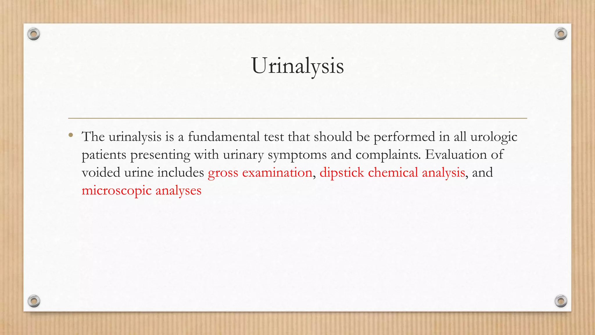
• The urinalysis is a fundamental test that should be performed in all urologic
patients presenting with urinary symptoms and complaints. Evaluation of
voided urine includes gross examination, dipstick chemical analysis, and
microscopic analyses
Collection of urinary samples
Males
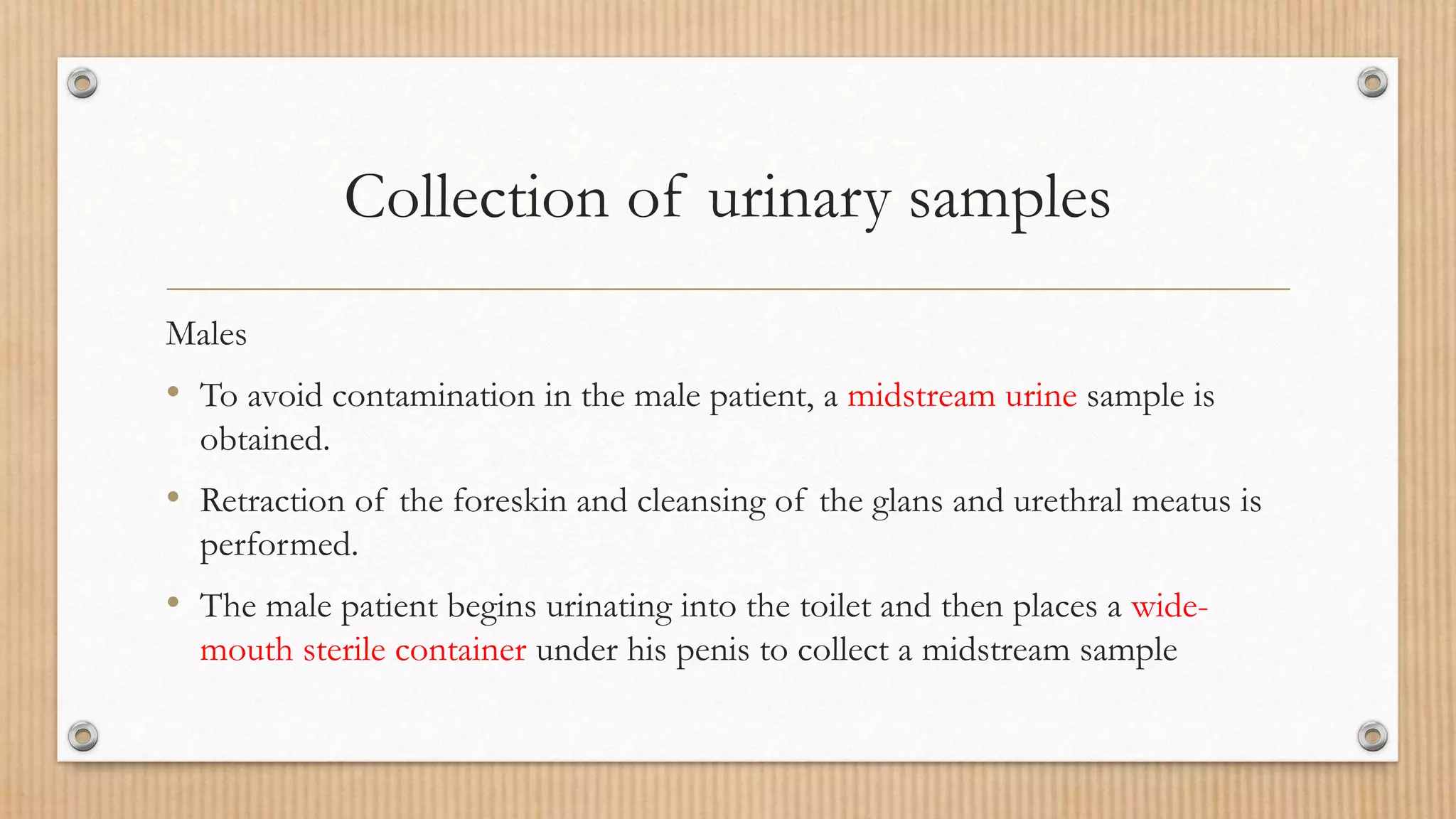
• To avoid contamination in the male patient, a midstream urine sample is
obtained.
• Retraction of the foreskin and cleansing of the glans and urethral meatus is
performed.
• The male patient begins urinating into the toilet and then places a wide-
mouth sterile container under his penis to collect a midstream sample
Collection of urinary samples
• A variety of methods are available to identify a potential source of infection
if not clearly from the bladder.
• One traditional test is the use of four aliquots to differentiate bacteria from
the bladder, urethra, and prostate
Collection of urinary samples
• The Voided Bladder 1 specimen is the initial 5 to 10 mL of urine voided.
• Voided Bladder 2 specimen is the midstream urine.
• Expressed Prostatic Secretions is the secretions obtained after gentle
prostatic massage.
• Voiding Bladder 3 specimen is the initial 2 to 3 mL of urine obtained after
prostatic massage.
Collection of urinary samples
• The value of these cultures for localization of urinary tract infections (UTIs)
is that the VB1 sample represents urethral flora, the VB2 sample represents
bladder flora, and the EPS and VB3 samples represent prostatic flora.
• The VB3 sample is particularly helpful when little or no prostatic fluid is
obtained by massage
Collection of urinary samples
Females
• Obtaining a sterile voided urine collection can be a challenge in the female patient.
• The standard midstream urinalysis in the female patient should be obtained after
separating the labia and cleansing the vaginal introitus and external urethral meatus.
• In the female patient with suspected recurrent UTIs or a history of antibiotic-
resistant infections, a catheterized urine sample should always be obtained.
Collection of urinary samples
Neonates and Infants
• The usual way to obtain a urine sample in a neonate or infant is to place a
sterile plastic bag with an adhesive collar over the infant’s genitalia.
Collection of urinary samples
• Whenever possible, all urine samples should be examined within 1 hour of
collection and plated for culture and sensitivity if indicated.
• If urine is allowed to stand at room temperature for longer periods, bacterial
overgrowth may occur, the pH may change, and red and white blood cell
casts may disintegrate. If it is not possible to examine the urine promptly, it
should be refrigerated at 5°C.
• When appropriate, a collection of urine via suprapubic aspiration may be
obtained.
Physical and Gross Examination of Urine
• The visual and physical examination of the urine includes an evaluation of
color and turbidity.
Physical and Gross Examination of Urine
Color
• Typical, normal urine color is pale yellow as a result of the presence of the pigment
urochrome.
• Urine color varies most commonly because of concentration, but many foods,
medications, metabolic products, and infections may produce abnormal urine color.
• For example, bright red color or pink suggests blood that may be active bleeding,
and purple or brown urine may indicate an old hemorrhage or a retained clot in
the bladder.
• Brown or cola-colored urine can be a sign of glomerular bleeding and disease.
Physical and Gross Examination of Urine
Turbidity
• Cloudy urine is commonly caused by phosphaturia, a benign process in which
excess phosphate crystals precipitate in alkaline urine. Phosphaturia is often
intermittent and may occur after meals, and many patients are otherwise
asymptomatic.
• The diagnosis of phosphaturia can be accomplished either by acidifying the urine
with acetic acid, which will result in immediate clearing, or by performing a
microscopic analysis, which will reveal large amounts of amorphous phosphate
crystals.
Physical and Gross Examination of Urine
Turbidity
• Pyuria is another common cause of cloudy urine. The large numbers of
white blood cells cause the urine to become turbid.
• Pyuria is readily distinguished from phosphaturia either by smelling the urine
(infected urine has a characteristic pungent odor) or by urine dipstick or
microscopic examination.
Physical and Gross Examination of Urine
• Rare causes of cloudy urine include chyluria (in which there is an abnormal
communication between the lymphatic system and the urinary tract
resulting in lymph fluid being mixed with urine), lipiduria, hyperoxaluria, and
hyperuricosuria
Chemical examination of urine
• Urine dipsticks provide a quick and inexpensive method for detecting
abnormal substances within the urine.
• Dipsticks are short, plastic strips with small marker pads that are
impregnated with different chemical reagents that react with abnormal
substances in the urine to produce a colorimetric change.
• Dipstick tests include those for specific gravity, pH, blood, protein,
glucose, ketones, urobilinogen, leukocyte esterase, and nitrites
Chemical examination of urine
• A common medication that may interfere with the dipstick analysis is
phenazopyridine (Pyridium). Phenazopyridine turns the urine bright orange
and makes dipstick evaluation of the urine unreliable
Chemical examination of urine
• The technique of obtaining an accurate dipstick includes
1- Completely immersing it in a fresh uncentrifuged urine specimen.
2- Withdrawing it quickly while drawing the edge along the rim of the
container to remove excess urine.
3- The dipstick should be held horizontally until the appropriate time for
reading and then compared with the color chart.
Chemical examination of urine
False-negative results for glucose and bilirubin may be seen in the presence of
elevated ascorbic acid concentrations in the urine.
Highly buffered alkaline urine may cause falsely low readings for specific
gravity and may lead to false-negative results for urinary protein.
Other common causes of false results with dipstick testing are outdated test
strips and exposure of the sticks, leading to damage to the reagents.
Chemical examination of urine
Specific Gravity and Osmolality
• Specific gravity of urine is easily determined from a urinary dipstick and usually
varies from 1.001 to 1.035.
• Specific gravity usually reflects the patient’s state of hydration but also affords
some idea of renal concentrating ability.
• A specific gravity less than 1.008 is regarded as dilute, and a specific gravity greater
than 1.020 is considered concentrated.
• Acute or chronic renal insufficiency can be associated with a specific gravity of
1.010.
Chemical examination of urine
• Conditions that decrease specific gravity include diuretics, increased fluid
intake, diabetes insipidus, and other causes of decreased renal concentrating
ability.
• Conversely, specific gravity can be increased with uncontrolled diabetes
mellitus caused by glycosuria, inappropriate secretion of antidiuretic
hormone, and any condition causing dehydration. It should be noted that
intravenous injection of some iodinated contrasts and administration of
dextran can cause a rise in specific gravity above 1.035.
Chemical examination of urine
• Osmolality is a measure of the amount of material dissolved in the urine
and usually varies between 50 and 1200 mOsm/L.
• Urine osmolality can be an indicator of renal function.
• Furthermore, with alterations in urine osmolality such as very dilute urine
(below 308 mOsm), reliable assessment of red blood cells in urine can be
compromised due to lysis
Chemical examination of urine
pH
• The dipstick test strip incorporates two colorimetric indicators, methyl red
and bromothymol blue, which yield clearly distinguishable colors over the
pH range from 5 to 9.
• Urinary pH may vary from 4.5 to 8; the average pH varies between 5.5 and
6.5
• A urinary pH between 4.5 and 5.5 is considered acidic, whereas a pH
between 6.5 and 8 is considered alkaline.
Chemical examination of urine
• In general, the urinary pH reflects the pH in the serum.
• In patients with metabolic or respiratory acidosis, the urine is usually
acidic; conversely, in patients with metabolic or respiratory alkalosis, the
urine is alkaline.
• Renal tubular acidosis (RTA) presents an exception to this rule. In patients
with both type I and II RTA, the serum is acidemic, but the urine is alkalotic
because of continued loss of bicarbonate in the urine.
Chemical examination of urine
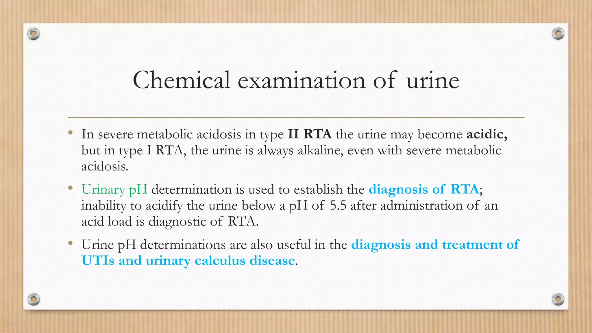
• In severe metabolic acidosis in type II RTA the urine may become acidic,
but in type I RTA, the urine is always alkaline, even with severe metabolic
acidosis.
• Urinary pH determination is used to establish the diagnosis of RTA;
inability to acidify the urine below a pH of 5.5 after administration of an
acid load is diagnostic of RTA.
• Urine pH determinations are also useful in the diagnosis and treatment of
UTIs and urinary calculus disease.
Chemical examination of urine
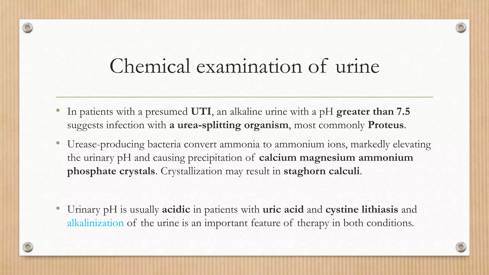
• In patients with a presumed UTI, an alkaline urine with a pH greater than 7.5
suggests infection with a urea-splitting organism, most commonly Proteus.
• Urease-producing bacteria convert ammonia to ammonium ions, markedly elevating
the urinary pH and causing precipitation of calcium magnesium ammonium
phosphate crystals. Crystallization may result in staghorn calculi.
• Urinary pH is usually acidic in patients with uric acid and cystine lithiasis and
alkalinization of the urine is an important feature of therapy in both conditions.
Chemical examination of urine
Blood/Hematuria
• Normal urine should contain less than 3 erythrocytes per HPF.
• A positive dipstick for blood in the urine indicates either hematuria,
hemoglobinuria, or myoglobinuria.
• The chemical detection of blood in the urine is based on the peroxidase-like
activity of hemoglobin.
Chemical examination of urine
• Hematuria can be distinguished from hemoglobinuria and myoglobinuria by
microscopic examination of the centrifuged urine; the presence of a large
number of erythrocytes establishes the diagnosis of hematuria.
• If erythrocytes are absent, examination of the serum will distinguish
hemoglobinuria and myoglobinuria.
Chemical examination of urine
• The sensitivity of urinary dipsticks in identifying microscopic hematuria,
defined as greater than or equal to 3 erythrocytes per HPF of centrifuged
sediment examined microscopically, is higher than 90%. Conversely, the
specificity of the dipstick for hematuria compared with microscopy is
somewhat lower, reflecting a higher false-positive rate with the dipstick.
• The dipstick result should be confirmed with a microscopic examination of
the centrifuged urinary sediment.
Differential Diagnosis and Evaluation of
Hematuria
• Identifying the hematuria as nephrologic versus urologic and glomerular
versus nonglomerular is one of the first steps in the analysis.
• Hematuria of nephrologic origin is frequently associated with casts in the
urine and almost always associated with significant proteinuria
• Erythrocytes arising from glomerular disease are typically dysmorphic and
show a wide range of morphologic alterations. Conversely, erythrocytes
arising from tubulointerstitial renal disease and of urologic origin have a
uniformly round shape.
Nonglomerular Hematuria
Medical/Nonsurgical
• Medical causes of nonglomerular hematuria of renal origin are secondary to either
tubulointerstitial, renovascular, or systemic disorders.
• The urinalysis in nonglomerular hematuria is distinguished from that of glomerular
hematuria by the presence of circular erythrocytes and the absence of erythrocyte
casts.
• It is frequently associated with significant proteinuria, which distinguishes
these nephrologic diseases from urologic diseases in which the degree of
proteinuria is usually minimal, even with heavy bleeding.
Nonglomerular Hematuria
• A family history of hematuria or bleeding tendency suggests the diagnosis of a
blood dyscrasia.
• A family history of urolithiasis associated with intermittent hematuria may
indicate inherited stone disease.
• A family history of renal cystic disease should prompt further radiologic
evaluation for medullary sponge kidney and adult polycystic kidney disease.
• Papillary necrosis as a cause of hematuria should be considered in diabetics,
AfricanAmericans (secondary to sickle cell disease or trait), and suspected
analgesic abusers.
Nonglomerular Hematuria
• Anticoagulation at normal therapeutic levels, however, does not predispose patients
to hematuria.
• Exercise-induced hematuria can be observed in patients with a recent history of
vigorous exercise such as extreme or long-distance running (>10 km) and the
hematuria may be of renal or bladder origin.
• A vascular disease may also result in nonglomerular hematuria. Renal artery
embolism and thrombosis, arteriovenous fistulae, and renal vein thrombosis may all
result in hematuria.
• Microscopic hematuria has also been reported in Nutcracker syndrome associated
with pelvic congestion.
Nonglomerular Hematuria
Surgical
• Nonglomerular hematuria caused by surgical and urologic causes includes
pathology of the collecting system of the urinary tract. Common causes
include urologic tumors, stones, benign prostatic hyperplasia, and
urinary tract infections.
Nonglomerular Hematuria
• It should be remembered, however, that proteinuria is not always present in
glomerular or nonglomerular renal disease.
• Asymptomatic microscopic hematuria (AMH) “is defined as three or greater
RBC/HPF on a properly collected urinary specimen in the absence of an
obvious benign cause.
Chemical examination of urine
Proteinuria
• Healthy adults excrete 80 to 150 mg of protein in the urine daily.
• Proteinuria may be the first indication of renovascular, glomerular, or
tubulointerstitial renal disease, or it may represent the overflow of abnormal
proteins into the urine in conditions such as multiple myeloma.
• The protein concentration in the urine depends on the state of hydration,
but it seldom exceeds 20 mg/dL.
Chemical examination of urine
• Proteinuria should first be classified by its timing into transient,
intermittent, or persistent.
• Transient proteinuria occurs commonly, especially in the pediatric
population, and usually resolves spontaneously within a few.
• It may result from fever, exercise, or emotional stress. In older patients,
transient proteinuria may be caused by congestive heart failure.
• Proteinuria may also occur intermittently, and this is frequently related to
postural change
Chemical examination of urine
Glucose
• Urine testing for glucose and ketones is useful in screening patients for
diabetes mellitus.
• Normally, almost all of the glucose filtered by the glomeruli is reabsorbed
in the proximal tubules.
• This so-called renal threshold corresponds to serum glucose of about 180
mg/ dL; above this level, glucose will be detected in the urine.
Chemical examination of urine
Ketones
• Ketones are not normally found in the urine but will appear when the
carbohydrate supplies in the body are depleted and body fat breakdown
occurs.
• This happens most commonly in diabetic ketoacidosis but may also occur
during pregnancy and after periods of starvation or rapid weight reduction.
Chemical examination of urine
• Ketones excreted include acetoacetic acid, acetone, and β-hydroxybutyric
acid.
• Dipstick testing will identify acetoacetic acid at concentrations of 5 to 10
mg/dL but will not detect acetone or β-hydroxybutyric acid.
Chemical examination of urine
Bilirubin and Urobilinogen
• Normal urine contains no bilirubin and only small amounts of urobilinogen.
• There are two types of bilirubin: direct (conjugated) and indirect.
• Direct bilirubin is made in the hepatocyte, where bilirubin is conjugated with
glucuronic acid.
• Conjugated bilirubin has a low molecular weight, is water soluble, and
normally passes from the liver to the small intestine through the bile ducts, where it
is converted to urobilinogen.
Chemical examination of urine
• Therefore conjugated bilirubin does not appear in the urine except in
pathologic conditions in which there is intrinsic hepatic disease or
obstruction of the bile ducts.
• Indirect bilirubin is of high molecular weight and bound in the serum to
albumin. It is water insoluble and therefore does not appear in the urine,
even in pathologic conditions.
Chemical examination of urine
• Urobilinogen is the end product of conjugated bilirubin metabolism.
Conjugated bilirubin passes through the bile ducts, where it is metabolized
by normal intestinal bacteria to urobilinogen. Normally, about 50% of the
urobilinogen is excreted in the stool, and 50% is reabsorbed into the
enterohepatic circulation.
• A small amount of absorbed urobilinogen, about 1 to 4 mg/day, will escape
hepatic uptake and be excreted in the urine. Hemolysis and hepatocellular
diseases that lead to increased bile pigments can result in increased urinary
urobilinogen.
Chemical examination of urine
Leukocyte Esterase and Nitrite Tests
• Leukocyte esterase activity indicates the presence of white blood cells in
the urine. The presence of nitrites in the urine is strongly suggestive of
bacteriuria.
• If the dipstick is positive for leukocyte esterase but negative for nitrites,
noninfectious causes of inflammation should be considered and a
microscopic analysis and urine culture should be obtained before any
empirical antibiotic therapy should be prescribed.
Chemical examination of urine
• Leukocyte esterase is produced by neutrophils and the sensitivity of this test
subsequently decreases with time because of lysis of the leukocytes.
• False-negative results with leukocyte esterase testing include increased
urinary specific gravity, glycosuria, presence of urobilinogen,
medications that alter urine color, and ingestion of large amounts of
ascorbic acid.
• The major cause of false-positive leukocyte esterase tests is specimen
contamination.
Chemical examination of urine
• Nitrites are not normally found in the urine, but many species of gram-
negative bacteria can convert nitrates to nitrites.
• The specificity of the nitrite dipstick for detecting bacteriuria is higher
than 90%.
• The sensitivity of the test, however, is considerably less, varying from 35% to
85%.
• The nitrite test is less accurate in urine specimens containing fewer than 10 5
organisms/ mL.
Microscopy Technique
The urinary sediment should be examined microscopically for
(1) cells,
(2) casts,
(3) crystals,
(4) bacteria,
(5) yeast, and (6) parasites.
Cells
• Erythrocyte morphology may be determined under high-power
magnification.
• Circular erythrocytes generally have an even distribution of hemoglobin
with either a round or crenated contour,
• Whereas dysmorphic erythrocytes are irregularly shaped with minimal
hemoglobin and irregular distribution of cytoplasm.
Cells
• Leukocytes can generally be identified under low-power magnification and
definitively diagnosed under high-power magnification.
• It is normal to find 1 or 2 leukocytes per HPF in men and up to 5 per HPF in
women in whom the urine sample may be contaminated with vaginal
secretions.
• A greater number of leukocytes generally indicates infection or
inflammation in the urinary tract.
Cells
• It may be possible to distinguish old leukocytes, which have a characteristic
small and wrinkled appearance and which are commonly found in the
vaginal secretions of normal women, from fresh leukocytes, which are
generally indicative of urinary tract pathology.
• Fresh leukocytes are generally larger and rounder.
Cells
• Epithelial cells are commonly observed in the urinary sediment.
• Squamous cells are frequently detected in female urine specimens and are derived
from the lower portion of the urethra, the trigone of postpubertal females, and the
vagina.
• Transitional cells
• Malignant transitional cells
• Renal tubular cells are the least commonly observed epithelial cells in the urine
but are most significant because their presence in the urine is always indicative of
renal pathology.
Casts
A cast is a protein coagulum that is formed in the renal tubule
• Types
Hyaline casts contain only mucoproteins, after exercise or heat exposure but
may also be observed in pyelonephritis or chronic renal disease.
RBC casts contain entrapped erythrocytes and are diagnostic of glomerular
bleeding
Casts
White blood cell casts are observed in acute glomerulonephritis, acute
pyelonephritis, and acute tubulointerstitial nephritis
Granular and waxy casts result from further degeneration of cellular
elements.
Fatty casts are seen in nephrotic syndrome, lipiduria, and hypothyroidism.
Crystals
• Identification of crystals in the urine is particularly important in patients
with stone disease because it may help determine the etiology.
• The identification of cystine crystals establishes the diagnosis of cystinuria.
• Crystals precipitated in acidic urine include calcium oxalate, uric acid, and
cystine.
Crysrals
• Crystals precipitated in an alkaline urine include calcium phosphate and
triple-phosphate (struvite) crystals.
• Cholesterol crystals are rarely seen in the urine and are not related to urinary
pH.
Bacteria
• Normal urine should not contain bacteria.
• 5 bacteria per HPF reflects colony counts of about 100,000/mL.
• This is the standard concentration used to establish the diagnosis of a UTI
in a clean-catch specimen.
• The finding of any bacteria in a properly collected midstream specimen from
a male patient should be further evaluated with a urine culture.
Yeast
• The most common yeast cells found in urine are Candida albicans.
• Yeasts can be distinguished by their characteristic budding and hyphae.
• Yeasts are most commonly seen in the urine of patients with diabetes
mellitus or as contaminants in women with vaginal candidiasis.
Parasites
• Trichomonas vaginalis is a frequent cause of vaginitis in women and
occasionally of urethritis in men.
• Schistosoma hematobium is a urinary tract pathogen that is not found in the
United States but is extremely common in countries of the Middle East and
North Africa.
• Examination of the urine shows the characteristic parasitic ova with a
terminal spike.
Urinalysis
Urinalysis

More Related Content

What's hot

Urine analysis.ppt
Urine analysis.ppt Urine analysis.ppt
Urine analysis.ppt
Nam Ly
 
Urinalysis
UrinalysisUrinalysis
Urinalysis
Microbiology
 
Urinalysis
UrinalysisUrinalysis
Urinalysis
allerasic
 
Urine analysis.pptx
Urine analysis.pptxUrine analysis.pptx
Urine analysis.pptx
Prakash Mishra
 
Urine analysis Class I
Urine analysis   Class IUrine analysis   Class I
Urine analysis Class I
Dr. Varughese George
 
Urine analysis
Urine analysisUrine analysis
Urine analysis
AFiFi Faridz
 
Urine and urinalysis
Urine and urinalysisUrine and urinalysis
Urine and urinalysis
Liwayway Memije-Cruz
 
Urine analysis
Urine analysisUrine analysis
Urine analysis
9890888615
 
Urinalysis- Methods, observations and clinical significance
 Urinalysis- Methods, observations and clinical significance Urinalysis- Methods, observations and clinical significance
Urinalysis- Methods, observations and clinical significance
Namrata Chhabra
 
Urinalysis ( analysis of urine )
Urinalysis ( analysis of urine )Urinalysis ( analysis of urine )
Urinalysis ( analysis of urine )
Doctor AlTokhy
 
Fecal tests or stool tests
Fecal tests or stool testsFecal tests or stool tests
Semen analysis or seminal fluid analysis
Semen analysis or seminal fluid analysisSemen analysis or seminal fluid analysis
Semen analysis or seminal fluid analysis
Dr.vasantrao pawar medical college
 
Stool Examination Abridged What A Medical Graduate Should Know
Stool Examination Abridged   What A Medical Graduate Should KnowStool Examination Abridged   What A Medical Graduate Should Know
Stool Examination Abridged What A Medical Graduate Should Know
Dr. Varughese George
 
Examination of Body Fluids - Pathology - MLT, ATOT, RDT
Examination of Body Fluids - Pathology - MLT, ATOT, RDTExamination of Body Fluids - Pathology - MLT, ATOT, RDT
Examination of Body Fluids - Pathology - MLT, ATOT, RDT
Dr. Salman Ansari
 
Amniotic fluid,hcg, sputum, bal & sweat
Amniotic fluid,hcg, sputum, bal & sweatAmniotic fluid,hcg, sputum, bal & sweat
Amniotic fluid,hcg, sputum, bal & sweat
Princess Alen Aguilar-Cabunoc
 
URINALYSIS CHEM PATH.pptx
URINALYSIS CHEM PATH.pptxURINALYSIS CHEM PATH.pptx
URINALYSIS CHEM PATH.pptx
Xavier875943
 
Urine analysis REVISION NOTES
Urine analysis REVISION NOTES Urine analysis REVISION NOTES
Urine analysis REVISION NOTES
TONY SCARIA
 

What's hot (20)

Urine analysis.ppt
Urine analysis.ppt Urine analysis.ppt
Urine analysis.ppt
 
Urinalysis
UrinalysisUrinalysis
Urinalysis
 
Urinalysis
UrinalysisUrinalysis
Urinalysis
 
Complete urine examination
Complete urine examinationComplete urine examination
Complete urine examination
 
Urine analysis.pptx
Urine analysis.pptxUrine analysis.pptx
Urine analysis.pptx
 
Urine analysis Class I
Urine analysis   Class IUrine analysis   Class I
Urine analysis Class I
 
Urine analysis
Urine analysisUrine analysis
Urine analysis
 
Urine examination
Urine examinationUrine examination
Urine examination
 
Urine and urinalysis
Urine and urinalysisUrine and urinalysis
Urine and urinalysis
 
Urine analysis
Urine analysisUrine analysis
Urine analysis
 
Urinalysis- Methods, observations and clinical significance
 Urinalysis- Methods, observations and clinical significance Urinalysis- Methods, observations and clinical significance
Urinalysis- Methods, observations and clinical significance
 
Urinalysis ( analysis of urine )
Urinalysis ( analysis of urine )Urinalysis ( analysis of urine )
Urinalysis ( analysis of urine )
 
Renal calculi
Renal calculiRenal calculi
Renal calculi
 
Fecal tests or stool tests
Fecal tests or stool testsFecal tests or stool tests
Fecal tests or stool tests
 
Semen analysis or seminal fluid analysis
Semen analysis or seminal fluid analysisSemen analysis or seminal fluid analysis
Semen analysis or seminal fluid analysis
 
Stool Examination Abridged What A Medical Graduate Should Know
Stool Examination Abridged   What A Medical Graduate Should KnowStool Examination Abridged   What A Medical Graduate Should Know
Stool Examination Abridged What A Medical Graduate Should Know
 
Examination of Body Fluids - Pathology - MLT, ATOT, RDT
Examination of Body Fluids - Pathology - MLT, ATOT, RDTExamination of Body Fluids - Pathology - MLT, ATOT, RDT
Examination of Body Fluids - Pathology - MLT, ATOT, RDT
 
Amniotic fluid,hcg, sputum, bal & sweat
Amniotic fluid,hcg, sputum, bal & sweatAmniotic fluid,hcg, sputum, bal & sweat
Amniotic fluid,hcg, sputum, bal & sweat
 
URINALYSIS CHEM PATH.pptx
URINALYSIS CHEM PATH.pptxURINALYSIS CHEM PATH.pptx
URINALYSIS CHEM PATH.pptx
 
Urine analysis REVISION NOTES
Urine analysis REVISION NOTES Urine analysis REVISION NOTES
Urine analysis REVISION NOTES
 

Similar to Urinalysis

URINE & STOOL EXAMINATION.pptx
URINE & STOOL EXAMINATION.pptxURINE & STOOL EXAMINATION.pptx
URINE & STOOL EXAMINATION.pptx
amanjotkaursidhu
 
Renal function tests
Renal function testsRenal function tests
Renal function tests
Sunita Patil
 
Urine examination , urine, chemical examination
Urine examination , urine, chemical examinationUrine examination , urine, chemical examination
Urine examination , urine, chemical examination
sandeep singh
 
UNDERSTANDING URINALYSIS.pptx
UNDERSTANDING URINALYSIS.pptxUNDERSTANDING URINALYSIS.pptx
UNDERSTANDING URINALYSIS.pptx
NicholasQuaysie1
 
Urine analysis pnnnnpt-MG.pptx
Urine analysis pnnnnpt-MG.pptxUrine analysis pnnnnpt-MG.pptx
Urine analysis pnnnnpt-MG.pptx
NabdNabd
 
Urine Examination (Practical).pdf
Urine Examination (Practical).pdfUrine Examination (Practical).pdf
Urine Examination (Practical).pdf
JYOTIMAYBARUAH
 
Urine routine and Microscopy.pptx
Urine routine and Microscopy.pptxUrine routine and Microscopy.pptx
Urine routine and Microscopy.pptx
ssuser2961ab
 
Analysis of Normal Urine.pptx
Analysis of Normal Urine.pptxAnalysis of Normal Urine.pptx
Analysis of Normal Urine.pptx
VivekRathi30
 
Body fluid ( the Urine ) Urinalysis
Body fluid ( the Urine ) Urinalysis Body fluid ( the Urine ) Urinalysis
Body fluid ( the Urine ) Urinalysis
Amany Elsayed
 
Urine examination
Urine examinationUrine examination
Urine examination
Malini Garg
 
urinalysis [Autosaved].pptx how to interpret
urinalysis [Autosaved].pptx how to interpreturinalysis [Autosaved].pptx how to interpret
urinalysis [Autosaved].pptx how to interpret
Lawrenceshamboko
 
Physical and chemical examination of urine
Physical and chemical examination of urinePhysical and chemical examination of urine
Physical and chemical examination of urine
Himil Parikh
 
sanjana.pptx
sanjana.pptxsanjana.pptx
sanjana.pptx
sanjananekkanti8787
 
Urine analysis physical
Urine analysis physicalUrine analysis physical
Urine analysis physical
Balaji Amit
 
urine analysis labortory diagnosisn work ppt
urine analysis labortory diagnosisn work ppturine analysis labortory diagnosisn work ppt
urine analysis labortory diagnosisn work ppt
moltanus
 
Urine analysis
Urine analysisUrine analysis
Urine analysis
Janani Mathialagan
 
Physical Examination of urine(analysis )part-1
Physical Examination of urine(analysis )part-1Physical Examination of urine(analysis )part-1
Physical Examination of urine(analysis )part-1
aamanimutakani
 
Applied aspects of Kidney and RFT by Dr. MP.pptx
Applied aspects of Kidney and RFT by Dr. MP.pptxApplied aspects of Kidney and RFT by Dr. MP.pptx
Applied aspects of Kidney and RFT by Dr. MP.pptx
Pandian M
 
Urine analysis
Urine analysisUrine analysis
Urine analysis
Vijyalaxmi Makwana
 

Similar to Urinalysis (20)

URINE & STOOL EXAMINATION.pptx
URINE & STOOL EXAMINATION.pptxURINE & STOOL EXAMINATION.pptx
URINE & STOOL EXAMINATION.pptx
 
Renal function tests
Renal function testsRenal function tests
Renal function tests
 
Urine examination , urine, chemical examination
Urine examination , urine, chemical examinationUrine examination , urine, chemical examination
Urine examination , urine, chemical examination
 
UNDERSTANDING URINALYSIS.pptx
UNDERSTANDING URINALYSIS.pptxUNDERSTANDING URINALYSIS.pptx
UNDERSTANDING URINALYSIS.pptx
 
Urine analysis pnnnnpt-MG.pptx
Urine analysis pnnnnpt-MG.pptxUrine analysis pnnnnpt-MG.pptx
Urine analysis pnnnnpt-MG.pptx
 
Urine Examination (Practical).pdf
Urine Examination (Practical).pdfUrine Examination (Practical).pdf
Urine Examination (Practical).pdf
 
Urine routine and Microscopy.pptx
Urine routine and Microscopy.pptxUrine routine and Microscopy.pptx
Urine routine and Microscopy.pptx
 
Analysis of Normal Urine.pptx
Analysis of Normal Urine.pptxAnalysis of Normal Urine.pptx
Analysis of Normal Urine.pptx
 
Body fluid ( the Urine ) Urinalysis
Body fluid ( the Urine ) Urinalysis Body fluid ( the Urine ) Urinalysis
Body fluid ( the Urine ) Urinalysis
 
Urine examination
Urine examinationUrine examination
Urine examination
 
urinalysis [Autosaved].pptx how to interpret
urinalysis [Autosaved].pptx how to interpreturinalysis [Autosaved].pptx how to interpret
urinalysis [Autosaved].pptx how to interpret
 
Physical and chemical examination of urine
Physical and chemical examination of urinePhysical and chemical examination of urine
Physical and chemical examination of urine
 
sanjana.pptx
sanjana.pptxsanjana.pptx
sanjana.pptx
 
Urine analysis physical
Urine analysis physicalUrine analysis physical
Urine analysis physical
 
urine analysis labortory diagnosisn work ppt
urine analysis labortory diagnosisn work ppturine analysis labortory diagnosisn work ppt
urine analysis labortory diagnosisn work ppt
 
Urine analysis
Urine analysisUrine analysis
Urine analysis
 
Kidney urine
Kidney urineKidney urine
Kidney urine
 
Physical Examination of urine(analysis )part-1
Physical Examination of urine(analysis )part-1Physical Examination of urine(analysis )part-1
Physical Examination of urine(analysis )part-1
 
Applied aspects of Kidney and RFT by Dr. MP.pptx
Applied aspects of Kidney and RFT by Dr. MP.pptxApplied aspects of Kidney and RFT by Dr. MP.pptx
Applied aspects of Kidney and RFT by Dr. MP.pptx
 
Urine analysis
Urine analysisUrine analysis
Urine analysis
 

Recently uploaded

Home assignment II on Spectroscopy 2024 Answers.pdf
Home assignment II on Spectroscopy 2024 Answers.pdfHome assignment II on Spectroscopy 2024 Answers.pdf
Home assignment II on Spectroscopy 2024 Answers.pdf
Tamralipta Mahavidyalaya
 
Phrasal Verbs.XXXXXXXXXXXXXXXXXXXXXXXXXX
Phrasal Verbs.XXXXXXXXXXXXXXXXXXXXXXXXXXPhrasal Verbs.XXXXXXXXXXXXXXXXXXXXXXXXXX
Phrasal Verbs.XXXXXXXXXXXXXXXXXXXXXXXXXX
MIRIAMSALINAS13
 
June 3, 2024 Anti-Semitism Letter Sent to MIT President Kornbluth and MIT Cor...
June 3, 2024 Anti-Semitism Letter Sent to MIT President Kornbluth and MIT Cor...June 3, 2024 Anti-Semitism Letter Sent to MIT President Kornbluth and MIT Cor...
June 3, 2024 Anti-Semitism Letter Sent to MIT President Kornbluth and MIT Cor...
Levi Shapiro
 
Instructions for Submissions thorugh G- Classroom.pptx
Instructions for Submissions thorugh G- Classroom.pptxInstructions for Submissions thorugh G- Classroom.pptx
Instructions for Submissions thorugh G- Classroom.pptx
Jheel Barad
 
Chapter 3 - Islamic Banking Products and Services.pptx
Chapter 3 - Islamic Banking Products and Services.pptxChapter 3 - Islamic Banking Products and Services.pptx
Chapter 3 - Islamic Banking Products and Services.pptx
Mohd Adib Abd Muin, Senior Lecturer at Universiti Utara Malaysia
 
1.4 modern child centered education - mahatma gandhi-2.pptx
1.4 modern child centered education - mahatma gandhi-2.pptx1.4 modern child centered education - mahatma gandhi-2.pptx
1.4 modern child centered education - mahatma gandhi-2.pptx
JosvitaDsouza2
 
special B.ed 2nd year old paper_20240531.pdf
special B.ed 2nd year old paper_20240531.pdfspecial B.ed 2nd year old paper_20240531.pdf
special B.ed 2nd year old paper_20240531.pdf
Special education needs
 
The Challenger.pdf DNHS Official Publication
The Challenger.pdf DNHS Official PublicationThe Challenger.pdf DNHS Official Publication
The Challenger.pdf DNHS Official Publication
Delapenabediema
 
Language Across the Curriculm LAC B.Ed.
Language Across the  Curriculm LAC B.Ed.Language Across the  Curriculm LAC B.Ed.
Language Across the Curriculm LAC B.Ed.
Atul Kumar Singh
 
Acetabularia Information For Class 9 .docx
Acetabularia Information For Class 9  .docxAcetabularia Information For Class 9  .docx
Acetabularia Information For Class 9 .docx
vaibhavrinwa19
 
Model Attribute Check Company Auto Property
Model Attribute  Check Company Auto PropertyModel Attribute  Check Company Auto Property
Model Attribute Check Company Auto Property
Celine George
 
Sha'Carri Richardson Presentation 202345
Sha'Carri Richardson Presentation 202345Sha'Carri Richardson Presentation 202345
Sha'Carri Richardson Presentation 202345
beazzy04
 
Biological Screening of Herbal Drugs in detailed.
Biological Screening of Herbal Drugs in detailed.Biological Screening of Herbal Drugs in detailed.
Biological Screening of Herbal Drugs in detailed.
Ashokrao Mane college of Pharmacy Peth-Vadgaon
 
Mule 4.6 & Java 17 Upgrade | MuleSoft Mysore Meetup #46
Mule 4.6 & Java 17 Upgrade | MuleSoft Mysore Meetup #46Mule 4.6 & Java 17 Upgrade | MuleSoft Mysore Meetup #46
Mule 4.6 & Java 17 Upgrade | MuleSoft Mysore Meetup #46
MysoreMuleSoftMeetup
 
Embracing GenAI - A Strategic Imperative
Embracing GenAI - A Strategic ImperativeEmbracing GenAI - A Strategic Imperative
Embracing GenAI - A Strategic Imperative
Peter Windle
 
TESDA TM1 REVIEWER FOR NATIONAL ASSESSMENT WRITTEN AND ORAL QUESTIONS WITH A...
TESDA TM1 REVIEWER  FOR NATIONAL ASSESSMENT WRITTEN AND ORAL QUESTIONS WITH A...TESDA TM1 REVIEWER  FOR NATIONAL ASSESSMENT WRITTEN AND ORAL QUESTIONS WITH A...
TESDA TM1 REVIEWER FOR NATIONAL ASSESSMENT WRITTEN AND ORAL QUESTIONS WITH A...
EugeneSaldivar
 
The Roman Empire A Historical Colossus.pdf
The Roman Empire A Historical Colossus.pdfThe Roman Empire A Historical Colossus.pdf
The Roman Empire A Historical Colossus.pdf
kaushalkr1407
 
Thesis Statement for students diagnonsed withADHD.ppt
Thesis Statement for students diagnonsed withADHD.pptThesis Statement for students diagnonsed withADHD.ppt
Thesis Statement for students diagnonsed withADHD.ppt
EverAndrsGuerraGuerr
 
CACJapan - GROUP Presentation 1- Wk 4.pdf
CACJapan - GROUP Presentation 1- Wk 4.pdfCACJapan - GROUP Presentation 1- Wk 4.pdf
CACJapan - GROUP Presentation 1- Wk 4.pdf
camakaiclarkmusic
 
A Strategic Approach: GenAI in Education
A Strategic Approach: GenAI in EducationA Strategic Approach: GenAI in Education
A Strategic Approach: GenAI in Education
Peter Windle
 

Recently uploaded (20)

Home assignment II on Spectroscopy 2024 Answers.pdf
Home assignment II on Spectroscopy 2024 Answers.pdfHome assignment II on Spectroscopy 2024 Answers.pdf
Home assignment II on Spectroscopy 2024 Answers.pdf
 
Phrasal Verbs.XXXXXXXXXXXXXXXXXXXXXXXXXX
Phrasal Verbs.XXXXXXXXXXXXXXXXXXXXXXXXXXPhrasal Verbs.XXXXXXXXXXXXXXXXXXXXXXXXXX
Phrasal Verbs.XXXXXXXXXXXXXXXXXXXXXXXXXX
 
June 3, 2024 Anti-Semitism Letter Sent to MIT President Kornbluth and MIT Cor...
June 3, 2024 Anti-Semitism Letter Sent to MIT President Kornbluth and MIT Cor...June 3, 2024 Anti-Semitism Letter Sent to MIT President Kornbluth and MIT Cor...
June 3, 2024 Anti-Semitism Letter Sent to MIT President Kornbluth and MIT Cor...
 
Instructions for Submissions thorugh G- Classroom.pptx
Instructions for Submissions thorugh G- Classroom.pptxInstructions for Submissions thorugh G- Classroom.pptx
Instructions for Submissions thorugh G- Classroom.pptx
 
Chapter 3 - Islamic Banking Products and Services.pptx
Chapter 3 - Islamic Banking Products and Services.pptxChapter 3 - Islamic Banking Products and Services.pptx
Chapter 3 - Islamic Banking Products and Services.pptx
 
1.4 modern child centered education - mahatma gandhi-2.pptx
1.4 modern child centered education - mahatma gandhi-2.pptx1.4 modern child centered education - mahatma gandhi-2.pptx
1.4 modern child centered education - mahatma gandhi-2.pptx
 
special B.ed 2nd year old paper_20240531.pdf
special B.ed 2nd year old paper_20240531.pdfspecial B.ed 2nd year old paper_20240531.pdf
special B.ed 2nd year old paper_20240531.pdf
 
The Challenger.pdf DNHS Official Publication
The Challenger.pdf DNHS Official PublicationThe Challenger.pdf DNHS Official Publication
The Challenger.pdf DNHS Official Publication
 
Language Across the Curriculm LAC B.Ed.
Language Across the  Curriculm LAC B.Ed.Language Across the  Curriculm LAC B.Ed.
Language Across the Curriculm LAC B.Ed.
 
Acetabularia Information For Class 9 .docx
Acetabularia Information For Class 9  .docxAcetabularia Information For Class 9  .docx
Acetabularia Information For Class 9 .docx
 
Model Attribute Check Company Auto Property
Model Attribute  Check Company Auto PropertyModel Attribute  Check Company Auto Property
Model Attribute Check Company Auto Property
 
Sha'Carri Richardson Presentation 202345
Sha'Carri Richardson Presentation 202345Sha'Carri Richardson Presentation 202345
Sha'Carri Richardson Presentation 202345
 
Biological Screening of Herbal Drugs in detailed.
Biological Screening of Herbal Drugs in detailed.Biological Screening of Herbal Drugs in detailed.
Biological Screening of Herbal Drugs in detailed.
 
Mule 4.6 & Java 17 Upgrade | MuleSoft Mysore Meetup #46
Mule 4.6 & Java 17 Upgrade | MuleSoft Mysore Meetup #46Mule 4.6 & Java 17 Upgrade | MuleSoft Mysore Meetup #46
Mule 4.6 & Java 17 Upgrade | MuleSoft Mysore Meetup #46
 
Embracing GenAI - A Strategic Imperative
Embracing GenAI - A Strategic ImperativeEmbracing GenAI - A Strategic Imperative
Embracing GenAI - A Strategic Imperative
 
TESDA TM1 REVIEWER FOR NATIONAL ASSESSMENT WRITTEN AND ORAL QUESTIONS WITH A...
TESDA TM1 REVIEWER  FOR NATIONAL ASSESSMENT WRITTEN AND ORAL QUESTIONS WITH A...TESDA TM1 REVIEWER  FOR NATIONAL ASSESSMENT WRITTEN AND ORAL QUESTIONS WITH A...
TESDA TM1 REVIEWER FOR NATIONAL ASSESSMENT WRITTEN AND ORAL QUESTIONS WITH A...
 
The Roman Empire A Historical Colossus.pdf
The Roman Empire A Historical Colossus.pdfThe Roman Empire A Historical Colossus.pdf
The Roman Empire A Historical Colossus.pdf
 
Thesis Statement for students diagnonsed withADHD.ppt
Thesis Statement for students diagnonsed withADHD.pptThesis Statement for students diagnonsed withADHD.ppt
Thesis Statement for students diagnonsed withADHD.ppt
 
CACJapan - GROUP Presentation 1- Wk 4.pdf
CACJapan - GROUP Presentation 1- Wk 4.pdfCACJapan - GROUP Presentation 1- Wk 4.pdf
CACJapan - GROUP Presentation 1- Wk 4.pdf
 
A Strategic Approach: GenAI in Education
A Strategic Approach: GenAI in EducationA Strategic Approach: GenAI in Education
A Strategic Approach: GenAI in Education
 

Urinalysis

  • 2. Urinalysis • The urinalysis is a fundamental test that should be performed in all urologic patients presenting with urinary symptoms and complaints. Evaluation of voided urine includes gross examination, dipstick chemical analysis, and microscopic analyses
  • 3. Collection of urinary samples Males • To avoid contamination in the male patient, a midstream urine sample is obtained. • Retraction of the foreskin and cleansing of the glans and urethral meatus is performed. • The male patient begins urinating into the toilet and then places a wide- mouth sterile container under his penis to collect a midstream sample
  • 4. Collection of urinary samples • A variety of methods are available to identify a potential source of infection if not clearly from the bladder. • One traditional test is the use of four aliquots to differentiate bacteria from the bladder, urethra, and prostate
  • 5. Collection of urinary samples • The Voided Bladder 1 specimen is the initial 5 to 10 mL of urine voided. • Voided Bladder 2 specimen is the midstream urine. • Expressed Prostatic Secretions is the secretions obtained after gentle prostatic massage. • Voiding Bladder 3 specimen is the initial 2 to 3 mL of urine obtained after prostatic massage.
  • 6. Collection of urinary samples • The value of these cultures for localization of urinary tract infections (UTIs) is that the VB1 sample represents urethral flora, the VB2 sample represents bladder flora, and the EPS and VB3 samples represent prostatic flora. • The VB3 sample is particularly helpful when little or no prostatic fluid is obtained by massage
  • 7. Collection of urinary samples Females • Obtaining a sterile voided urine collection can be a challenge in the female patient. • The standard midstream urinalysis in the female patient should be obtained after separating the labia and cleansing the vaginal introitus and external urethral meatus. • In the female patient with suspected recurrent UTIs or a history of antibiotic- resistant infections, a catheterized urine sample should always be obtained.
  • 8. Collection of urinary samples Neonates and Infants • The usual way to obtain a urine sample in a neonate or infant is to place a sterile plastic bag with an adhesive collar over the infant’s genitalia.
  • 9. Collection of urinary samples • Whenever possible, all urine samples should be examined within 1 hour of collection and plated for culture and sensitivity if indicated. • If urine is allowed to stand at room temperature for longer periods, bacterial overgrowth may occur, the pH may change, and red and white blood cell casts may disintegrate. If it is not possible to examine the urine promptly, it should be refrigerated at 5°C. • When appropriate, a collection of urine via suprapubic aspiration may be obtained.
  • 10. Physical and Gross Examination of Urine • The visual and physical examination of the urine includes an evaluation of color and turbidity.
  • 11. Physical and Gross Examination of Urine Color • Typical, normal urine color is pale yellow as a result of the presence of the pigment urochrome. • Urine color varies most commonly because of concentration, but many foods, medications, metabolic products, and infections may produce abnormal urine color. • For example, bright red color or pink suggests blood that may be active bleeding, and purple or brown urine may indicate an old hemorrhage or a retained clot in the bladder. • Brown or cola-colored urine can be a sign of glomerular bleeding and disease.
  • 12.
  • 13. Physical and Gross Examination of Urine Turbidity • Cloudy urine is commonly caused by phosphaturia, a benign process in which excess phosphate crystals precipitate in alkaline urine. Phosphaturia is often intermittent and may occur after meals, and many patients are otherwise asymptomatic. • The diagnosis of phosphaturia can be accomplished either by acidifying the urine with acetic acid, which will result in immediate clearing, or by performing a microscopic analysis, which will reveal large amounts of amorphous phosphate crystals.
  • 14. Physical and Gross Examination of Urine Turbidity • Pyuria is another common cause of cloudy urine. The large numbers of white blood cells cause the urine to become turbid. • Pyuria is readily distinguished from phosphaturia either by smelling the urine (infected urine has a characteristic pungent odor) or by urine dipstick or microscopic examination.
  • 15. Physical and Gross Examination of Urine • Rare causes of cloudy urine include chyluria (in which there is an abnormal communication between the lymphatic system and the urinary tract resulting in lymph fluid being mixed with urine), lipiduria, hyperoxaluria, and hyperuricosuria
  • 16. Chemical examination of urine • Urine dipsticks provide a quick and inexpensive method for detecting abnormal substances within the urine. • Dipsticks are short, plastic strips with small marker pads that are impregnated with different chemical reagents that react with abnormal substances in the urine to produce a colorimetric change. • Dipstick tests include those for specific gravity, pH, blood, protein, glucose, ketones, urobilinogen, leukocyte esterase, and nitrites
  • 17. Chemical examination of urine • A common medication that may interfere with the dipstick analysis is phenazopyridine (Pyridium). Phenazopyridine turns the urine bright orange and makes dipstick evaluation of the urine unreliable
  • 18. Chemical examination of urine • The technique of obtaining an accurate dipstick includes 1- Completely immersing it in a fresh uncentrifuged urine specimen. 2- Withdrawing it quickly while drawing the edge along the rim of the container to remove excess urine. 3- The dipstick should be held horizontally until the appropriate time for reading and then compared with the color chart.
  • 19. Chemical examination of urine False-negative results for glucose and bilirubin may be seen in the presence of elevated ascorbic acid concentrations in the urine. Highly buffered alkaline urine may cause falsely low readings for specific gravity and may lead to false-negative results for urinary protein. Other common causes of false results with dipstick testing are outdated test strips and exposure of the sticks, leading to damage to the reagents.
  • 20.
  • 21. Chemical examination of urine Specific Gravity and Osmolality • Specific gravity of urine is easily determined from a urinary dipstick and usually varies from 1.001 to 1.035. • Specific gravity usually reflects the patient’s state of hydration but also affords some idea of renal concentrating ability. • A specific gravity less than 1.008 is regarded as dilute, and a specific gravity greater than 1.020 is considered concentrated. • Acute or chronic renal insufficiency can be associated with a specific gravity of 1.010.
  • 22. Chemical examination of urine • Conditions that decrease specific gravity include diuretics, increased fluid intake, diabetes insipidus, and other causes of decreased renal concentrating ability. • Conversely, specific gravity can be increased with uncontrolled diabetes mellitus caused by glycosuria, inappropriate secretion of antidiuretic hormone, and any condition causing dehydration. It should be noted that intravenous injection of some iodinated contrasts and administration of dextran can cause a rise in specific gravity above 1.035.
  • 23. Chemical examination of urine • Osmolality is a measure of the amount of material dissolved in the urine and usually varies between 50 and 1200 mOsm/L. • Urine osmolality can be an indicator of renal function. • Furthermore, with alterations in urine osmolality such as very dilute urine (below 308 mOsm), reliable assessment of red blood cells in urine can be compromised due to lysis
  • 24. Chemical examination of urine pH • The dipstick test strip incorporates two colorimetric indicators, methyl red and bromothymol blue, which yield clearly distinguishable colors over the pH range from 5 to 9. • Urinary pH may vary from 4.5 to 8; the average pH varies between 5.5 and 6.5 • A urinary pH between 4.5 and 5.5 is considered acidic, whereas a pH between 6.5 and 8 is considered alkaline.
  • 25. Chemical examination of urine • In general, the urinary pH reflects the pH in the serum. • In patients with metabolic or respiratory acidosis, the urine is usually acidic; conversely, in patients with metabolic or respiratory alkalosis, the urine is alkaline. • Renal tubular acidosis (RTA) presents an exception to this rule. In patients with both type I and II RTA, the serum is acidemic, but the urine is alkalotic because of continued loss of bicarbonate in the urine.
  • 26. Chemical examination of urine • In severe metabolic acidosis in type II RTA the urine may become acidic, but in type I RTA, the urine is always alkaline, even with severe metabolic acidosis. • Urinary pH determination is used to establish the diagnosis of RTA; inability to acidify the urine below a pH of 5.5 after administration of an acid load is diagnostic of RTA. • Urine pH determinations are also useful in the diagnosis and treatment of UTIs and urinary calculus disease.
  • 27. Chemical examination of urine • In patients with a presumed UTI, an alkaline urine with a pH greater than 7.5 suggests infection with a urea-splitting organism, most commonly Proteus. • Urease-producing bacteria convert ammonia to ammonium ions, markedly elevating the urinary pH and causing precipitation of calcium magnesium ammonium phosphate crystals. Crystallization may result in staghorn calculi. • Urinary pH is usually acidic in patients with uric acid and cystine lithiasis and alkalinization of the urine is an important feature of therapy in both conditions.
  • 28. Chemical examination of urine Blood/Hematuria • Normal urine should contain less than 3 erythrocytes per HPF. • A positive dipstick for blood in the urine indicates either hematuria, hemoglobinuria, or myoglobinuria. • The chemical detection of blood in the urine is based on the peroxidase-like activity of hemoglobin.
  • 29. Chemical examination of urine • Hematuria can be distinguished from hemoglobinuria and myoglobinuria by microscopic examination of the centrifuged urine; the presence of a large number of erythrocytes establishes the diagnosis of hematuria. • If erythrocytes are absent, examination of the serum will distinguish hemoglobinuria and myoglobinuria.
  • 30. Chemical examination of urine • The sensitivity of urinary dipsticks in identifying microscopic hematuria, defined as greater than or equal to 3 erythrocytes per HPF of centrifuged sediment examined microscopically, is higher than 90%. Conversely, the specificity of the dipstick for hematuria compared with microscopy is somewhat lower, reflecting a higher false-positive rate with the dipstick. • The dipstick result should be confirmed with a microscopic examination of the centrifuged urinary sediment.
  • 31. Differential Diagnosis and Evaluation of Hematuria • Identifying the hematuria as nephrologic versus urologic and glomerular versus nonglomerular is one of the first steps in the analysis. • Hematuria of nephrologic origin is frequently associated with casts in the urine and almost always associated with significant proteinuria • Erythrocytes arising from glomerular disease are typically dysmorphic and show a wide range of morphologic alterations. Conversely, erythrocytes arising from tubulointerstitial renal disease and of urologic origin have a uniformly round shape.
  • 32.
  • 33.
  • 34. Nonglomerular Hematuria Medical/Nonsurgical • Medical causes of nonglomerular hematuria of renal origin are secondary to either tubulointerstitial, renovascular, or systemic disorders. • The urinalysis in nonglomerular hematuria is distinguished from that of glomerular hematuria by the presence of circular erythrocytes and the absence of erythrocyte casts. • It is frequently associated with significant proteinuria, which distinguishes these nephrologic diseases from urologic diseases in which the degree of proteinuria is usually minimal, even with heavy bleeding.
  • 35. Nonglomerular Hematuria • A family history of hematuria or bleeding tendency suggests the diagnosis of a blood dyscrasia. • A family history of urolithiasis associated with intermittent hematuria may indicate inherited stone disease. • A family history of renal cystic disease should prompt further radiologic evaluation for medullary sponge kidney and adult polycystic kidney disease. • Papillary necrosis as a cause of hematuria should be considered in diabetics, AfricanAmericans (secondary to sickle cell disease or trait), and suspected analgesic abusers.
  • 36. Nonglomerular Hematuria • Anticoagulation at normal therapeutic levels, however, does not predispose patients to hematuria. • Exercise-induced hematuria can be observed in patients with a recent history of vigorous exercise such as extreme or long-distance running (>10 km) and the hematuria may be of renal or bladder origin. • A vascular disease may also result in nonglomerular hematuria. Renal artery embolism and thrombosis, arteriovenous fistulae, and renal vein thrombosis may all result in hematuria. • Microscopic hematuria has also been reported in Nutcracker syndrome associated with pelvic congestion.
  • 37. Nonglomerular Hematuria Surgical • Nonglomerular hematuria caused by surgical and urologic causes includes pathology of the collecting system of the urinary tract. Common causes include urologic tumors, stones, benign prostatic hyperplasia, and urinary tract infections.
  • 38. Nonglomerular Hematuria • It should be remembered, however, that proteinuria is not always present in glomerular or nonglomerular renal disease. • Asymptomatic microscopic hematuria (AMH) “is defined as three or greater RBC/HPF on a properly collected urinary specimen in the absence of an obvious benign cause.
  • 39. Chemical examination of urine Proteinuria • Healthy adults excrete 80 to 150 mg of protein in the urine daily. • Proteinuria may be the first indication of renovascular, glomerular, or tubulointerstitial renal disease, or it may represent the overflow of abnormal proteins into the urine in conditions such as multiple myeloma. • The protein concentration in the urine depends on the state of hydration, but it seldom exceeds 20 mg/dL.
  • 40. Chemical examination of urine • Proteinuria should first be classified by its timing into transient, intermittent, or persistent. • Transient proteinuria occurs commonly, especially in the pediatric population, and usually resolves spontaneously within a few. • It may result from fever, exercise, or emotional stress. In older patients, transient proteinuria may be caused by congestive heart failure. • Proteinuria may also occur intermittently, and this is frequently related to postural change
  • 41. Chemical examination of urine Glucose • Urine testing for glucose and ketones is useful in screening patients for diabetes mellitus. • Normally, almost all of the glucose filtered by the glomeruli is reabsorbed in the proximal tubules. • This so-called renal threshold corresponds to serum glucose of about 180 mg/ dL; above this level, glucose will be detected in the urine.
  • 42. Chemical examination of urine Ketones • Ketones are not normally found in the urine but will appear when the carbohydrate supplies in the body are depleted and body fat breakdown occurs. • This happens most commonly in diabetic ketoacidosis but may also occur during pregnancy and after periods of starvation or rapid weight reduction.
  • 43. Chemical examination of urine • Ketones excreted include acetoacetic acid, acetone, and β-hydroxybutyric acid. • Dipstick testing will identify acetoacetic acid at concentrations of 5 to 10 mg/dL but will not detect acetone or β-hydroxybutyric acid.
  • 44. Chemical examination of urine Bilirubin and Urobilinogen • Normal urine contains no bilirubin and only small amounts of urobilinogen. • There are two types of bilirubin: direct (conjugated) and indirect. • Direct bilirubin is made in the hepatocyte, where bilirubin is conjugated with glucuronic acid. • Conjugated bilirubin has a low molecular weight, is water soluble, and normally passes from the liver to the small intestine through the bile ducts, where it is converted to urobilinogen.
  • 45. Chemical examination of urine • Therefore conjugated bilirubin does not appear in the urine except in pathologic conditions in which there is intrinsic hepatic disease or obstruction of the bile ducts. • Indirect bilirubin is of high molecular weight and bound in the serum to albumin. It is water insoluble and therefore does not appear in the urine, even in pathologic conditions.
  • 46. Chemical examination of urine • Urobilinogen is the end product of conjugated bilirubin metabolism. Conjugated bilirubin passes through the bile ducts, where it is metabolized by normal intestinal bacteria to urobilinogen. Normally, about 50% of the urobilinogen is excreted in the stool, and 50% is reabsorbed into the enterohepatic circulation. • A small amount of absorbed urobilinogen, about 1 to 4 mg/day, will escape hepatic uptake and be excreted in the urine. Hemolysis and hepatocellular diseases that lead to increased bile pigments can result in increased urinary urobilinogen.
  • 47. Chemical examination of urine Leukocyte Esterase and Nitrite Tests • Leukocyte esterase activity indicates the presence of white blood cells in the urine. The presence of nitrites in the urine is strongly suggestive of bacteriuria. • If the dipstick is positive for leukocyte esterase but negative for nitrites, noninfectious causes of inflammation should be considered and a microscopic analysis and urine culture should be obtained before any empirical antibiotic therapy should be prescribed.
  • 48. Chemical examination of urine • Leukocyte esterase is produced by neutrophils and the sensitivity of this test subsequently decreases with time because of lysis of the leukocytes. • False-negative results with leukocyte esterase testing include increased urinary specific gravity, glycosuria, presence of urobilinogen, medications that alter urine color, and ingestion of large amounts of ascorbic acid. • The major cause of false-positive leukocyte esterase tests is specimen contamination.
  • 49. Chemical examination of urine • Nitrites are not normally found in the urine, but many species of gram- negative bacteria can convert nitrates to nitrites. • The specificity of the nitrite dipstick for detecting bacteriuria is higher than 90%. • The sensitivity of the test, however, is considerably less, varying from 35% to 85%. • The nitrite test is less accurate in urine specimens containing fewer than 10 5 organisms/ mL.
  • 50. Microscopy Technique The urinary sediment should be examined microscopically for (1) cells, (2) casts, (3) crystals, (4) bacteria, (5) yeast, and (6) parasites.
  • 51. Cells • Erythrocyte morphology may be determined under high-power magnification. • Circular erythrocytes generally have an even distribution of hemoglobin with either a round or crenated contour, • Whereas dysmorphic erythrocytes are irregularly shaped with minimal hemoglobin and irregular distribution of cytoplasm.
  • 52.
  • 53.
  • 54.
  • 55. Cells • Leukocytes can generally be identified under low-power magnification and definitively diagnosed under high-power magnification. • It is normal to find 1 or 2 leukocytes per HPF in men and up to 5 per HPF in women in whom the urine sample may be contaminated with vaginal secretions. • A greater number of leukocytes generally indicates infection or inflammation in the urinary tract.
  • 56. Cells • It may be possible to distinguish old leukocytes, which have a characteristic small and wrinkled appearance and which are commonly found in the vaginal secretions of normal women, from fresh leukocytes, which are generally indicative of urinary tract pathology. • Fresh leukocytes are generally larger and rounder.
  • 57. Cells • Epithelial cells are commonly observed in the urinary sediment. • Squamous cells are frequently detected in female urine specimens and are derived from the lower portion of the urethra, the trigone of postpubertal females, and the vagina. • Transitional cells • Malignant transitional cells • Renal tubular cells are the least commonly observed epithelial cells in the urine but are most significant because their presence in the urine is always indicative of renal pathology.
  • 58.
  • 59. Casts A cast is a protein coagulum that is formed in the renal tubule • Types Hyaline casts contain only mucoproteins, after exercise or heat exposure but may also be observed in pyelonephritis or chronic renal disease. RBC casts contain entrapped erythrocytes and are diagnostic of glomerular bleeding
  • 60.
  • 61.
  • 62. Casts White blood cell casts are observed in acute glomerulonephritis, acute pyelonephritis, and acute tubulointerstitial nephritis Granular and waxy casts result from further degeneration of cellular elements. Fatty casts are seen in nephrotic syndrome, lipiduria, and hypothyroidism.
  • 63. Crystals • Identification of crystals in the urine is particularly important in patients with stone disease because it may help determine the etiology. • The identification of cystine crystals establishes the diagnosis of cystinuria. • Crystals precipitated in acidic urine include calcium oxalate, uric acid, and cystine.
  • 64. Crysrals • Crystals precipitated in an alkaline urine include calcium phosphate and triple-phosphate (struvite) crystals. • Cholesterol crystals are rarely seen in the urine and are not related to urinary pH.
  • 65.
  • 66. Bacteria • Normal urine should not contain bacteria. • 5 bacteria per HPF reflects colony counts of about 100,000/mL. • This is the standard concentration used to establish the diagnosis of a UTI in a clean-catch specimen. • The finding of any bacteria in a properly collected midstream specimen from a male patient should be further evaluated with a urine culture.
  • 67.
  • 68.
  • 69. Yeast • The most common yeast cells found in urine are Candida albicans. • Yeasts can be distinguished by their characteristic budding and hyphae. • Yeasts are most commonly seen in the urine of patients with diabetes mellitus or as contaminants in women with vaginal candidiasis.
  • 70.
  • 71. Parasites • Trichomonas vaginalis is a frequent cause of vaginitis in women and occasionally of urethritis in men. • Schistosoma hematobium is a urinary tract pathogen that is not found in the United States but is extremely common in countries of the Middle East and North Africa. • Examination of the urine shows the characteristic parasitic ova with a terminal spike.