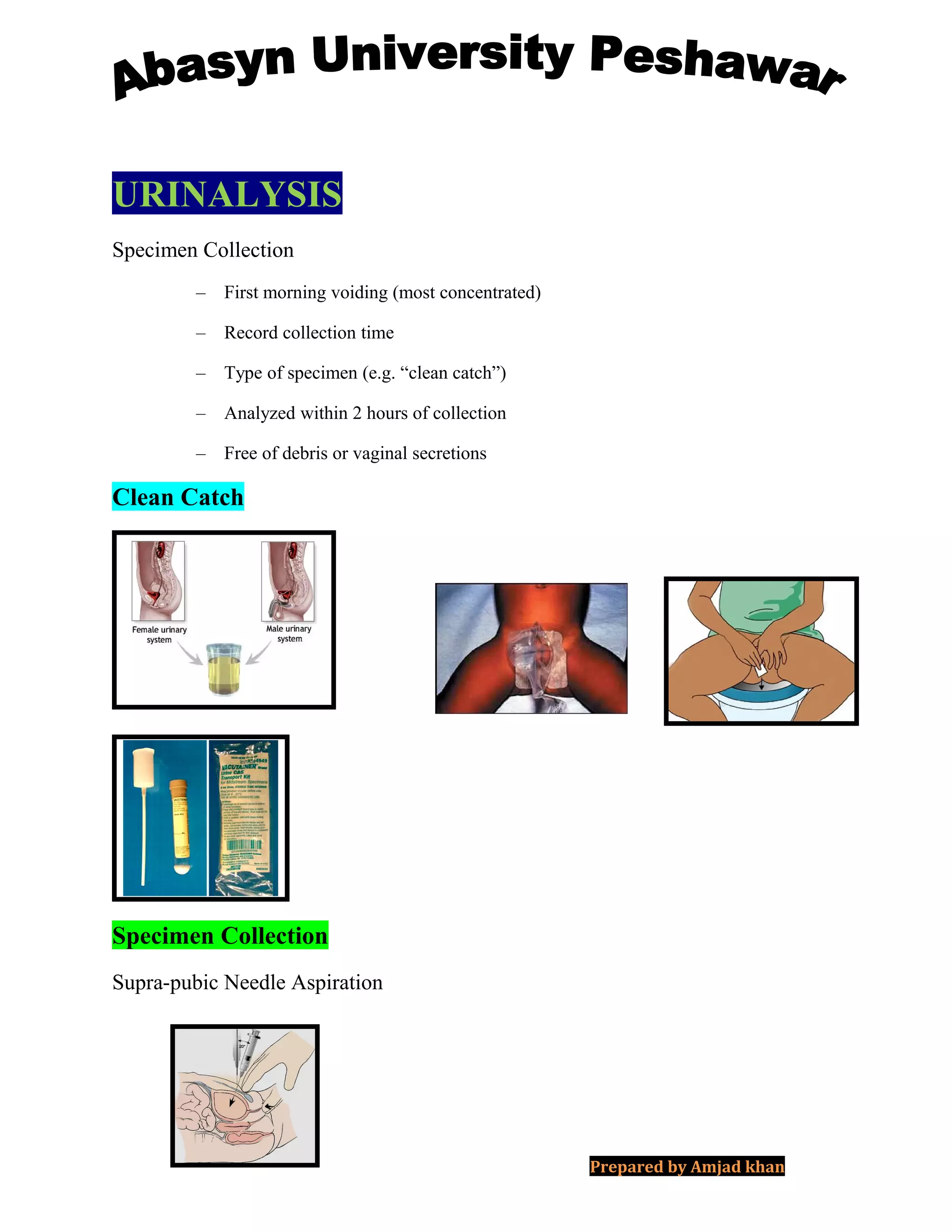
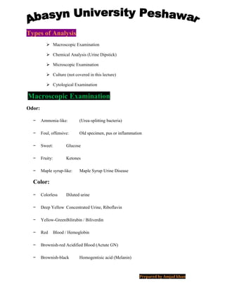
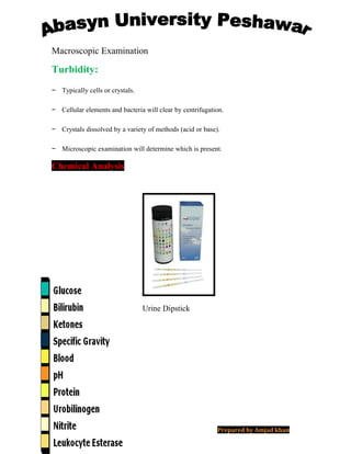
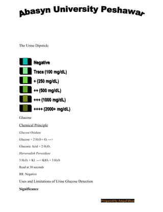
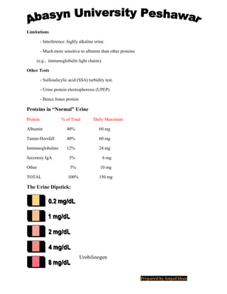
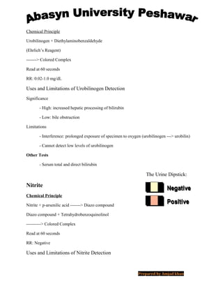
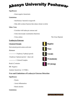
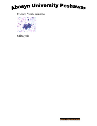
URINALYSIS
Specimen Collection
– First morning voiding (most concentrated)
– Record collection time
– Type of specimen (e.g. “clean catch”)
– Analyzed within 2 hours of collection
– Free of debris or vaginal secretions
Clean Catch
Specimen Collection
Supra-pubic Needle Aspiration
Prepared by Amjad khan
Types of Analysis
 Macroscopic Examination
 Chemical Analysis (Urine Dipstick)
 Microscopic Examination
 Culture (not covered in this lecture)
 Cytological Examination
Macroscopic Examination
Odor:
− Ammonia-like: (Urea-splitting bacteria)
− Foul, offensive: Old specimen, pus or inflammation
− Sweet: Glucose
− Fruity: Ketones
− Maple syrup-like: Maple Syrup Urine Disease
Color:
− Colorless Diluted urine
− Deep Yellow Concentrated Urine, Riboflavin
− Yellow-GreenBilirubin / Biliverdin
− Red Blood / Hemoglobin
− Brownish-red Acidified Blood (Actute GN)
− Brownish-black Homogentisic acid (Melanin)
Prepared by Amjad khan
Macroscopic Examination
Turbidity:
− Typically cells or crystals.
− Cellular elements and bacteria will clear by centrifugation.
− Crystals dissolved by a variety of methods (acid or base).
− Microscopic examination will determine which is present.
Chemical Analysis
Urine Dipstick
Prepared by Amjad khan
The Urine Dipstick:
Glucose
Chemical Principle
Glucose Oxidase
Glucose + 2 H2O+ O2 --->
Gluconic Acid + 2 H2O2
Horseradish Peroxidase
3 H2O2 + KI ---> KIO3 + 3 H2O
Read at 30 seconds
RR: Negative
Uses and Limitations of Urine Glucose Detection
Significance
Prepared by Amjad khan
– Diabetes mellitus.
– Renal glycosuria.
Limitations
– Interference: reducing agents, ketones.
– Only measures glucose and not other sugars.
– Renal threshold must be passed in order for glucose to spill into the urine.
Other Tests
– CuSO4 test for reducing sugars.
Detection of Reducing Sugars* by CuSO4
Sugar Disease(s)
- Galactose Galactosemias
- Fructose Fructosuria, Fructose Intolerance, etc.
- Lactose Lactase Deficiency
- Pentoses Essential Pentosuria
- Maltose Non-pathogenic
* NOT Sucrose because it is not a reducing sugar
The Urine Dipstick:
Bilirrubin
Chemical Principle
Bilirubin + Diazo salt ---------> Azobilirubin
Read at 30 seconds
RR: Negative
Prepared by Amjad khan
Uses and Limitations of Urine Bilirrubin Detection
Significance
- Increased direct bilirubin (correlates with urobilinogen and serum bilirubin)
Limitations
- Interference: prolonged exposure of sample to light
- Only measures direct bilirubin--will not pick up indirect bilirubin
Other Tests
- Ictotest (more sensitive tablet version of same assay)
- Serum test for total and direct bilirubin is more informative
The Urine Dipstick:
Ketones
Chemical Principle
Acetoacetic Acid + Nitroprusside
------> Colored Complex
Read at 40 seconds
RR: Negative
Uses and Limitations of Urine Ketone Detection
Significance
- Diabetic ketoacidosis
- Prolonged fasting
Limitations
- Interference: expired reagents (degradation with exposure to moisture in air)
Prepared by Amjad khan
- Only measures acetoacetate not other ketone bodies (such as in rebound ketosis).
Other Tests
- Ketostix (more sensitive tablet version of same assay)
- Serum glucose measurement to confirm DKA
The Urine Dipstick:
Specific Gravity
Chemical Principle
X+
+ Polymethyl vinyl ether / maleic anhydride
--------------->
X+
-Polymethyl vinyl ether / maleic anhydride + H+
H+
interacts with a Bromthymol Blue indicator to
form a colored complex.
Read up to 2 minutes
RR: 1.003-1.035
Uses and Limitations of Urine Specific Gravity
Significance
- Diabetes insipidus
Limitations
- Interference: alkaline urine
- Does not measure non-ionized solutes (e.g. glucose)
Prepared by Amjad khan
Other Tests
- Refractometry
- Hydrometer
- Osmolality measurement (typically used with water deprivation test)
The Urine Dipstick:
Negative
Trace (non-hemolyzed)
Moderate (non-hemolyzed)
+ (weak)
++ (moderate)
+++ (strong)
Hemolyzed
Blood
Chemical Principle
Lysing agent to lyse red blood cells
Diisopropylbenzene dihydroperoxide +
Tetramethylbenzidine
Prepared by Amjad khan
------------> Colored Complex
Read at 60 seconds
RR: Negative
Analytic Sensitivity: 10 RBCs
Uses and Limitations of Urine Blood Detection
Significance
- Hematuria (nephritis, trauma, etc)
- Hemoglobinuria (hemolysis, etc)
- Myoglobinuria (rhabdomyolysis, etc)
Limitations
- Interference: reducing agents, microbial peroxidases
- Cannot distinguish between the above disease processes
Other Tests
- Urine microscopic examination
- Urine cytology
The Urine Dipstick:
pH
Chemical Principle
Methyl Red (at high concentration; low pH) and
H+
interacts with:
Bromthymol Blue (at low concentration; high pH), to form a colored
complexes
(dual indicator system)
Read up to 2 minutes
R.R.: 4.5-8.0
Prepared by Amjad khan
Uses and Limitations of Urine pH Detection
Significance
- Acidic (less than 4.5): metabolic acidosis, high-protein diet
- Alkaline (greater than 8.0): renal tubular acidosis (>5.5)
Limitations
- Interference: bacterial overgrowth (alkaline or acidic),
“run over effect” effect of protein pad on pH indicator pad
Other Tests
- Titrable acidity
- Blood gases to determine acid-base status
PH Run Over Effect
Buffers from the protein area of the strip (pH 3.0) spill over to the pH
area of the strip and make the pH of the sample appear more acidic than it really is.
Prepared by Amjad khan
The Urine Dipstick:
Protein
Chemical Principle
Prepared by Amjad khan
Causes of Proteinuria
Functional Renal
- Severe muscular exertion - Glomerulonephritis
- Pregnancy - Nephrotic syndrome
- Orthostatic proteinuria - Renal tumor or infection
Pre-Renal Post-Renal
- Fever - Cystitis
- Renal hypoxia - Urethritis or prostatitis
- Hypertension - Contamination with vaginal
secretions
Nephrotic Syndrome (> 3.5 g/dL in 24 h)
Primary
- Lipoid nephrosis (severe)
- Membranous glomerulonephritis
- Membranoproliferative glomerulonephritis
Secondary
- Diabetes mellitus (Kimmelsteil-Wilson lesions)
- Systemic lupus erythematosus
- Amyloidosis and other infiltrative diseases
- Renal vein thrombosis
Uses and Limitations of Urine Protein Detection
Significance
- Proteinuria and the nephrotic syndrome.
Prepared by Amjad khan
Limitations
- Interference: highly alkaline urine.
- Much more sensitive to albumin than other proteins
(e.g., immunoglobulin light chains).
Other Tests
- Sulfosalicylic acid (SSA) turbidity test.
- Urine protein electrophoresis (UPEP)
- Bence Jones protein
Proteins in “Normal” Urine
Protein % of Total Daily Maximum
Albumin 40% 60 mg
Tamm-Horsfall 40% 60 mg
Immunoglobulins 12% 24 mg
Secretory IgA 3% 6 mg
Other 5% 10 mg
TOTAL 100% 150 mg
The Urine Dipstick:
Urobilinogen
Prepared by Amjad khan
Chemical Principle
Urobilinogen + Diethylaminobenzaldehyde
(Ehrlich’s Reagent)
-------> Colored Complex
Read at 60 seconds
RR: 0.02-1.0 mg/dL
Uses and Limitations of Urobilinogen Detection
Significance
- High: increased hepatic processing of bilirubin
- Low: bile obstruction
Limitations
- Interference: prolonged exposure of specimen to oxygen (urobilinogen ---> urobilin)
- Cannot detect low levels of urobilinogen
Other Tests
- Serum total and direct bilirubin
The Urine Dipstick:
Nitrite
Chemical Principle
Nitrite + p-arsenilic acid -------> Diazo compound
Diazo compound + Tetrahydrobenzoquinolinol
----------> Colored Complex
Read at 60 seconds
RR: Negative
Uses and Limitations of Nitrite Detection
Prepared by Amjad khan
Significance
- Gram negative bacteriuria
Limitations
- Interference: bacterial overgrowth
- Only able to detect bacteria that reduce nitrate to nitrite
Other Tests
- Correlate with leukocyte esterase and
- Urine microscopic examination (bacteria)
- Urine culture The Urine Dipstick
Leukocyte Esterase
Chemical Principle
Derivatized pyrrole amino acid ester
Esterases
------------> 3-hydroxy-5-phenyl pyrrole
3-hydroxy-5-phenyl pyrrole + diazo salt
-------------> Colored Complex
Read at 2 minutes
RR: Negative
Analytic Sensitivity: 3-5 WBCs
Uses and Limitations of Leukocyte Esterase Detection
Significance
- Pyuria
- Acute inflammation
- Renal calculus
Limitations
Prepared by Amjad khan
- Interference: oxidizing agents, menstrual contamination
Other Tests
- Urine microscopic examination (WBCs and bacteria)
- Urine culture
Microscopic Examination
General Aspects
Preservation
- Cells and casts begin to disintegrate in 1 - 3 hrs. at room temp.
- Refrigeration for up to 48 hours (little loss of cells).
Specimen concentration
- Ten to twenty-fold concentration by centrifugation.
Types of microscopy
- Phase contrast microscopy
- Polarized microscopy
- Bright field microscopy with special staining
(e.g., Sternheimer-Malbin stain)
Microscopic Examination
Abnormal Findings
Per High Power Field (HPF) (400x)
– > 3 erythrocytes
– > 5 leukocytes
– > 2 renal tubular cells
– > 10 bacteria
Per Low Power Field (LPF) (200x)
Prepared by Amjad khan
– > 3 hyaline casts or > 1 granular cast
– > 10 squamous cells (indicative of contaminated specimen)
– Any other cast (RBCs, WBCs)
Presence of:
– Fungal hyphae or yeast, parasite, viral inclusions
– Pathological crystals (cystine, leucine, tyrosine)
– Large number of uric acid or calcium oxalate crystals
Microscopic Examination
CELLS
Erythrocytes
- “Dysmorphic” vs. “normal” (> 10 per HPF)
Leukocytes
- Neutrophils (glitter cells) More than 1 per 3 HPF
- Eosinophils Hansel test (special stain)
Epithelial Cells
- Squamous cells Indicate level of contamination
- Renal tubular epithelial cells Few are normal
- Transitional epithelial cells Few are normal
- Oval fat bodies Abnormal, indicate Nephrosis
Microscopic Examination
RBCs
Prepared by Amjad khan
WBCs
Squamous Cells
Tubular Epithelial Cells
Transitional Cells
Prepared by Amjad khan
Oval Fat Body
LE Cell
Microscopic Examination
Bacteria & Yeasts
Bacteria
- Bacteriuria More than 10 per HPF
Yeasts
- Candidiasis Most likely a contaminant
Prepared by Amjad khan
but should correlate with
clinical picture.
Viruses
- CMV inclusions Probable viral cystitis.
Bacteria
Yeasts
Cytomegalovirus
Microscopic Examination
Casts
Prepared by Amjad khan
Erythrocyte Casts:Glomerular diseases
Leukocyte Casts: Pyuria, glomerular disease
Degenerating Casts:
- Granular casts Nonspecific (Tamm-Horsfall protein)
- Hyaline castsNonspecific (Tamm-Horsfall protein)
- Waxy casts Nonspecific
- Fatty casts Nephrotic syndrome
(oval fat body casts)
RBCs Cast
RBCs Cast - Histology
Prepared by Amjad khan
WBCs Cast
Tubular Epith. Cast
Granular Cast Hyaline Cast
Waxy Cast
Prepared by Amjad khan
Fatty Cast
Significance of Cellular Casts
Microscopic Examination
Crystals
Prepared by Amjad khan
- Urate
Ammonium biurate
Uric acid
- Triple Phosphate
- Calcium Oxalate
- Amino Acids
Cystine
Leucine
Tyrosine
- Sulfonamide
Calcium Oxalate Crystals
Triple Phosphate Crystals
Urate Crystals
Prepared by Amjad khan
Leucine Crystals
Cystine Crystals
Ammonium Biurate Crystals
Cholesterol Crystals
Prepared by Amjad khan
Cytological Examination
Staining:
– Papanicolau
– Wright’s
– Immunoperoxidase
– Immunofluorescence
Cytology: Normal
Cytology: Reactive
Cytology: Reactive
Prepared by Amjad khan
Cytology: Polyoma (Decoy Cell)
Immunoperoxidase to SV40 ag
Cytology: TCC Low Grade
Cytology: TCC High Grade
Prepared by Amjad khan
Cytology: Squamous Cell Ca.
Cytology: Renal Cell Ca.
Prepared by Amjad khan
Cytology: Prostatic Carcinoma
Urinalysis
Prepared by Amjad khan

Urinalysis

  • 1.
    URINALYSIS Specimen Collection – Firstmorning voiding (most concentrated) – Record collection time – Type of specimen (e.g. “clean catch”) – Analyzed within 2 hours of collection – Free of debris or vaginal secretions Clean Catch Specimen Collection Supra-pubic Needle Aspiration Prepared by Amjad khan
  • 2.
    Types of Analysis Macroscopic Examination  Chemical Analysis (Urine Dipstick)  Microscopic Examination  Culture (not covered in this lecture)  Cytological Examination Macroscopic Examination Odor: − Ammonia-like: (Urea-splitting bacteria) − Foul, offensive: Old specimen, pus or inflammation − Sweet: Glucose − Fruity: Ketones − Maple syrup-like: Maple Syrup Urine Disease Color: − Colorless Diluted urine − Deep Yellow Concentrated Urine, Riboflavin − Yellow-GreenBilirubin / Biliverdin − Red Blood / Hemoglobin − Brownish-red Acidified Blood (Actute GN) − Brownish-black Homogentisic acid (Melanin) Prepared by Amjad khan
  • 3.
    Macroscopic Examination Turbidity: − Typicallycells or crystals. − Cellular elements and bacteria will clear by centrifugation. − Crystals dissolved by a variety of methods (acid or base). − Microscopic examination will determine which is present. Chemical Analysis Urine Dipstick Prepared by Amjad khan
  • 4.
    The Urine Dipstick: Glucose ChemicalPrinciple Glucose Oxidase Glucose + 2 H2O+ O2 ---> Gluconic Acid + 2 H2O2 Horseradish Peroxidase 3 H2O2 + KI ---> KIO3 + 3 H2O Read at 30 seconds RR: Negative Uses and Limitations of Urine Glucose Detection Significance Prepared by Amjad khan
  • 5.
    – Diabetes mellitus. –Renal glycosuria. Limitations – Interference: reducing agents, ketones. – Only measures glucose and not other sugars. – Renal threshold must be passed in order for glucose to spill into the urine. Other Tests – CuSO4 test for reducing sugars. Detection of Reducing Sugars* by CuSO4 Sugar Disease(s) - Galactose Galactosemias - Fructose Fructosuria, Fructose Intolerance, etc. - Lactose Lactase Deficiency - Pentoses Essential Pentosuria - Maltose Non-pathogenic * NOT Sucrose because it is not a reducing sugar The Urine Dipstick: Bilirrubin Chemical Principle Bilirubin + Diazo salt ---------> Azobilirubin Read at 30 seconds RR: Negative Prepared by Amjad khan
  • 6.
    Uses and Limitationsof Urine Bilirrubin Detection Significance - Increased direct bilirubin (correlates with urobilinogen and serum bilirubin) Limitations - Interference: prolonged exposure of sample to light - Only measures direct bilirubin--will not pick up indirect bilirubin Other Tests - Ictotest (more sensitive tablet version of same assay) - Serum test for total and direct bilirubin is more informative The Urine Dipstick: Ketones Chemical Principle Acetoacetic Acid + Nitroprusside ------> Colored Complex Read at 40 seconds RR: Negative Uses and Limitations of Urine Ketone Detection Significance - Diabetic ketoacidosis - Prolonged fasting Limitations - Interference: expired reagents (degradation with exposure to moisture in air) Prepared by Amjad khan
  • 7.
    - Only measuresacetoacetate not other ketone bodies (such as in rebound ketosis). Other Tests - Ketostix (more sensitive tablet version of same assay) - Serum glucose measurement to confirm DKA The Urine Dipstick: Specific Gravity Chemical Principle X+ + Polymethyl vinyl ether / maleic anhydride ---------------> X+ -Polymethyl vinyl ether / maleic anhydride + H+ H+ interacts with a Bromthymol Blue indicator to form a colored complex. Read up to 2 minutes RR: 1.003-1.035 Uses and Limitations of Urine Specific Gravity Significance - Diabetes insipidus Limitations - Interference: alkaline urine - Does not measure non-ionized solutes (e.g. glucose) Prepared by Amjad khan
  • 8.
    Other Tests - Refractometry -Hydrometer - Osmolality measurement (typically used with water deprivation test) The Urine Dipstick: Negative Trace (non-hemolyzed) Moderate (non-hemolyzed) + (weak) ++ (moderate) +++ (strong) Hemolyzed Blood Chemical Principle Lysing agent to lyse red blood cells Diisopropylbenzene dihydroperoxide + Tetramethylbenzidine Prepared by Amjad khan
  • 9.
    ------------> Colored Complex Readat 60 seconds RR: Negative Analytic Sensitivity: 10 RBCs Uses and Limitations of Urine Blood Detection Significance - Hematuria (nephritis, trauma, etc) - Hemoglobinuria (hemolysis, etc) - Myoglobinuria (rhabdomyolysis, etc) Limitations - Interference: reducing agents, microbial peroxidases - Cannot distinguish between the above disease processes Other Tests - Urine microscopic examination - Urine cytology The Urine Dipstick: pH Chemical Principle Methyl Red (at high concentration; low pH) and H+ interacts with: Bromthymol Blue (at low concentration; high pH), to form a colored complexes (dual indicator system) Read up to 2 minutes R.R.: 4.5-8.0 Prepared by Amjad khan
  • 10.
    Uses and Limitationsof Urine pH Detection Significance - Acidic (less than 4.5): metabolic acidosis, high-protein diet - Alkaline (greater than 8.0): renal tubular acidosis (>5.5) Limitations - Interference: bacterial overgrowth (alkaline or acidic), “run over effect” effect of protein pad on pH indicator pad Other Tests - Titrable acidity - Blood gases to determine acid-base status PH Run Over Effect Buffers from the protein area of the strip (pH 3.0) spill over to the pH area of the strip and make the pH of the sample appear more acidic than it really is. Prepared by Amjad khan
  • 11.
    The Urine Dipstick: Protein ChemicalPrinciple Prepared by Amjad khan
  • 12.
    Causes of Proteinuria FunctionalRenal - Severe muscular exertion - Glomerulonephritis - Pregnancy - Nephrotic syndrome - Orthostatic proteinuria - Renal tumor or infection Pre-Renal Post-Renal - Fever - Cystitis - Renal hypoxia - Urethritis or prostatitis - Hypertension - Contamination with vaginal secretions Nephrotic Syndrome (> 3.5 g/dL in 24 h) Primary - Lipoid nephrosis (severe) - Membranous glomerulonephritis - Membranoproliferative glomerulonephritis Secondary - Diabetes mellitus (Kimmelsteil-Wilson lesions) - Systemic lupus erythematosus - Amyloidosis and other infiltrative diseases - Renal vein thrombosis Uses and Limitations of Urine Protein Detection Significance - Proteinuria and the nephrotic syndrome. Prepared by Amjad khan
  • 13.
    Limitations - Interference: highlyalkaline urine. - Much more sensitive to albumin than other proteins (e.g., immunoglobulin light chains). Other Tests - Sulfosalicylic acid (SSA) turbidity test. - Urine protein electrophoresis (UPEP) - Bence Jones protein Proteins in “Normal” Urine Protein % of Total Daily Maximum Albumin 40% 60 mg Tamm-Horsfall 40% 60 mg Immunoglobulins 12% 24 mg Secretory IgA 3% 6 mg Other 5% 10 mg TOTAL 100% 150 mg The Urine Dipstick: Urobilinogen Prepared by Amjad khan
  • 14.
    Chemical Principle Urobilinogen +Diethylaminobenzaldehyde (Ehrlich’s Reagent) -------> Colored Complex Read at 60 seconds RR: 0.02-1.0 mg/dL Uses and Limitations of Urobilinogen Detection Significance - High: increased hepatic processing of bilirubin - Low: bile obstruction Limitations - Interference: prolonged exposure of specimen to oxygen (urobilinogen ---> urobilin) - Cannot detect low levels of urobilinogen Other Tests - Serum total and direct bilirubin The Urine Dipstick: Nitrite Chemical Principle Nitrite + p-arsenilic acid -------> Diazo compound Diazo compound + Tetrahydrobenzoquinolinol ----------> Colored Complex Read at 60 seconds RR: Negative Uses and Limitations of Nitrite Detection Prepared by Amjad khan
  • 15.
    Significance - Gram negativebacteriuria Limitations - Interference: bacterial overgrowth - Only able to detect bacteria that reduce nitrate to nitrite Other Tests - Correlate with leukocyte esterase and - Urine microscopic examination (bacteria) - Urine culture The Urine Dipstick Leukocyte Esterase Chemical Principle Derivatized pyrrole amino acid ester Esterases ------------> 3-hydroxy-5-phenyl pyrrole 3-hydroxy-5-phenyl pyrrole + diazo salt -------------> Colored Complex Read at 2 minutes RR: Negative Analytic Sensitivity: 3-5 WBCs Uses and Limitations of Leukocyte Esterase Detection Significance - Pyuria - Acute inflammation - Renal calculus Limitations Prepared by Amjad khan
  • 16.
    - Interference: oxidizingagents, menstrual contamination Other Tests - Urine microscopic examination (WBCs and bacteria) - Urine culture Microscopic Examination General Aspects Preservation - Cells and casts begin to disintegrate in 1 - 3 hrs. at room temp. - Refrigeration for up to 48 hours (little loss of cells). Specimen concentration - Ten to twenty-fold concentration by centrifugation. Types of microscopy - Phase contrast microscopy - Polarized microscopy - Bright field microscopy with special staining (e.g., Sternheimer-Malbin stain) Microscopic Examination Abnormal Findings Per High Power Field (HPF) (400x) – > 3 erythrocytes – > 5 leukocytes – > 2 renal tubular cells – > 10 bacteria Per Low Power Field (LPF) (200x) Prepared by Amjad khan
  • 17.
    – > 3hyaline casts or > 1 granular cast – > 10 squamous cells (indicative of contaminated specimen) – Any other cast (RBCs, WBCs) Presence of: – Fungal hyphae or yeast, parasite, viral inclusions – Pathological crystals (cystine, leucine, tyrosine) – Large number of uric acid or calcium oxalate crystals Microscopic Examination CELLS Erythrocytes - “Dysmorphic” vs. “normal” (> 10 per HPF) Leukocytes - Neutrophils (glitter cells) More than 1 per 3 HPF - Eosinophils Hansel test (special stain) Epithelial Cells - Squamous cells Indicate level of contamination - Renal tubular epithelial cells Few are normal - Transitional epithelial cells Few are normal - Oval fat bodies Abnormal, indicate Nephrosis Microscopic Examination RBCs Prepared by Amjad khan
  • 18.
    WBCs Squamous Cells Tubular EpithelialCells Transitional Cells Prepared by Amjad khan
  • 19.
    Oval Fat Body LECell Microscopic Examination Bacteria & Yeasts Bacteria - Bacteriuria More than 10 per HPF Yeasts - Candidiasis Most likely a contaminant Prepared by Amjad khan
  • 20.
    but should correlatewith clinical picture. Viruses - CMV inclusions Probable viral cystitis. Bacteria Yeasts Cytomegalovirus Microscopic Examination Casts Prepared by Amjad khan
  • 21.
    Erythrocyte Casts:Glomerular diseases LeukocyteCasts: Pyuria, glomerular disease Degenerating Casts: - Granular casts Nonspecific (Tamm-Horsfall protein) - Hyaline castsNonspecific (Tamm-Horsfall protein) - Waxy casts Nonspecific - Fatty casts Nephrotic syndrome (oval fat body casts) RBCs Cast RBCs Cast - Histology RBCs Cast - Histology Prepared by Amjad khan
  • 22.
    WBCs Cast Tubular Epith.Cast Granular Cast Hyaline Cast Waxy Cast Prepared by Amjad khan
  • 23.
    Fatty Cast Significance ofCellular Casts Microscopic Examination Crystals Prepared by Amjad khan
  • 24.
    - Urate Ammonium biurate Uricacid - Triple Phosphate - Calcium Oxalate - Amino Acids Cystine Leucine Tyrosine - Sulfonamide Calcium Oxalate Crystals Triple Phosphate Crystals Urate Crystals Prepared by Amjad khan
  • 25.
    Leucine Crystals Cystine Crystals AmmoniumBiurate Crystals Cholesterol Crystals Prepared by Amjad khan
  • 26.
    Cytological Examination Staining: – Papanicolau –Wright’s – Immunoperoxidase – Immunofluorescence Cytology: Normal Cytology: Reactive Cytology: Reactive Prepared by Amjad khan
  • 27.
    Cytology: Polyoma (DecoyCell) Cytology: Polyoma (Decoy Cell) Immunoperoxidase to SV40 ag Cytology: TCC Low Grade Cytology: TCC High Grade Prepared by Amjad khan
  • 28.
    Cytology: Squamous CellCa. Cytology: Renal Cell Ca. Prepared by Amjad khan
  • 29.