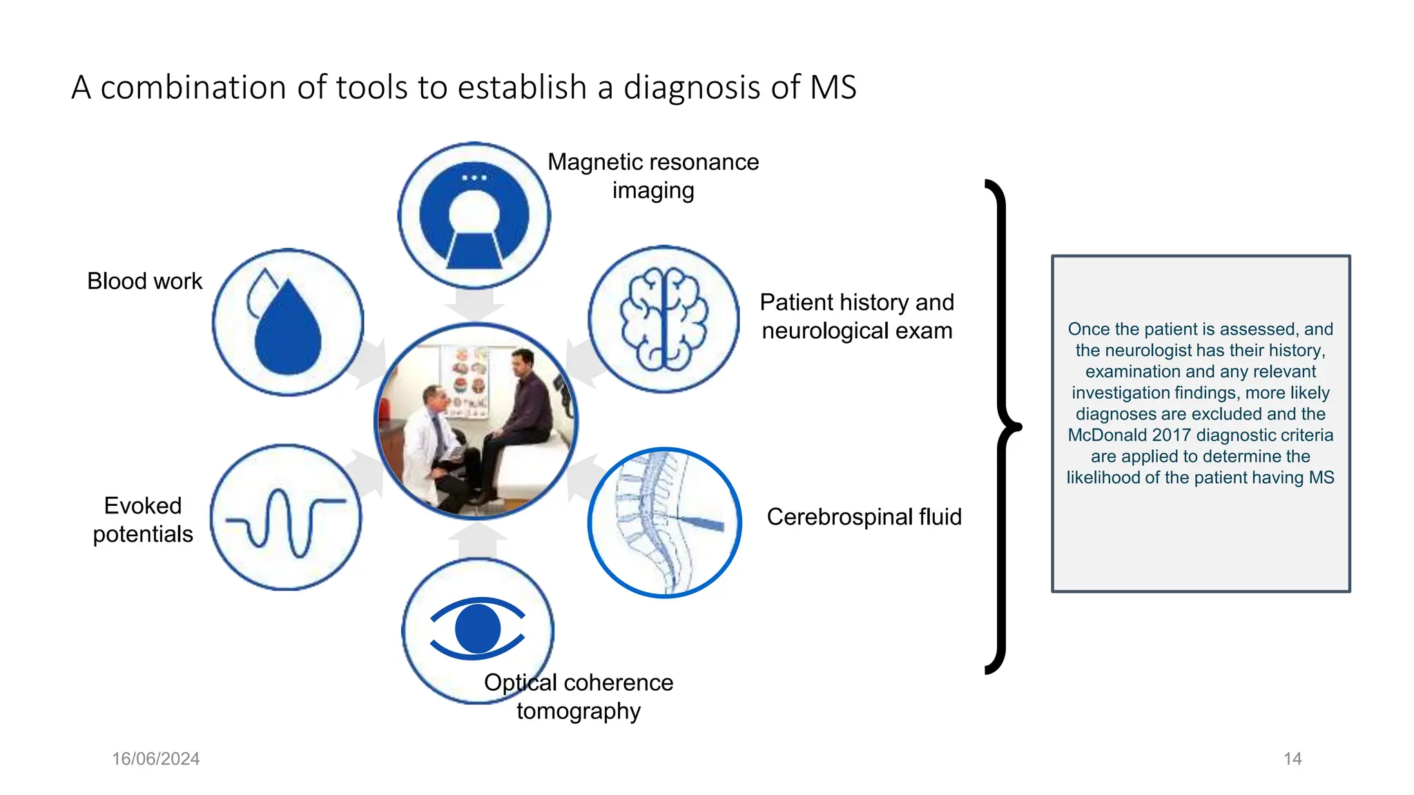
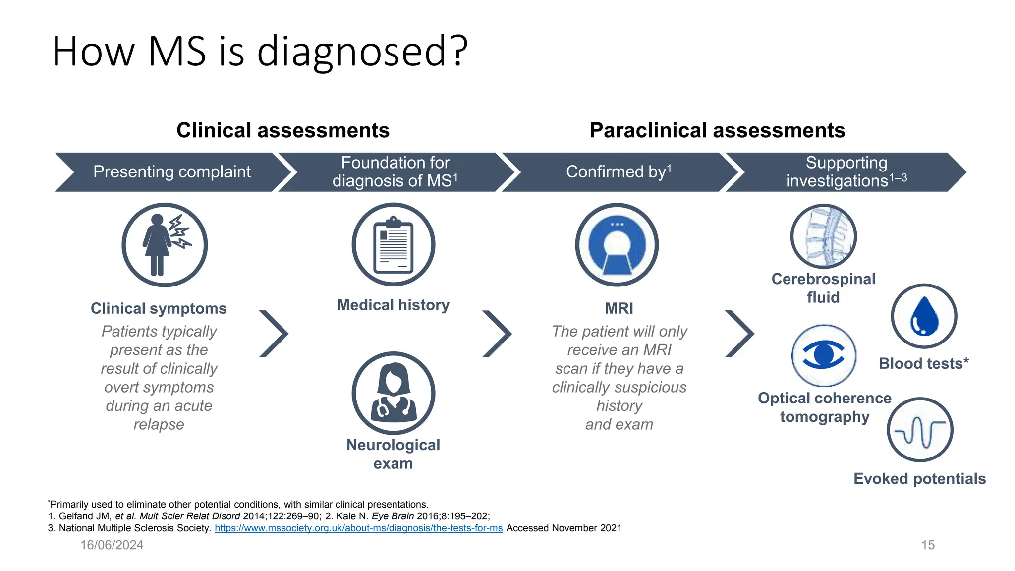
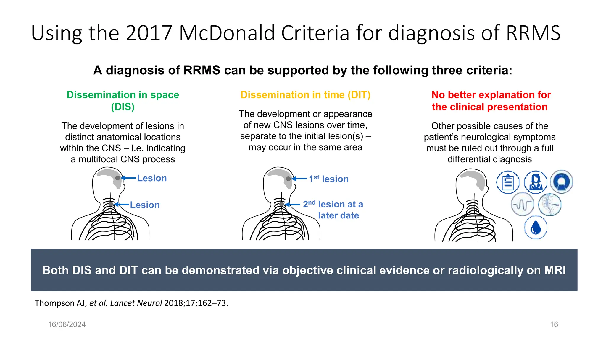
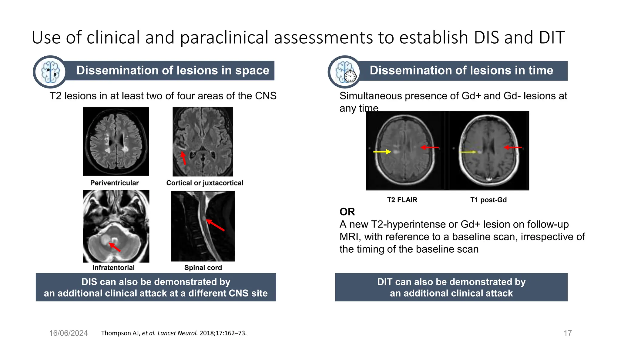
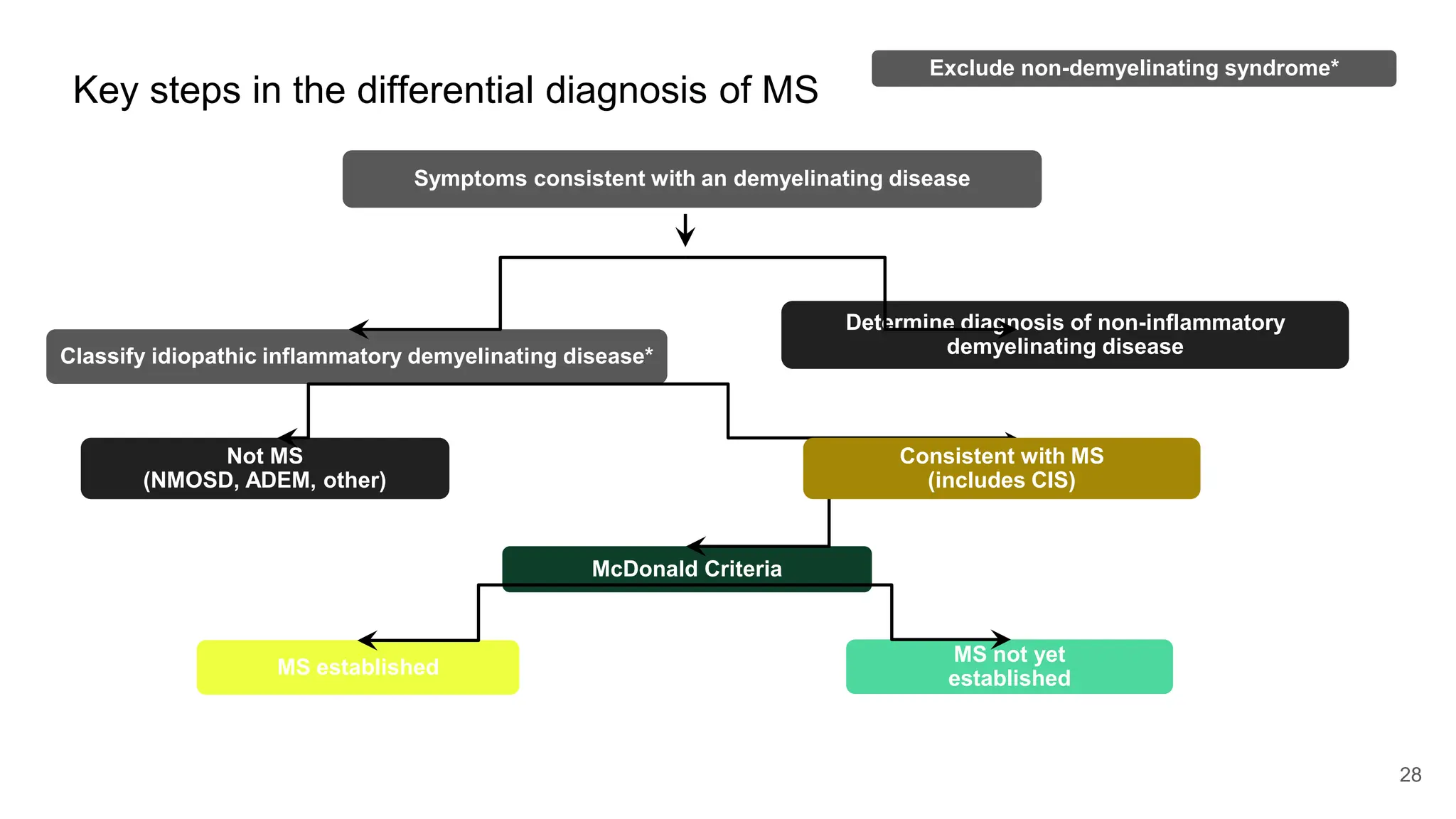
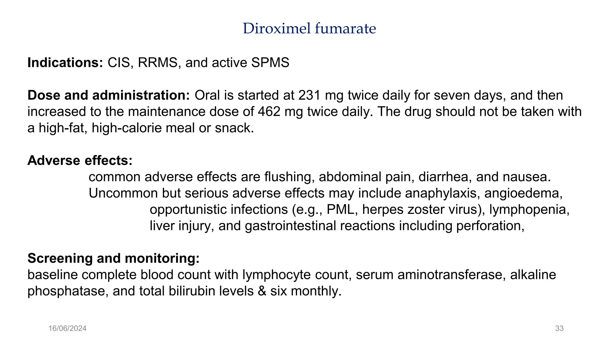
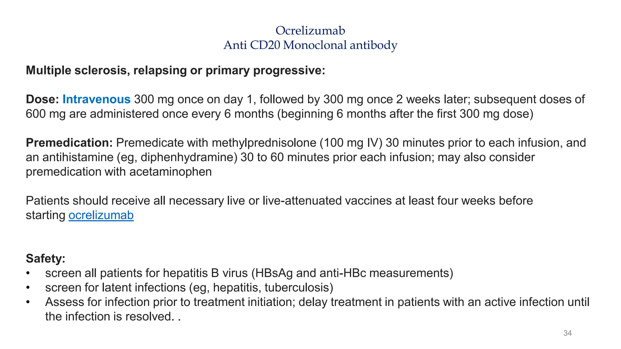
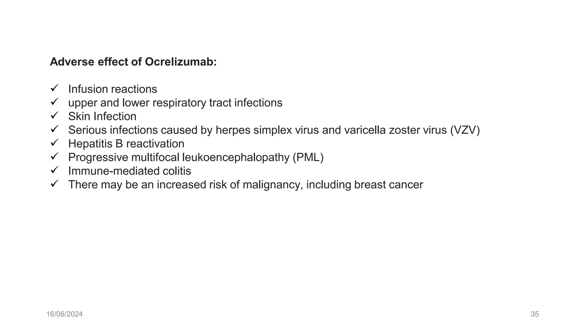
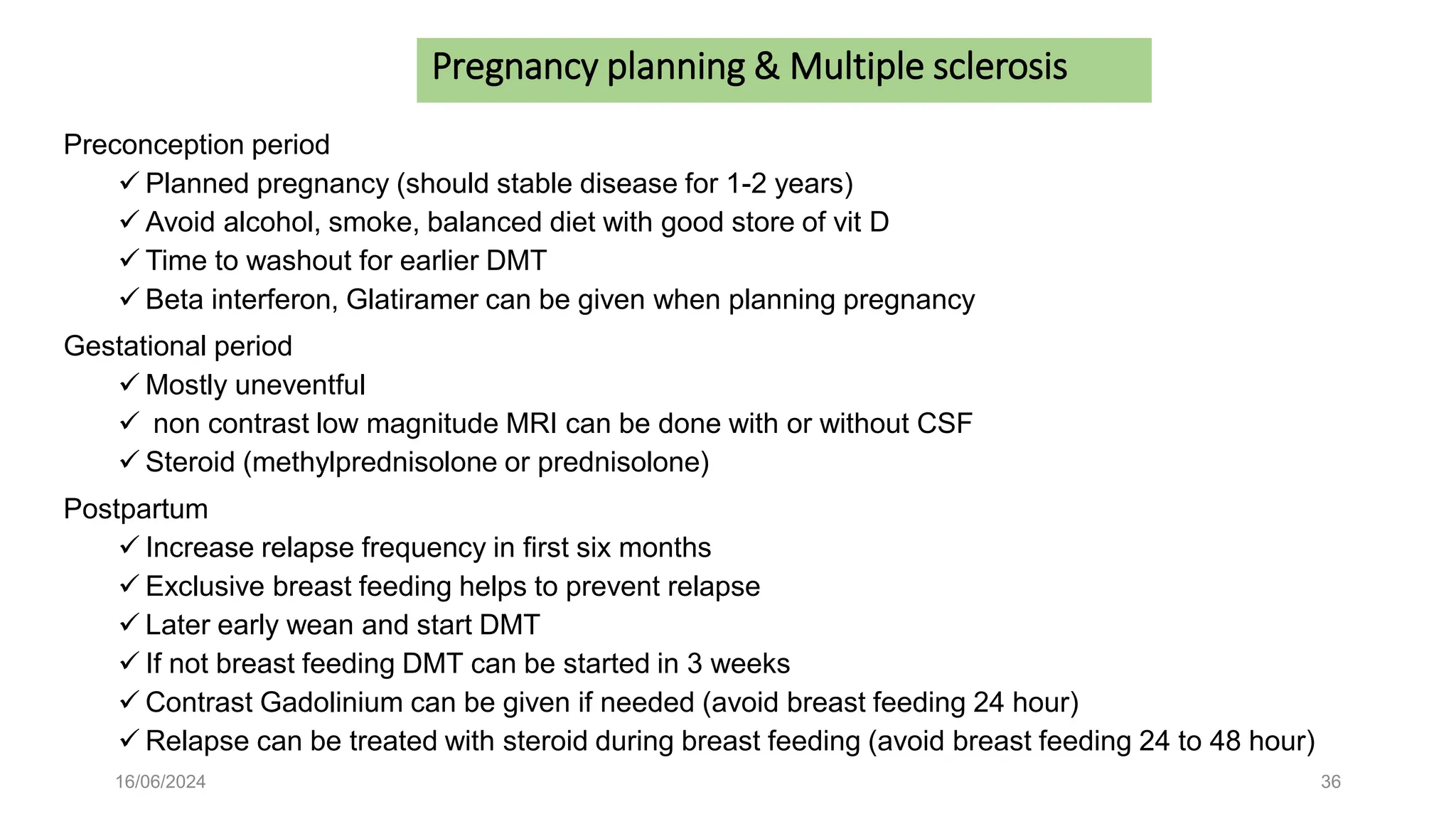
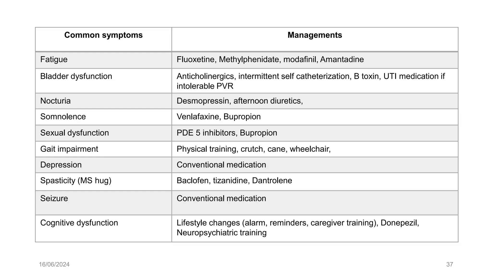
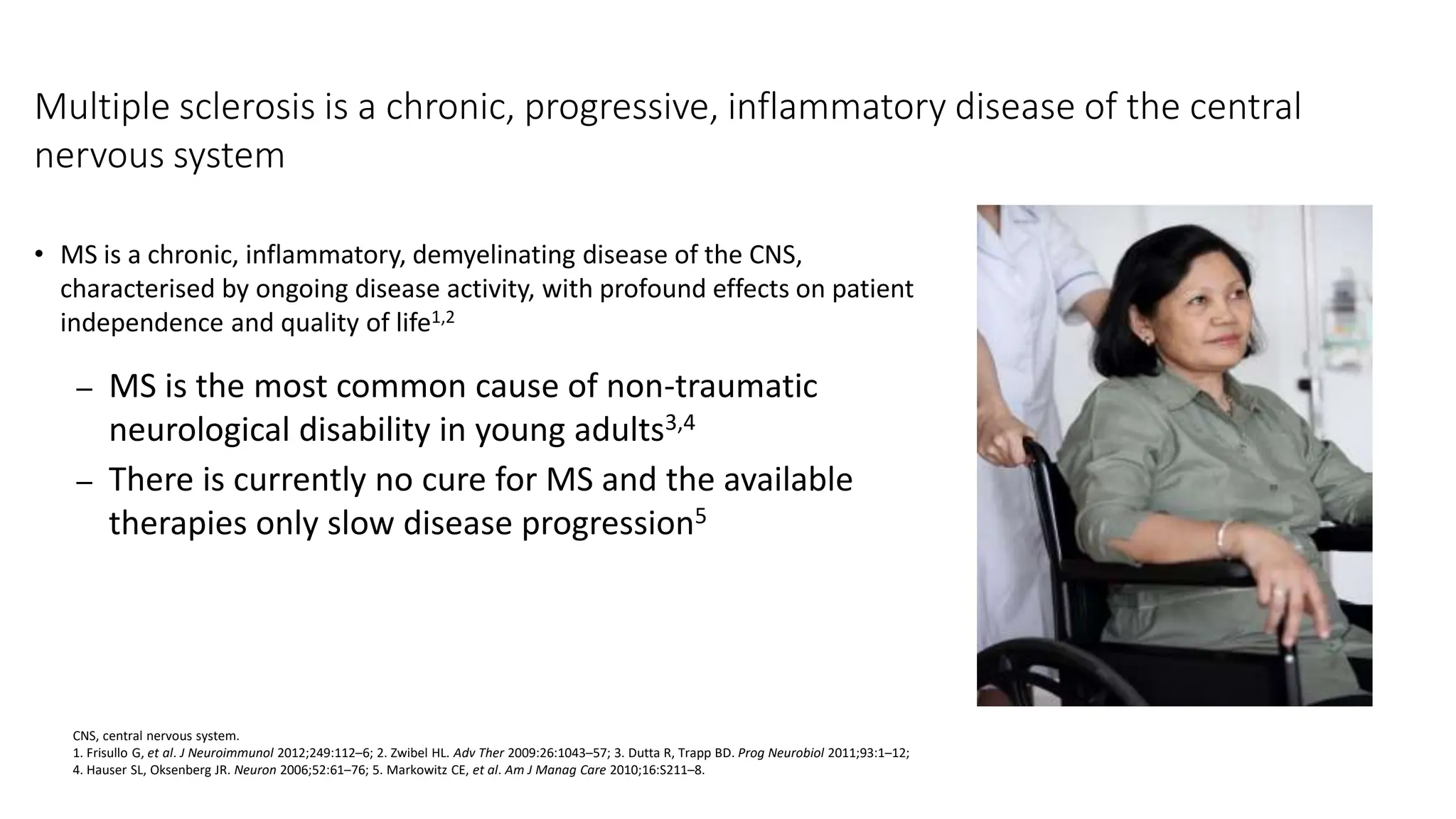
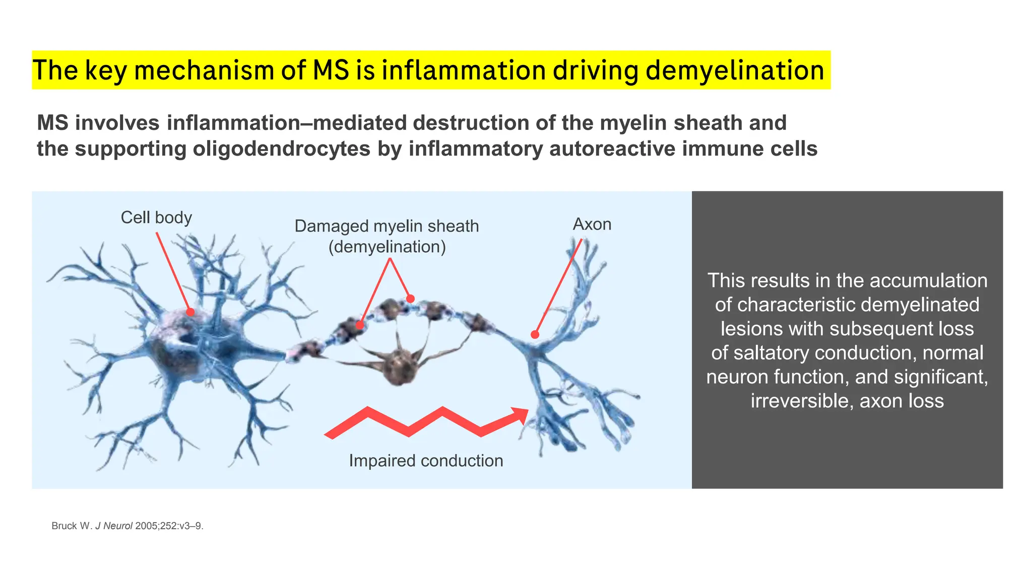
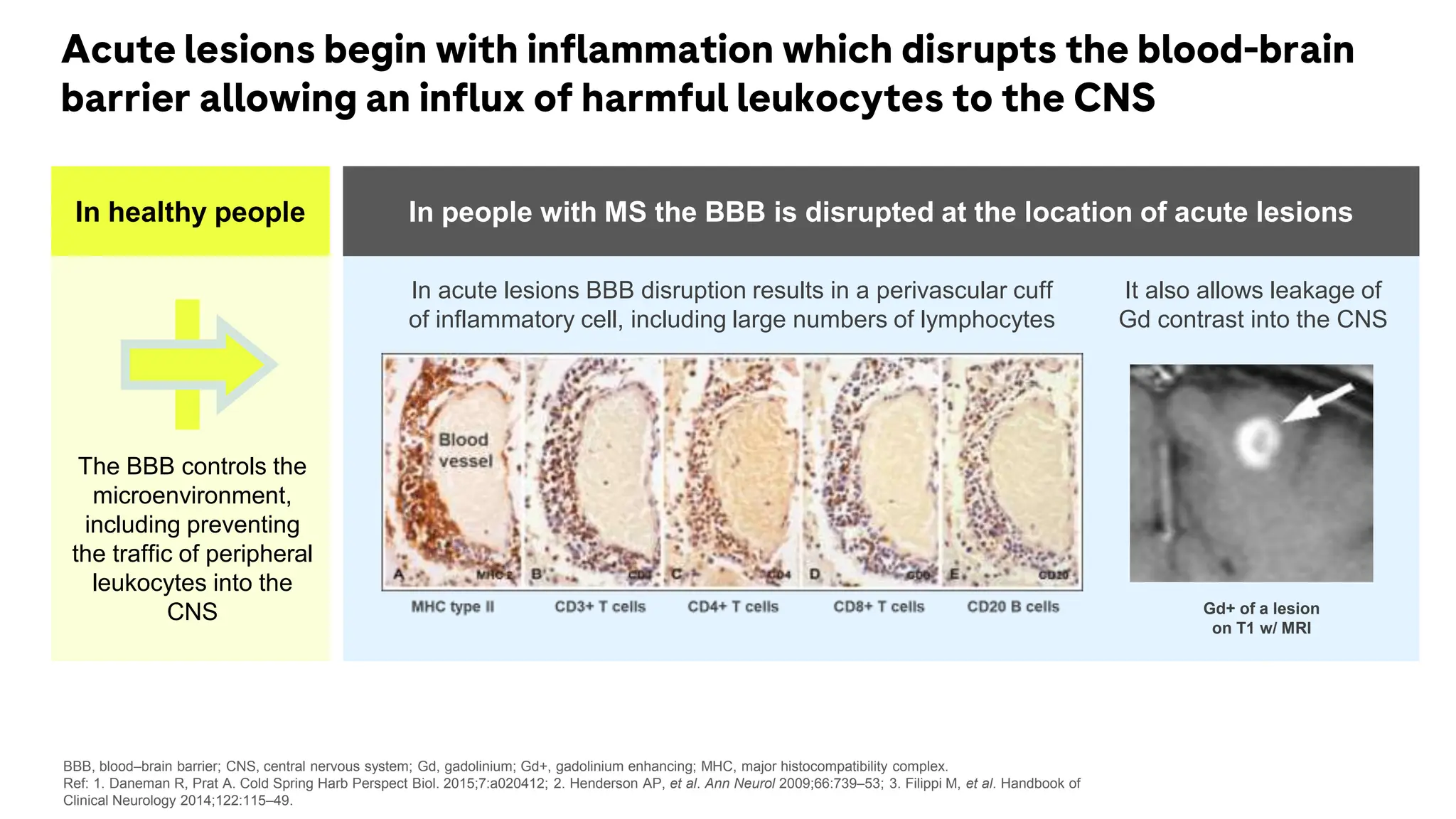
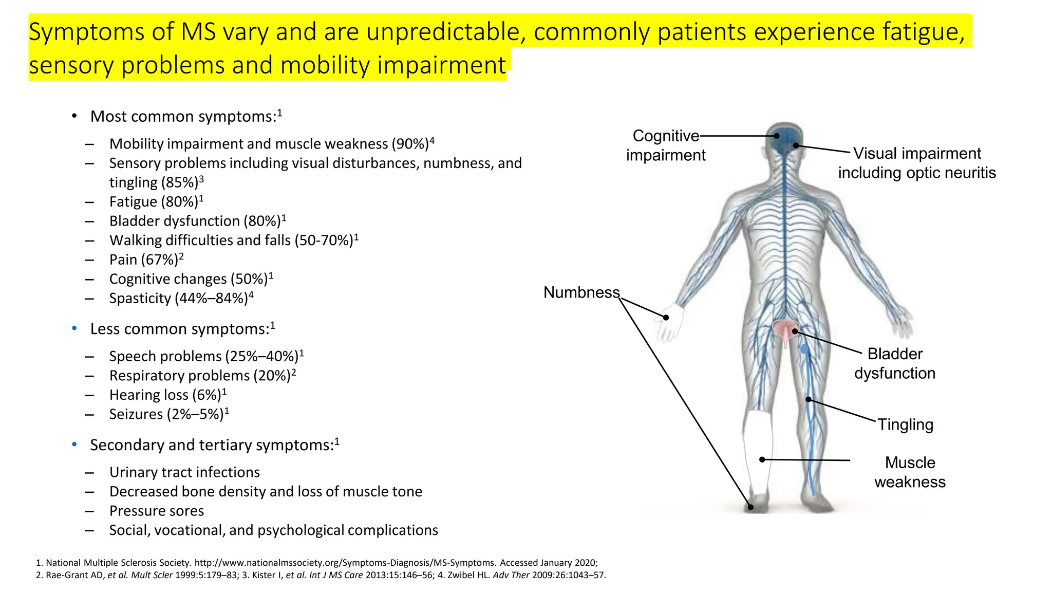
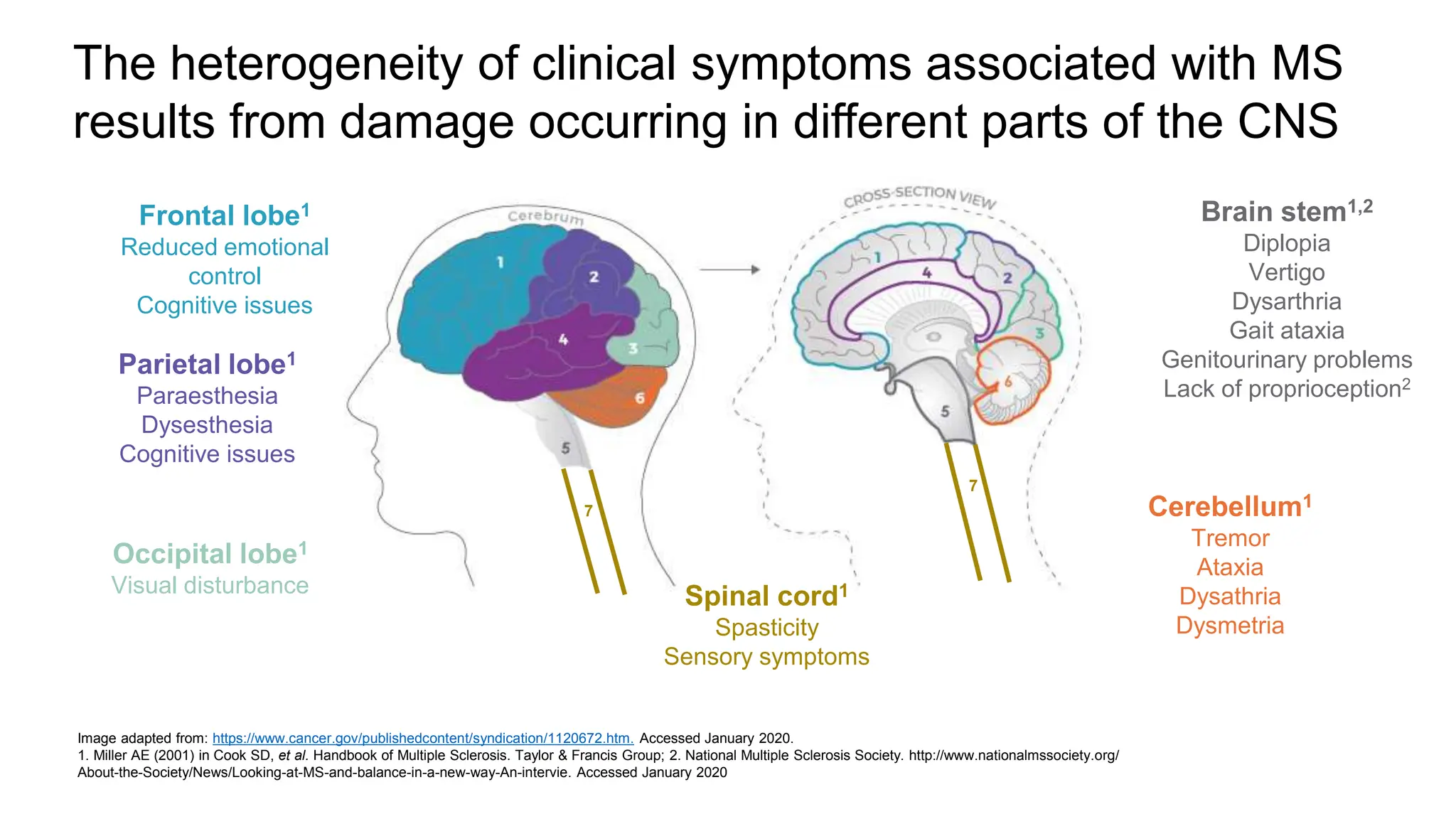
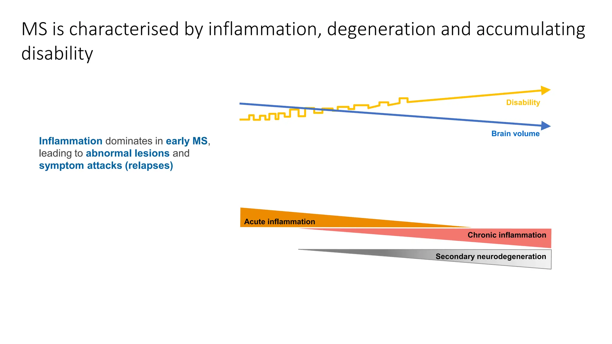
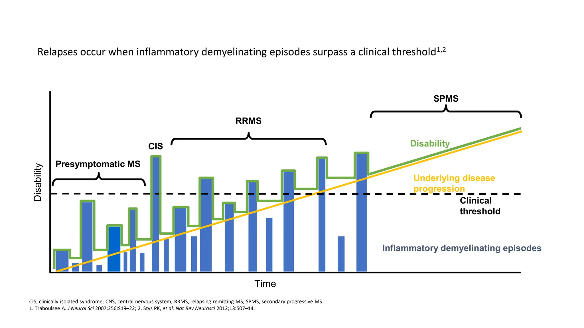
Multiple sclerosis (MS) is a chronic, inflammatory disease affecting the central nervous system characterized by demyelination, leading to significant disability in young adults. There is no cure for MS, with current treatments only slowing its progression, while symptoms vary widely, impacting mobility, sensory functions, and cognitive processes. Diagnosis involves a combination of clinical assessments and imaging techniques, guided by the 2017 McDonald criteria.
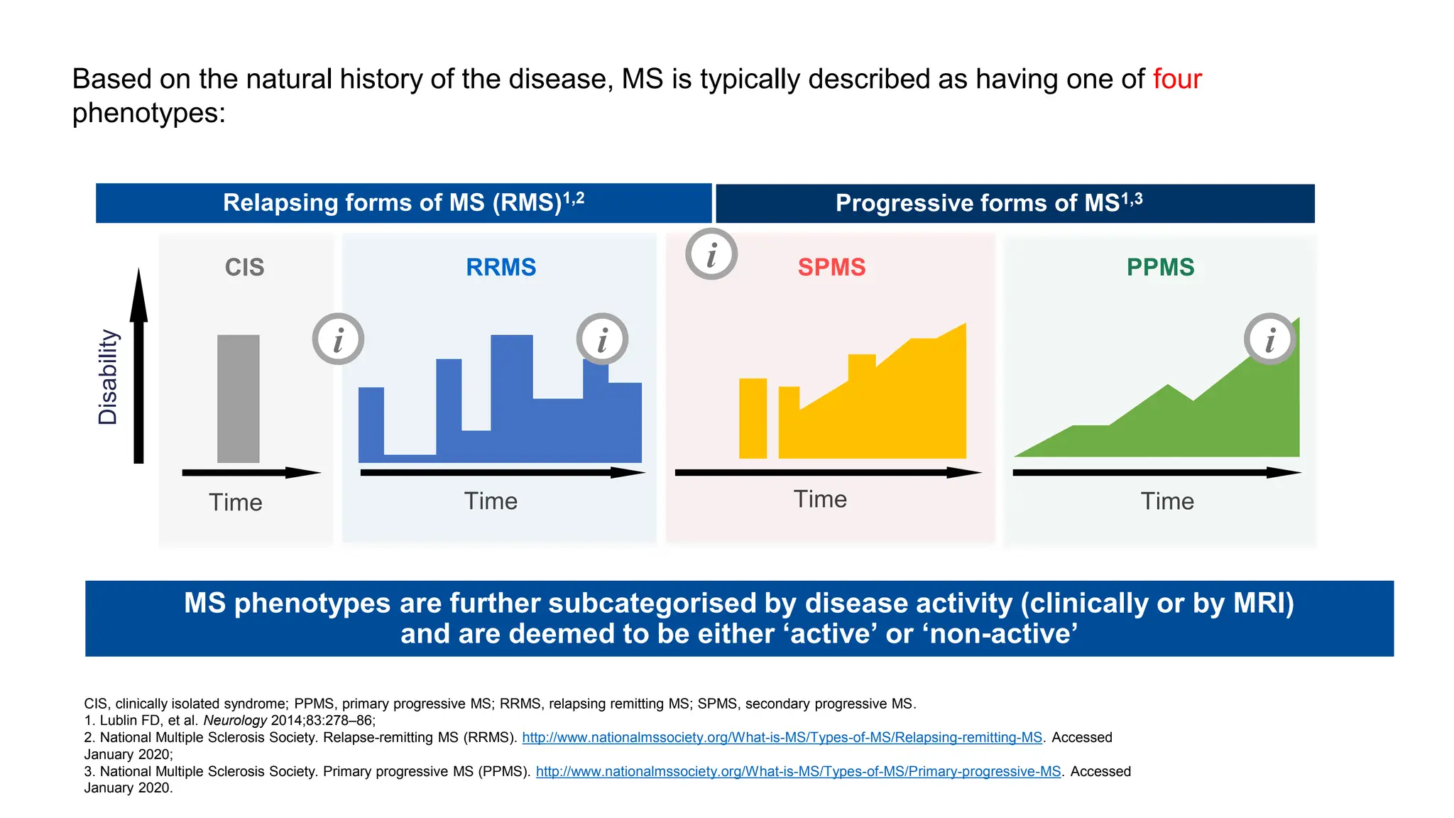
![Relapsing-remitting MS typically has an earlier age of onset than PPMS, sex bias
and different initial presentation
NARCOMS, North American Research Committee on Multiple Sclerosis; PPMS, primary progressive MS; RMS, relapsing MS.
1. Rice CM, et al. J Neurol Neurosurg Psychiatry 2013;84:1100–6; 2. Antel J, et al. Acta Neuropathol 2012;123:627–38; 3. Salter A, et al. Mult Scler 2017 [Epub ahead of print];
4. Cottrell DA, et al. Brain 1999;122:625‒39; 5. Sola P, et al. Mult Scler J 2011;17:303–11; 6. Compston A, Coles A. Lancet 2008;372:1502–17.
PPMS
RRMS
Minimal sex bias1–3*
Higher incidence in females
(3:1 ratio, female to male)1,2
Sensory symptoms
predominate, followed by
visual symptoms1,4‒6
Motor symptoms predominate,
followed by sensory symptoms1,4‒6
~30 years of age1
~45 years of age3](https://image.slidesharecdn.com/updateonms-240616002837-3b931c9f/75/Understanding-Multiple-Sclerosis-and-Management-Updates-11-2048.jpg)
![Disability progression is faster in patients with PPMS than RMS
Curves are not age adjusted.
EDSS, Expanded Disability Status Scale; PPMS, primary progressive MS; RMS, relapsing-onset MS; RRMS, relapsing-remitting MS; SPMS, secondary progressive MS.
1. Sola P, et al. Mult Scler 2011;17:303–11; 2. Musella A, et al. Frontiers Aging Neurosci. 2018;10:238; 3. Salter A, et al. Mult Scler 2017 [Epub ahead of print].
Kaplan–Meier curve for time to EDSS 41
PPMS (n=45)
RMS (RRMS [n=69]
SPMS [n=35])
0.00
0.25
0.50
0.75
1.00
Probability
of
not
reaching
EDSS
of
4
312
Time from diagnosis (months)
0 24 48 72 96 120 144 168 192 216 240 264 288
p<0.0001](https://image.slidesharecdn.com/updateonms-240616002837-3b931c9f/75/Understanding-Multiple-Sclerosis-and-Management-Updates-12-2048.jpg)