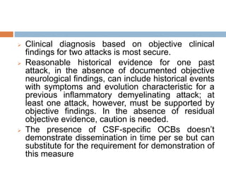
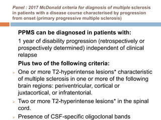
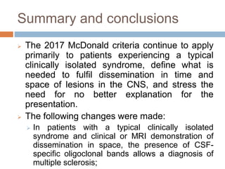
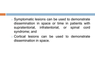
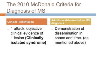
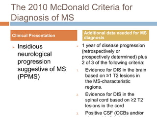
This document summarizes the revisions made to the McDonald criteria for diagnosing multiple sclerosis over time. The 2017 revisions aimed to simplify and clarify the 2010 criteria to facilitate earlier diagnosis when MS is likely but not definitively diagnosed. Key changes include allowing symptomatic and asymptomatic lesions to demonstrate dissemination in space and time, and cortical/juxtacortical lesions to fulfill MRI criteria. CSF oligoclonal bands alone can now establish a diagnosis. The revisions were motivated by new data on diagnostic accuracy in diverse populations and distinguishing MS from similar conditions.
This presentation discusses the revised McDonald's criteria (2017) for the diagnosis of multiple sclerosis. Major changes from the last diagnostic criteria proposed in 2010 have been discussed. Clinical and MRI criteria for dissemination in space and time have been discussed.
This PPT focuses on the diagnosis and treatment of the primary headache disorders, with special emphasis on migraine, the headache most likely to bring patients to physicians and pharmacists. warning signs of the ominous headache, which, although rare, can herald a life-threatening condition. Clinical characteristics of the primary headache types, migraine, tension-type headache, and cluster headache, are described
Magnetic resonance imaging (MRI) is an essential tool for multiple sclerosis (MS) diagnosis and treatment, understanding MS natural history and pathophysiology, and as an outcome measure in clinical trials. This review will provide descriptions of the features, Pathophysiological substrates, and clinical utility of MRI measures of MS including T2-weighted, proton density (PD), and fluid-attenuated inversion recovery (FLAIR) hyperintense lesions, T1-weighted hypointense lesions, gadolinium-enhancing lesions, and measures of brain atrophy. Lesion presence and atrophy within both the brain and spinal cord will be described. This review will also provide a description of non-conventional MRI markers including diffusion tensor imaging (DTI), functional MRI (fMRI), magnetization transfer ratio(MTR) imaging, relaxometry/quantitative magnetic susceptibility (QS) mapping, and magnetic resonance spectroscopy (MRS). Basic descriptions of how these measures are obtained, the pathological substrates, clinical correlates (e.g. with physical disability, cognition, fatigue, etc.) and advantages/ drawbacks of each technique will be reviewed. Conclusions will be drawn on the overall clinical utility and future directions for use of MRI in MS.
Neuroradiology in multiple sclerosis
MRI in diagnosis of MS
MRI in D.D. of MS
MRI in monitoring disease progression and response to DMT
New imaging techniques
NVBDCP.pptx Nation vector borne disease control programSapna Thakur
NVBDCP was launched in 2003-2004 . Vector-Borne Disease: Disease that results from an infection transmitted to humans and other animals by blood-feeding arthropods, such as mosquitoes, ticks, and fleas. Examples of vector-borne diseases include Dengue fever, West Nile Virus, Lyme disease, and malaria.
Couples presenting to the infertility clinic- Do they really have infertility...Sujoy Dasgupta
Dr Sujoy Dasgupta presented the study on "Couples presenting to the infertility clinic- Do they really have infertility? – The unexplored stories of non-consummation" in the 13th Congress of the Asia Pacific Initiative on Reproduction (ASPIRE 2024) at Manila on 24 May, 2024.
New Directions in Targeted Therapeutic Approaches for Older Adults With Mantl...i3 Health
i3 Health is pleased to make the speaker slides from this activity available for use as a non-accredited self-study or teaching resource.
This slide deck presented by Dr. Kami Maddocks, Professor-Clinical in the Division of Hematology and
Associate Division Director for Ambulatory Operations
The Ohio State University Comprehensive Cancer Center, will provide insight into new directions in targeted therapeutic approaches for older adults with mantle cell lymphoma.
STATEMENT OF NEED
Mantle cell lymphoma (MCL) is a rare, aggressive B-cell non-Hodgkin lymphoma (NHL) accounting for 5% to 7% of all lymphomas. Its prognosis ranges from indolent disease that does not require treatment for years to very aggressive disease, which is associated with poor survival (Silkenstedt et al, 2021). Typically, MCL is diagnosed at advanced stage and in older patients who cannot tolerate intensive therapy (NCCN, 2022). Although recent advances have slightly increased remission rates, recurrence and relapse remain very common, leading to a median overall survival between 3 and 6 years (LLS, 2021). Though there are several effective options, progress is still needed towards establishing an accepted frontline approach for MCL (Castellino et al, 2022). Treatment selection and management of MCL are complicated by the heterogeneity of prognosis, advanced age and comorbidities of patients, and lack of an established standard approach for treatment, making it vital that clinicians be familiar with the latest research and advances in this area. In this activity chaired by Michael Wang, MD, Professor in the Department of Lymphoma & Myeloma at MD Anderson Cancer Center, expert faculty will discuss prognostic factors informing treatment, the promising results of recent trials in new therapeutic approaches, and the implications of treatment resistance in therapeutic selection for MCL.
Target Audience
Hematology/oncology fellows, attending faculty, and other health care professionals involved in the treatment of patients with mantle cell lymphoma (MCL).
Learning Objectives
1.) Identify clinical and biological prognostic factors that can guide treatment decision making for older adults with MCL
2.) Evaluate emerging data on targeted therapeutic approaches for treatment-naive and relapsed/refractory MCL and their applicability to older adults
3.) Assess mechanisms of resistance to targeted therapies for MCL and their implications for treatment selection
Ethanol (CH3CH2OH), or beverage alcohol, is a two-carbon alcohol
that is rapidly distributed in the body and brain. Ethanol alters many
neurochemical systems and has rewarding and addictive properties. It
is the oldest recreational drug and likely contributes to more morbidity,
mortality, and public health costs than all illicit drugs combined. The
5th edition of the Diagnostic and Statistical Manual of Mental Disorders
(DSM-5) integrates alcohol abuse and alcohol dependence into a single
disorder called alcohol use disorder (AUD), with mild, moderate,
and severe subclassifications (American Psychiatric Association, 2013).
In the DSM-5, all types of substance abuse and dependence have been
combined into a single substance use disorder (SUD) on a continuum
from mild to severe. A diagnosis of AUD requires that at least two of
the 11 DSM-5 behaviors be present within a 12-month period (mild
AUD: 2–3 criteria; moderate AUD: 4–5 criteria; severe AUD: 6–11 criteria).
The four main behavioral effects of AUD are impaired control over
drinking, negative social consequences, risky use, and altered physiological
effects (tolerance, withdrawal). This chapter presents an overview
of the prevalence and harmful consequences of AUD in the U.S.,
the systemic nature of the disease, neurocircuitry and stages of AUD,
comorbidities, fetal alcohol spectrum disorders, genetic risk factors, and
pharmacotherapies for AUD.
The prostate is an exocrine gland of the male mammalian reproductive system
It is a walnut-sized gland that forms part of the male reproductive system and is located in front of the rectum and just below the urinary bladder
Function is to store and secrete a clear, slightly alkaline fluid that constitutes 10-30% of the volume of the seminal fluid that along with the spermatozoa, constitutes semen
A healthy human prostate measures (4cm-vertical, by 3cm-horizontal, 2cm ant-post ).
It surrounds the urethra just below the urinary bladder. It has anterior, median, posterior and two lateral lobes
It’s work is regulated by androgens which are responsible for male sex characteristics
Generalised disease of the prostate due to hormonal derangement which leads to non malignant enlargement of the gland (increase in the number of epithelial cells and stromal tissue)to cause compression of the urethra leading to symptoms (LUTS
These simplified slides by Dr. Sidra Arshad present an overview of the non-respiratory functions of the respiratory tract.
Learning objectives:
1. Enlist the non-respiratory functions of the respiratory tract
2. Briefly explain how these functions are carried out
3. Discuss the significance of dead space
4. Differentiate between minute ventilation and alveolar ventilation
5. Describe the cough and sneeze reflexes
Study Resources:
1. Chapter 39, Guyton and Hall Textbook of Medical Physiology, 14th edition
2. Chapter 34, Ganong’s Review of Medical Physiology, 26th edition
3. Chapter 17, Human Physiology by Lauralee Sherwood, 9th edition
4. Non-respiratory functions of the lungs https://academic.oup.com/bjaed/article/13/3/98/278874
Lung Cancer: Artificial Intelligence, Synergetics, Complex System Analysis, S...Oleg Kshivets
RESULTS: Overall life span (LS) was 2252.1±1742.5 days and cumulative 5-year survival (5YS) reached 73.2%, 10 years – 64.8%, 20 years – 42.5%. 513 LCP lived more than 5 years (LS=3124.6±1525.6 days), 148 LCP – more than 10 years (LS=5054.4±1504.1 days).199 LCP died because of LC (LS=562.7±374.5 days). 5YS of LCP after bi/lobectomies was significantly superior in comparison with LCP after pneumonectomies (78.1% vs.63.7%, P=0.00001 by log-rank test). AT significantly improved 5YS (66.3% vs. 34.8%) (P=0.00000 by log-rank test) only for LCP with N1-2. Cox modeling displayed that 5YS of LCP significantly depended on: phase transition (PT) early-invasive LC in terms of synergetics, PT N0—N12, cell ratio factors (ratio between cancer cells- CC and blood cells subpopulations), G1-3, histology, glucose, AT, blood cell circuit, prothrombin index, heparin tolerance, recalcification time (P=0.000-0.038). Neural networks, genetic algorithm selection and bootstrap simulation revealed relationships between 5YS and PT early-invasive LC (rank=1), PT N0—N12 (rank=2), thrombocytes/CC (3), erythrocytes/CC (4), eosinophils/CC (5), healthy cells/CC (6), lymphocytes/CC (7), segmented neutrophils/CC (8), stick neutrophils/CC (9), monocytes/CC (10); leucocytes/CC (11). Correct prediction of 5YS was 100% by neural networks computing (area under ROC curve=1.0; error=0.0).
CONCLUSIONS: 5YS of LCP after radical procedures significantly depended on: 1) PT early-invasive cancer; 2) PT N0--N12; 3) cell ratio factors; 4) blood cell circuit; 5) biochemical factors; 6) hemostasis system; 7) AT; 8) LC characteristics; 9) LC cell dynamics; 10) surgery type: lobectomy/pneumonectomy; 11) anthropometric data. Optimal diagnosis and treatment strategies for LC are: 1) screening and early detection of LC; 2) availability of experienced thoracic surgeons because of complexity of radical procedures; 3) aggressive en block surgery and adequate lymph node dissection for completeness; 4) precise prediction; 5) adjuvant chemoimmunoradiotherapy for LCP with unfavorable prognosis.
Acute scrotum is a general term referring to an emergency condition affecting the contents or the wall of the scrotum.
There are a number of conditions that present acutely, predominantly with pain and/or swelling
A careful and detailed history and examination, and in some cases, investigations allow differentiation between these diagnoses. A prompt diagnosis is essential as the patient may require urgent surgical intervention
Testicular torsion refers to twisting of the spermatic cord, causing ischaemia of the testicle.
Testicular torsion results from inadequate fixation of the testis to the tunica vaginalis producing ischemia from reduced arterial inflow and venous outflow obstruction.
The prevalence of testicular torsion in adult patients hospitalized with acute scrotal pain is approximately 25 to 50 percent
Recomendações da OMS sobre cuidados maternos e neonatais para uma experiência pós-natal positiva.
Em consonância com os ODS – Objetivos do Desenvolvimento Sustentável e a Estratégia Global para a Saúde das Mulheres, Crianças e Adolescentes, e aplicando uma abordagem baseada nos direitos humanos, os esforços de cuidados pós-natais devem expandir-se para além da cobertura e da simples sobrevivência, de modo a incluir cuidados de qualidade.
Estas diretrizes visam melhorar a qualidade dos cuidados pós-natais essenciais e de rotina prestados às mulheres e aos recém-nascidos, com o objetivo final de melhorar a saúde e o bem-estar materno e neonatal.
Uma “experiência pós-natal positiva” é um resultado importante para todas as mulheres que dão à luz e para os seus recém-nascidos, estabelecendo as bases para a melhoria da saúde e do bem-estar a curto e longo prazo. Uma experiência pós-natal positiva é definida como aquela em que as mulheres, pessoas que gestam, os recém-nascidos, os casais, os pais, os cuidadores e as famílias recebem informação consistente, garantia e apoio de profissionais de saúde motivados; e onde um sistema de saúde flexível e com recursos reconheça as necessidades das mulheres e dos bebês e respeite o seu contexto cultural.
Estas diretrizes consolidadas apresentam algumas recomendações novas e já bem fundamentadas sobre cuidados pós-natais de rotina para mulheres e neonatos que recebem cuidados no pós-parto em unidades de saúde ou na comunidade, independentemente dos recursos disponíveis.
É fornecido um conjunto abrangente de recomendações para cuidados durante o período puerperal, com ênfase nos cuidados essenciais que todas as mulheres e recém-nascidos devem receber, e com a devida atenção à qualidade dos cuidados; isto é, a entrega e a experiência do cuidado recebido. Estas diretrizes atualizam e ampliam as recomendações da OMS de 2014 sobre cuidados pós-natais da mãe e do recém-nascido e complementam as atuais diretrizes da OMS sobre a gestão de complicações pós-natais.
O estabelecimento da amamentação e o manejo das principais intercorrências é contemplada.
Recomendamos muito.
Vamos discutir essas recomendações no nosso curso de pós-graduação em Aleitamento no Instituto Ciclos.
Esta publicação só está disponível em inglês até o momento.
Prof. Marcus Renato de Carvalho
www.agostodourado.com
TEST BANK for Operations Management, 14th Edition by William J. Stevenson, Ve...kevinkariuki227
TEST BANK for Operations Management, 14th Edition by William J. Stevenson, Verified Chapters 1 - 19, Complete Newest Version.pdf
Explore natural remedies for syphilis treatment in Singapore. Discover alternative therapies, herbal remedies, and lifestyle changes that may complement conventional treatments. Learn about holistic approaches to managing syphilis symptoms and supporting overall health.
These lecture slides, by Dr Sidra Arshad, offer a quick overview of physiological basis of a normal electrocardiogram.
Learning objectives:
1. Define an electrocardiogram (ECG) and electrocardiography
2. Describe how dipoles generated by the heart produce the waveforms of the ECG
3. Describe the components of a normal electrocardiogram of a typical bipolar leads (limb II)
4. Differentiate between intervals and segments
5. Enlist some common indications for obtaining an ECG
Study Resources:
1. Chapter 11, Guyton and Hall Textbook of Medical Physiology, 14th edition
2. Chapter 9, Human Physiology - From Cells to Systems, Lauralee Sherwood, 9th edition
3. Chapter 29, Ganong’s Review of Medical Physiology, 26th edition
4. Electrocardiogram, StatPearls - https://www.ncbi.nlm.nih.gov/books/NBK549803/
5. ECG in Medical Practice by ABM Abdullah, 4th edition
6. ECG Basics, http://www.nataliescasebook.com/tag/e-c-g-basics
micro teaching on communication m.sc nursing.pdfAnurag Sharma
Microteaching is a unique model of practice teaching. It is a viable instrument for the. desired change in the teaching behavior or the behavior potential which, in specified types of real. classroom situations, tends to facilitate the achievement of specified types of objectives.
263778731218 Abortion Clinic /Pills In Harare ,sisternakatoto
263778731218 Abortion Clinic /Pills In Harare ,ABORTION WOMEN’S CLINIC +27730423979 IN women clinic we believe that every woman should be able to make choices in her pregnancy. Our job is to provide compassionate care, safety,affordable and confidential services. That’s why we have won the trust from all generations of women all over the world. we use non surgical method(Abortion pills) to terminate…Dr.LISA +27730423979women Clinic is committed to providing the highest quality of obstetrical and gynecological care to women of all ages. Our dedicated staff aim to treat each patient and her health concerns with compassion and respect.Our dedicated group ABORTION WOMEN’S CLINIC +27730423979 IN women clinic we believe that every woman should be able to make choices in her pregnancy. Our job is to provide compassionate care, safety,affordable and confidential services. That’s why we have won the trust from all generations of women all over the world. we use non surgical method(Abortion pills) to terminate…Dr.LISA +27730423979women Clinic is committed to providing the highest quality of obstetrical and gynecological care to women of all ages. Our dedicated staff aim to treat each patient and her health concerns with compassion and respect.Our dedicated group of receptionists, nurses, and physicians have worked together as a teamof receptionists, nurses, and physicians have worked together as a team wwww.lisywomensclinic.co.za/
2. This is a position
paper: Published in
The LANCET
Neurology on
December 21, 2017.
3. Diagnostic criteria for MS
(History)
SCHUMACHER CRITERIA:
First international recognized criteria.
Developed in 1965.
Development of the following designations
Clinically Definitive MS (CDMS)
Probable MS
Possible MS
The Schumacher criteria were updated in
1983 by the Poser Criteria.
4. POSER CRITERIA: developed in 1983.
The criteria can yield five conclusions:
Clinically definite MS: requires
two attacks (relapses) of more than 24 hours duration,
and more than one month apart, together with clinical
evidence of lesions in two places within the central
nervous system.
Laboratory supported definite MS: includes
evidence from a lumbar puncture showing oligoclonal
bands.
Clinically probable MS: the above combination of
clinical and paraclincial evidence but no oligoclonal
bands.
Laboratory supported probable MS: oligoclonal
banding without clinical or paraclinical evidence of
5. The McDonald Criteria:
In 2001, McDonald and colleagues ushered in
the modern era of MS diagnosis by proposing
new diagnostic criteria that permitted new
activity on follow-up MRIs to substitute for a
second clinical attack in order to meet
dissemination in space and time.
Importantly, these criteria maintained an
acceptable level of diagnostic sensitivity and
specificity despite less rigorous clinical
requirements.
6. They have been extensively revised several
times, as in 2005, 2010 and most recently in
2017.
The revisions are made by a panel of MS
experts who look at the most up-to-date
research on how MS appears and progresses
in patients.
10. The 2010 McDonald Criteria for
Diagnosis of MS
≥2 attacks; objective
clinical evidence of
≥2 lesions or
objective clinical
evidence of 1 lesion
with reasonable
historical evidence
of a prior attack.
None
*(no additional tests
are required)
Clinical Presentation
Additional data needed for MS
diagnosis
11. The 2010 McDonald Criteria for
Diagnosis of MS
≥2 attacks; objective
clinical evidence of
1 lesion
Dissemination in
space,
demonstrated by:
≥ 1T2 lesion in at
least 2 of 4 MS-
typical regions of the
CNS
Await a further
clinical attack
implicating a
Clinical Presentation
Additional data needed for MS
diagnosis
12. The 2010 McDonald Criteria for
Diagnosis of MS
≥1 attack; objective
clinical evidence of
2 lesions.
Dissemination in time,
demonstrated by:
Simultaneous presence of
asymptomatic gadolinium-
enhancing and
nonenhancing lesions at
any time; or
A new T2 and/or
gadolinium-enhancing
lesion(s) on follow-up MRI,
irrespective of its timing
with reference to a
baseline scan; or
Await a second clinical
attack
Clinical Presentation
Additional data needed for MS
diagnosis
13. The 2010 McDonald Criteria for
Diagnosis of MS
1 attack; objective
clinical evidence of
1 lesion (Clinically
isolated syndrome)
Demonstration of
dissemination in
space and time. (as
mentioned above)
Clinical Presentation
Additional data needed for MS
diagnosis
14. The 2010 McDonald Criteria for
Diagnosis of MS
Insidious
neurological
progression
suggestive of MS
(PPMS)
1 year of disease progression
(retrospectively or
prospectively determined) plus
2 of 3 of the following criteria:
1. Evidence for DIS in the brain
based on ≥1 T2 lesions in
the MS-characteristic
regions.
2. Evidence for DIS in the
spinal cord based on ≥2 T2
lesions in the cord
3. Positive CSF (OCBs and/or
Clinical Presentation
Additional data needed for MS
diagnosis
15. If the Criteria are fulfilled and there is no better
explanation for the clinical presentation, the
diagnosis is ‘‘MS’’;
If suspicious, but the Criteria are not
completely met, the diagnosis is ‘‘possible
MS’’;
If another diagnosis arises during the
evaluation that better explains the clinical
presentation, then the diagnosis is ‘‘not MS.’’
17. The magnetic resonance imaging in
multiple sclerosis (MAGNIMS), which is a
European collaborative research network,
published in 2016 new recommendations to
upgrade the imaging diagnosis criteria
for multiple sclerosis (MS).
These came as a consensus, based on
evidence-based and expert opinions, aiming to
improve on the previous McDonald diagnostic
criteria from 2010.
18. Dissemination in space:
The new 2016 MAGNIMS MRI criteria
establish disease dissemination in space, by
detecting involvement of at least two of the five
following areas of the CNS:
periventricular: ≥3 lesions
cortical-juxtacortical: ≥1 lesions
infratentorial: ≥1 lesions
spinal cord: ≥1 lesions
optic nerve: ≥1 lesions
19. Dissemination in time:
Dissemination in time can be established in
one of two ways:
A new lesion when compared to a previous scan
(irrespective of timing)
T2 bright lesion and/or gadolinium-enhancing
Presence of enhancing lesion and a non-
enhancing T2 bright lesion on any one scan.
21. Introduction
The 2010 McDonald criteria for the diagnosis
of multiple sclerosis are widely used in
research and clinical practice.
Scientific advances in the past 7 years
suggest that they might no longer provide the
most up-to-date guidance for clinicians and
researchers.
The International Panel on Diagnosis of
Multiple Sclerosis reviewed the 2010
McDonald criteria and recommended
revisions.
22. New data, emerging technology, and evolving
consensus necessitate a periodic re-examination
of diagnostic criteria and their usefulness.
The Panel reconvened under the auspices of the
International Advisory Committee on Clinical Trials
in Multiple Sclerosis (sponsored by the US
National Multiple Sclerosis Society and the
European Committee for Treatment and Research
in Multiple Sclerosis) for two meetings (Nov 2–5,
2016, in Philadelphia, PA, USA, and May 20–21,
2017, in Berlin, Germany)
23. Rationale and methods for the
2017 revisions
The Panel meetings to consider revisions to
the 2010 McDonald criteria were motivated by
new data in several areas:
The performance of the 2010 McDonald
criteria in diverse populations;
The distinction between multiple sclerosis and
other diseases with potentially overlapping
clinical and imaging features, such as
neuromyelitis optica spectrum disorders
(NMOSDs);
24. Challenges in making the diagnosis in individuals
with presentations other than a typical clinically
isolated syndrome;
The frequency and consequences of
misdiagnosis; and CSF and other paraclinical
tests that could be used to diagnose multiple
sclerosis.
The meetings were further informed by the
proposed 2016 revisions of MRI criteria for the
diagnosis of multiple sclerosis by the European
Magnetic Resonance Imaging in Multiple
Sclerosis (MAGNIMS) network.
25. The 2017 McDonald criteria are intended to
simplify or clarify components of the 2010
McDonald criteria to facilitate earlier diagnosis
when multiple sclerosis is likely but not
diagnosable with the 2010 McDonald criteria,
and to the specificity of the 2010 McDonald
criteria and promote their appropriate
application to reduce the frequency of
misdiagnosis.
26. PANEL 1: Glossary
Attack: Attack, relapse, exacerbation, and (when
it is the first episode) clinically isolated syndrome
are synonyms.
Clinically isolated syndrome: A monophasic
clinical episode with patient-reported symptoms
and objective findings reflecting a focal or
multifocal inflammatory demyelinating event in the
CNS, developing acutely or subacutely, with a
duration of at least 24 h, with or without recovery,
and in the absence of fever or infection; similar to
a typical multiple sclerosis relapse (attack and
exacerbation) but in a patient not known to have
multiple sclerosis.
27. Thus, if the patient is subsequently diagnosed
with multiple sclerosis (by fulfilling
dissemination in space and time, and ruling
out other diagnoses), the clinically isolated
syndrome was that patient’s first attack. A
clinically isolated syndrome can be monofocal
(reflecting pathology in a single location) or
multifocal; the specific manifestations of a
clinically isolated syndrome depend on the
anatomical location (or locations) of the
pathology.
28. Typical presentations include unilateral optic
neuritis, focal supratentorial syndrome, focal
brainstem or cerebellar syndrome, or partial
myelopathy;
Examples of atypical presentations include
bilateral optic neuritis, complete
ophthalmoplegia, complete myelopathy,
encephalopathy, headache, alteration of
consciousness, meningismus, or isolated
fatigue.
29. Radiologically isolated syndrome: MRI
findings strongly suggestive of multiple
sclerosis in a patient with no neurological
manifestations or other clear cut explanation.
30. Lesion: An area of hyperintensity on a T2-
weighted or proton-density weighted MRI scan
that is at least 3 mm in long axis.
Infratentorial MRI lesion: A T2-hyperintense
lesion in the brainstem (typically near the
surface), cerebellar peduncles, or cerebellum.
Juxtacortical MRI lesion: A T2-hyperintense
cerebral white matter lesion abutting the
cortex, and not separated from it by white
matter.
31. Spinal cord MRI lesion: A hyperintense
lesion in the cervical, thoracic, or lumbar spinal
cord seen on T2 plus short tau inversion
recovery, proton-density images, or other
appropriate sequences, or in two planes on T2
images.
Periventricular MRI lesion: A T2-
hyperintense cerebral white matter lesion
abutting the lateral ventricles without white
matter in between, including lesions in the
corpus callosum but excluding lesions in deep
grey matter structures.
32. Cortical MRI lesions: Lesions within the
cerebral cortex. Typically, special MRI
techniques such as double inversion recovery,
phase-sensitive inversion recovery, and
magnetisation-prepared rapid acquisition with
gradient echo sequences are required to
visualise these lesions. The lesions detected
by these techniques are primarily of the
leukocortical type; subpial lesions are rarely
detected. Care is needed to distinguish
potential cortical lesions from neuroimaging
artefacts.
33. Objective clinical or paraclinical evidence
(as it relates to a current or historical
attack): An abnormality on neurological
examination, imaging (MRI or optical
coherence tomography), or neurophysiological
testing (visual evoked potentials) that
corresponds to the anatomical location
suggested by the symptoms of the clinically
isolated syndrome--
34. Eg: optic disc pallor or a relative afferent
pupillary defect, optic nerve T2 hyperintensity
on MRI, retinal nerve fibre layer thinning on
optical coherence tomography, or P100
latency prolongation on visual evoked
potentials in a patient reporting a previous
episode of self-limited, painful, monocular
visual impairment. Caution should be
exercised in accepting symptoms
accompanied only by patient-reported
subjective alteration as evidence of a current
or previous attack.
35. Relapse: A monophasic clinical episode with
patient-reported symptoms and objective
findings typical of multiple sclerosis, reflecting
a focal or multifocal inflammatory
demyelinating event in the CNS, developing
acutely or subacutely, with a duration of at
least 24 h, with or without recovery, and in the
absence of fever or infection. Attack, relapse,
exacerbation, and (when it is the first
episode) clinically isolated syndrome are
synonyms.
36. Exacerbation: Attack, relapse, exacerbation,
and (when it is the first episode) clinically
isolated syndrome are synonyms.
Relapsing-remitting course: A multiple
sclerosis course characterised by relapses
with stable neurological disability between
episodes.
37. Progressive course: A multiple sclerosis
course characterised by steadily increasing
objectively documented neurological disability
independent of relapses. Fluctuations, periods
of stability, and superimposed relapses might
occur. Primary progressive multiple sclerosis
(a progressive course from disease onset) and
secondary progressive multiple sclerosis (a
progressive course following an initial
relapsing-remitting course) are distinguished.
38. Panel : 2017 revisions to the
McDonald criteria
In a patient with a typical clinically isolated
syndrome and fulfilment of clinical or MRI
criteria for dissemination in space and no
better explanation for the clinical presentation,
demonstration of CSF-specific oligoclonal
bands in the absence of other CSF findings
atypical of multiple sclerosis allows a
diagnosis of this disease to be made. This
recommendation is an addition to the 2010
McDonald criteria.
39. Symptomatic and asymptomatic MRI lesions
can be considered in the determination of
dissemination in space or time. MRI lesions in
the optic nerve in a patient presenting with
optic neuritis remain an exception and, owing
to insufficient evidence, cannot be used in
fulfilling the McDonald criteria.
In the 2010 McDonald criteria, the
symptomatic lesion in a patient presenting with
brainstem or spinal cord syndrome could not
be included as MRI evidence of dissemination
in space or time.
40. Cortical and juxtacortical lesions can be used in
fulfilling MRI criteria for dissemination in space.
Cortical lesions could not be used in fulfilling MRI
criteria for dissemination in space in the 2010
McDonald criteria.
The diagnostic criteria for primary progressive
multiple sclerosis in the 2017 McDonald criteria
remain the same as those outlined in the 2010
McDonald criteria, aside from removal of the
distinction between symptomatic and
asymptomatic MRI lesions and that cortical
lesions can be used.
41. At the time of diagnosis, a provisional disease
course should be specified (relapsing-
remitting, primary progressive, or secondary
progressive) and whether the course is active
or not, and progressive or not based on the
previous year’s history. The phenotype should
be periodically re-evaluated based on
accumulated information. This
recommendation is an addition to the 2010
McDonald criteria.
42. On the basis of these data, the Panel
recommended including symptomatic and
asymptomatic MRI lesions in the determination
of DIS and DIT (panel 5). An exception relates
to lesions in the optic nerve in a patient
presenting with optic neuritis, as the Panel felt
evidence was insufficient to support inclusion
of the optic nerve as a site to determine DIS in
these patients.
43. Panel : 2017 McDonald criteria for demonstration of
dissemination in space and time by MRI in a patient
with a clinically isolated syndrome
Dissemination in space can be demonstrated
by one or more T2-hyperintense lesions* that
are characteristic of multiple sclerosis in two or
more of four areas of the CNS:
periventricular†, cortical or juxtacortical, and
infratentorial brain regions, and the spinal
cord.
*Unlike the 2010 McDonald criteria, no distinction between symptomatic and
asymptomatic MRI lesions is required.
†For some patients—eg, individuals older than 50 years or those with vascular
risk factors—it might be prudent for the clinician to seek a higher number of
periventricular lesions.
44. Dissemination in time can be demonstrated
by the simultaneous presence of gadolinium-
enhancing and non-enhancing lesions* at any
time or by a new T2-hyperintense or
gadolinium-enhancing lesion on follow-up MRI,
with reference to a baseline scan, irrespective
of the timing of the baseline MRI.
* The presence of CSF-specific OCBs doesn’t
demonstrate dissemination in time per se but can
substitute for the requirement for demonstration of this
measure
45. 2017 McDonald criteria for MS:
Clinical
attack
No. of lesions with
objective clinical
evidence
Additional data needed
for a diagnosis of MS
≥2 ≥2 None
≥2 1 (as well as clear-cut
historical evidence of a
previous attack involving
a lesion in a distinct
anatomical location)
None
≥2 1 Dissemination in space
demonstrated by an
additional clinical attack
implicating a different
CNS site or by MRI
46. Clinica
l attack
No. of lesions
with objective
clinical
evidence
Additional data needed for a
diagnosis of MS
1 ≥2 Dissemination in time
demonstrated by an additional
clinical attack or by MRI OR
demonstration of CSF-specific
OCBs.
1 1 Dissemination in space
demonstrated by an additional
clinical attack implicating a different
CNS site or by MRI AND;
Dissemination in time
demonstrated by an additional
clinical attack or by MRI OR
demonstration of CSF-specific
OCBs.
47. If the 2017 McDonald Criteria are fulfilled and
there is no better explanation for the clinical
presentation, the diagnosis is multiple
sclerosis.
If multiple sclerosis is suspected by virtue of a
clinically isolated syndrome but the 2017
McDonald Criteria are not completely met, the
diagnosis is possible multiple sclerosis.
If another diagnosis arises during the
evaluation that better explains the clinical
presentation, the diagnosis is not multiple
sclerosis.
48. Unless MRI is not possible, brain MRI should be
obtained in all patients in whom the diagnosis of
multiple sclerosis is being considered.
In addition, spinal cord MRI or CSF examination
should be considered in patients with insufficient
clinical and MRI evidence supporting multiple
sclerosis, with a presentation other than a typical
clinically isolated syndrome, or with atypical
features.
If imaging or other tests (eg, CSF) are undertaken
and are negative, caution needs to be taken
before making a diagnosis of multiple sclerosis,
and alternative diagnoses should be considered.
49. Clinical diagnosis based on objective clinical
findings for two attacks is most secure.
Reasonable historical evidence for one past
attack, in the absence of documented objective
neurological findings, can include historical events
with symptoms and evolution characteristic for a
previous inflammatory demyelinating attack; at
least one attack, however, must be supported by
objective findings. In the absence of residual
objective evidence, caution is needed.
The presence of CSF-specific OCBs doesn’t
demonstrate dissemination in time per se but can
substitute for the requirement for demonstration of
this measure
50. Panel : 2017 McDonald criteria for diagnosis of multiple sclerosis
in patients with a disease course characterised by progression
from onset (primary progressive multiple sclerosis)
PPMS can be diagnosed in patients with:
1 year of disability progression (retrospectively or
prospectively determined) independent of clinical
relapse
Plus two of the following criteria:
One or more T2-hyperintense lesions* characteristic
of multiple sclerosis in one or more of the following
brain regions: periventricular, cortical or
juxtacortical, or infratentorial.
Two or more T2-hyperintense lesions* in the spinal
cord.
Presence of CSF-specific oligoclonal bands
51. Summary and conclusions
The 2017 McDonald criteria continue to apply
primarily to patients experiencing a typical
clinically isolated syndrome, define what is
needed to fulfil dissemination in time and
space of lesions in the CNS, and stress the
need for no better explanation for the
presentation.
The following changes were made:
In patients with a typical clinically isolated
syndrome and clinical or MRI demonstration of
dissemination in space, the presence of CSF-
specific oligoclonal bands allows a diagnosis of
multiple sclerosis;
52. Symptomatic lesions can be used to demonstrate
dissemination in space or time in patients with
supratentorial, infratentorial, or spinal cord
syndrome; and
Cortical lesions can be used to demonstrate
dissemination in space.
53. Although diagnosis of multiple sclerosis is
increasingly based on paraclinical tests,
optimal diagnosis requires the judgment of a
clinician with multiple sclerosis-related
expertise, aided by appropriate radiological
and other paraclinical assessments. The 2017
McDonald criteria are not treatment guidelines.
54. The goal is to make a rapid and accurate
diagnosis of multiple sclerosis to allow
appropriate management (initiation of
treatment or observation), keeping fully in mind
the potential dangers of misdiagnosis in an era
with increasing numbers of treatment options
for multiple sclerosis, which carry varying
degrees of risk.
55. The importance of correct diagnosis is further
heightened by the observation that certain
disease-modifying therapies for multiple
sclerosis are contraindicated in some of the
more common differential diagnoses (eg,
NMOSDs).
Research to further refine the criteria should
focus on optic nerve involvement, validation in
diverse populations, and incorporation of
advanced imaging, neurophysiological, and
body fluid markers.