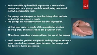
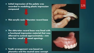
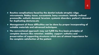
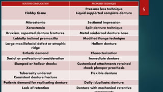
This document explores various unconventional approaches to fabricating complete dentures, highlighting the limitations of conventional methods in addressing complications faced by patients. It details techniques like complete dentures for flabby ridges, immediate dentures, tooth-supported overdentures, and innovative designs such as liquid-supported and hollow dentures. The document emphasizes the importance of adapting denture techniques to meet both functional and aesthetic patient needs.


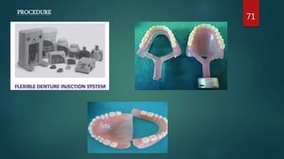
![ADVANTAGES
Translucency
Dentures can be made very thin and light weight.
Better accuracy.
Absolute biocompatibility.
Can be used as temporary dentures during the therapeutic episodes after
surgical reconstruction of jaw.
Reduced midline denture fractures.
Excellent mouldability, light weight to density ratio and high thermal strength.
reduces post-insertion complaints of denture-induced trauma [ulceration].
DISADVANTAGES
Flexibility is normally, not an advantage in complete dentures as retentive
peripheral seal can be broken in function.
Also a greater than normal shrinkage makes it difficult to fabricate
70](https://image.slidesharecdn.com/unconventionaldentures4-201121054704/85/UNCONVENTIONAL-DENTURES-70-320.jpg)