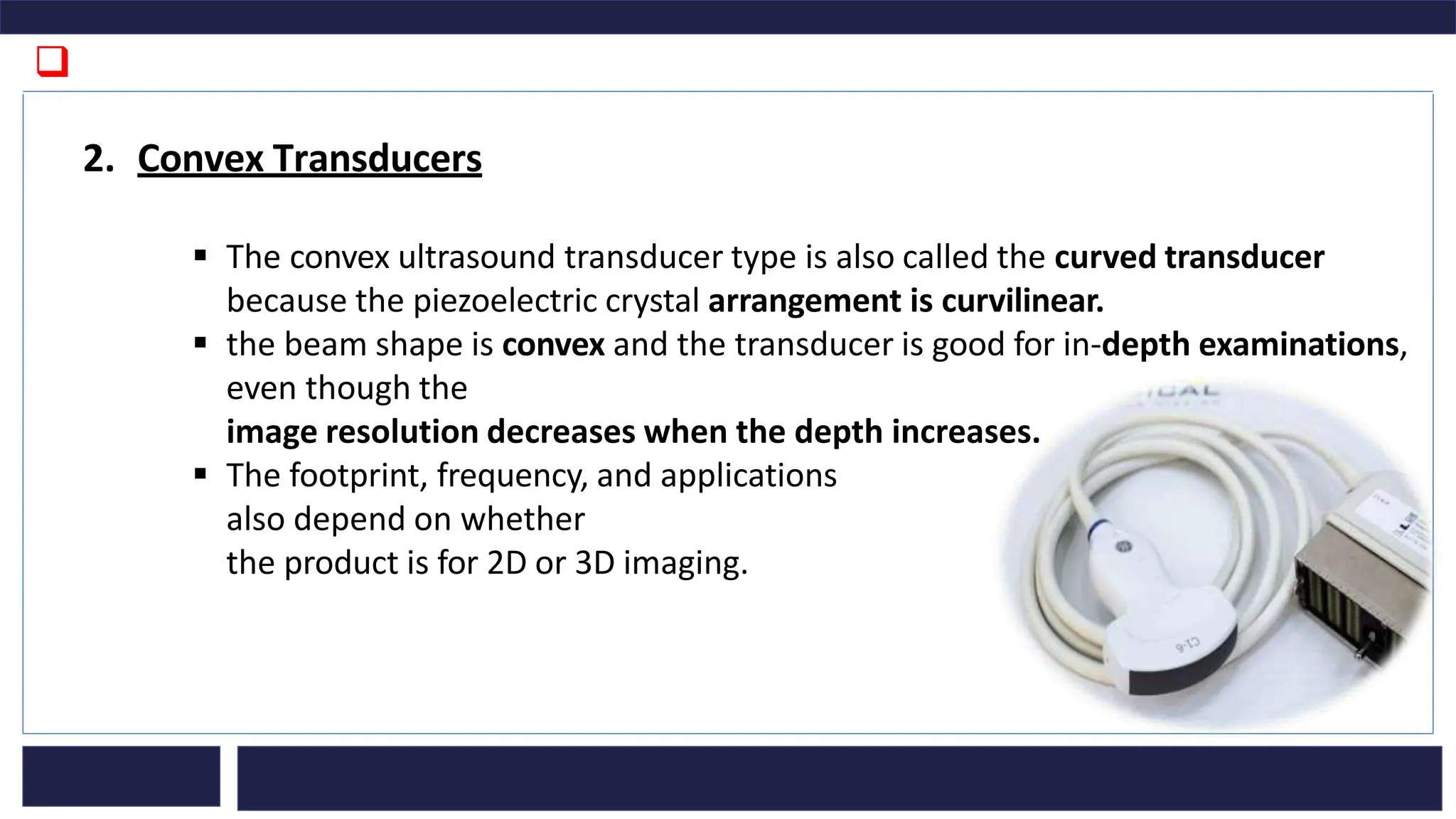
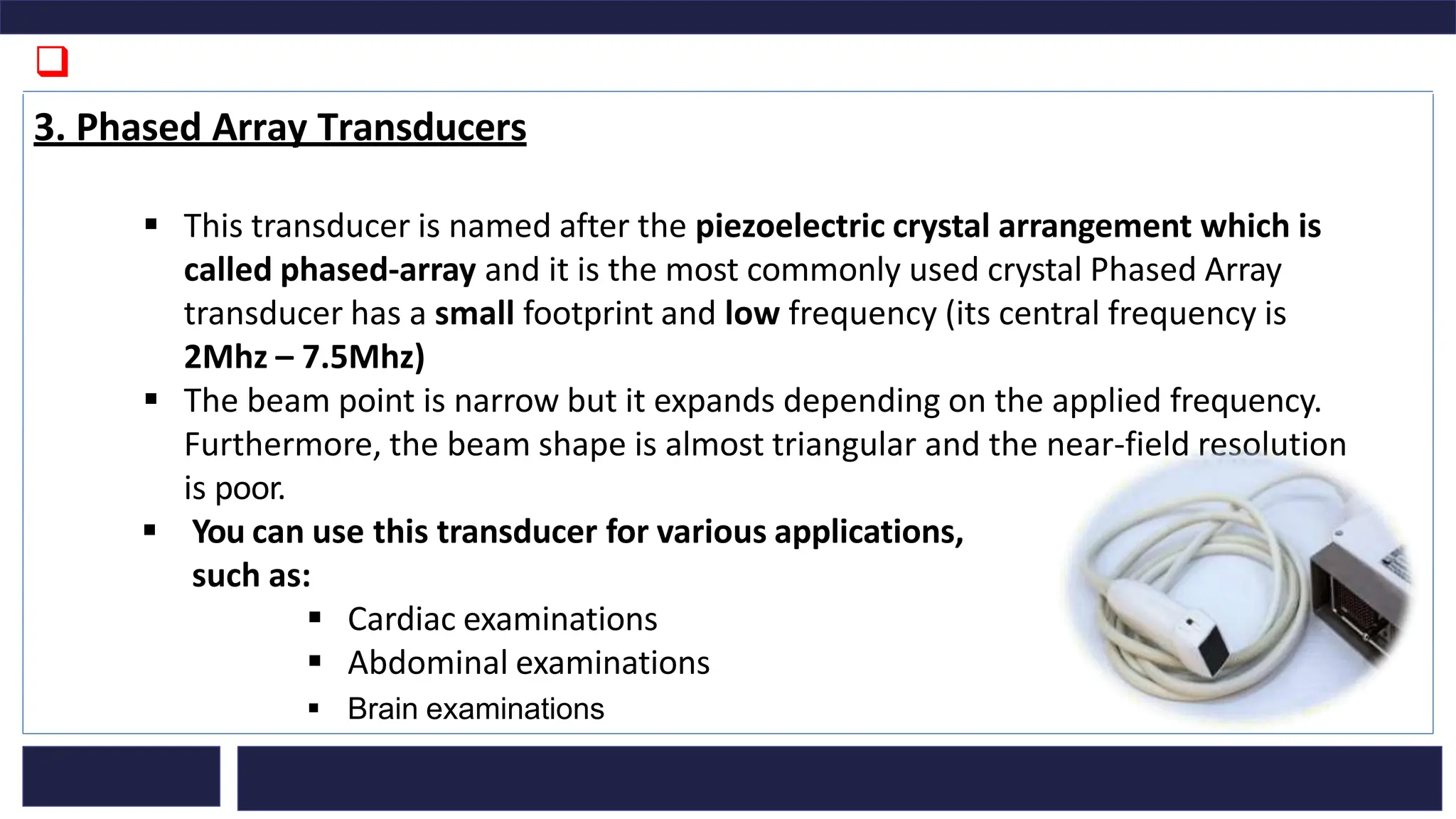
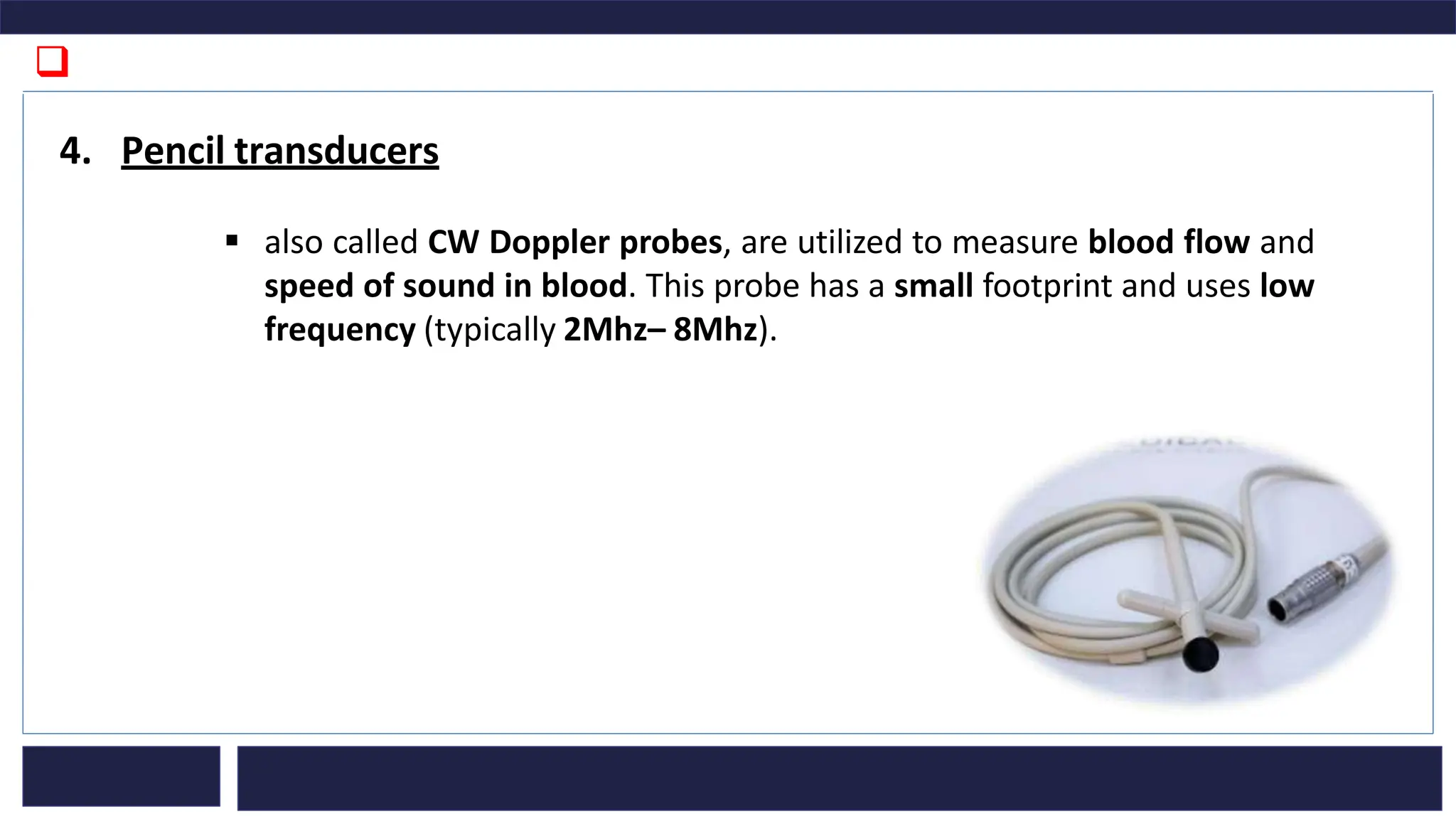
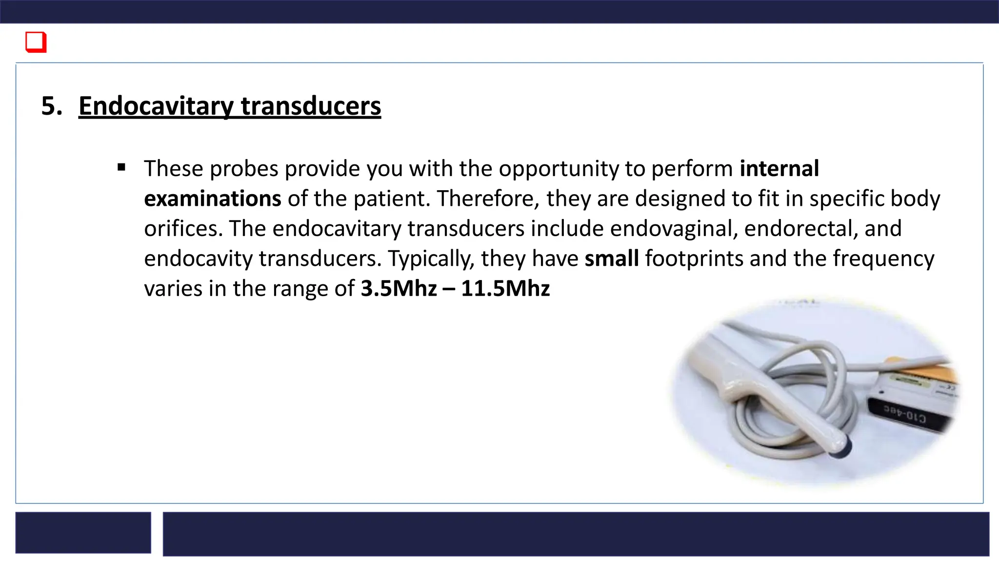
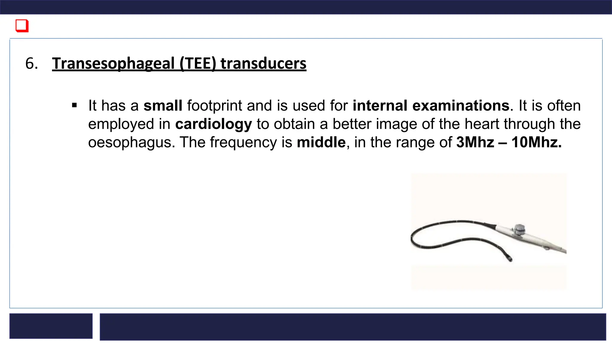
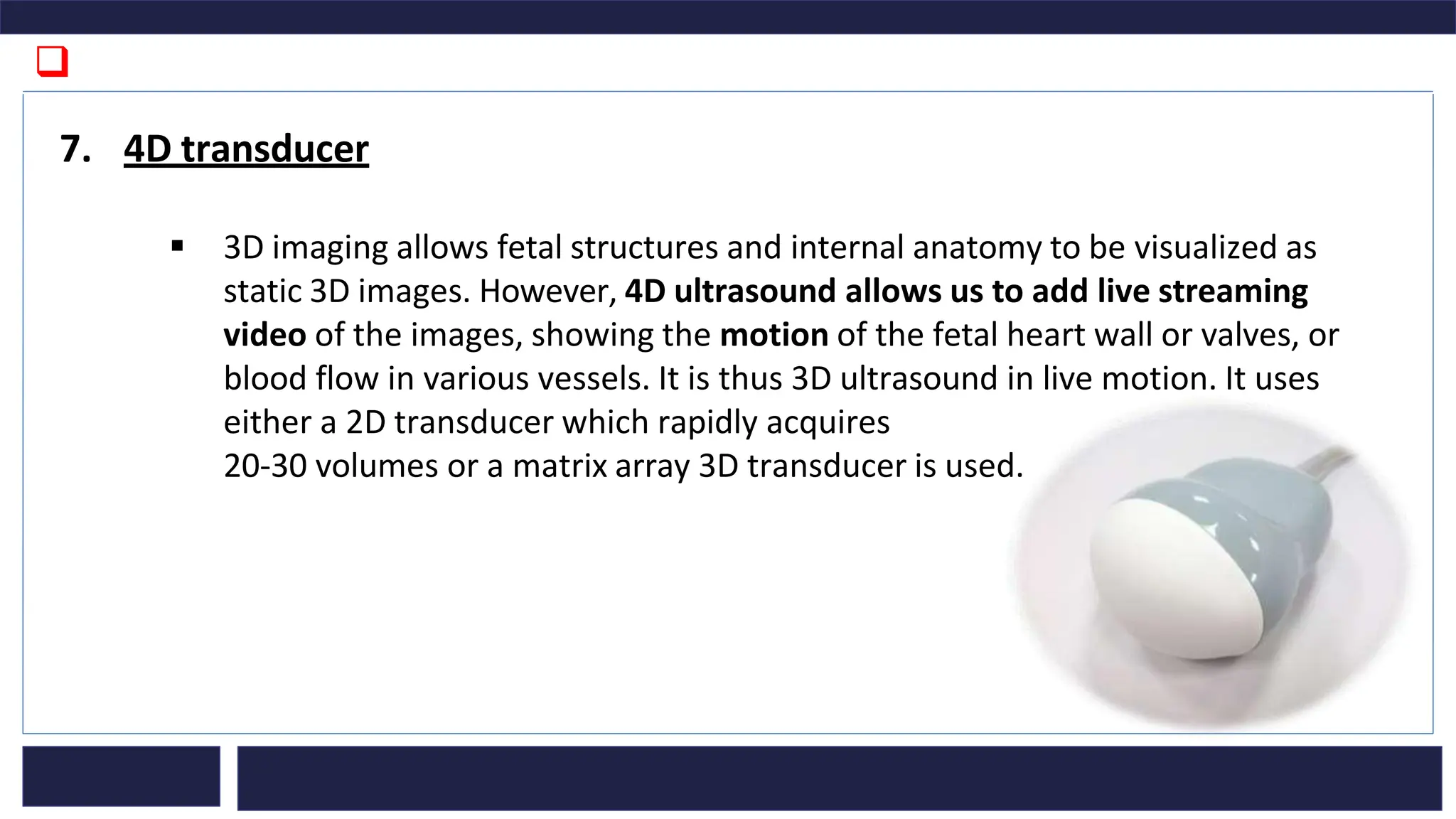
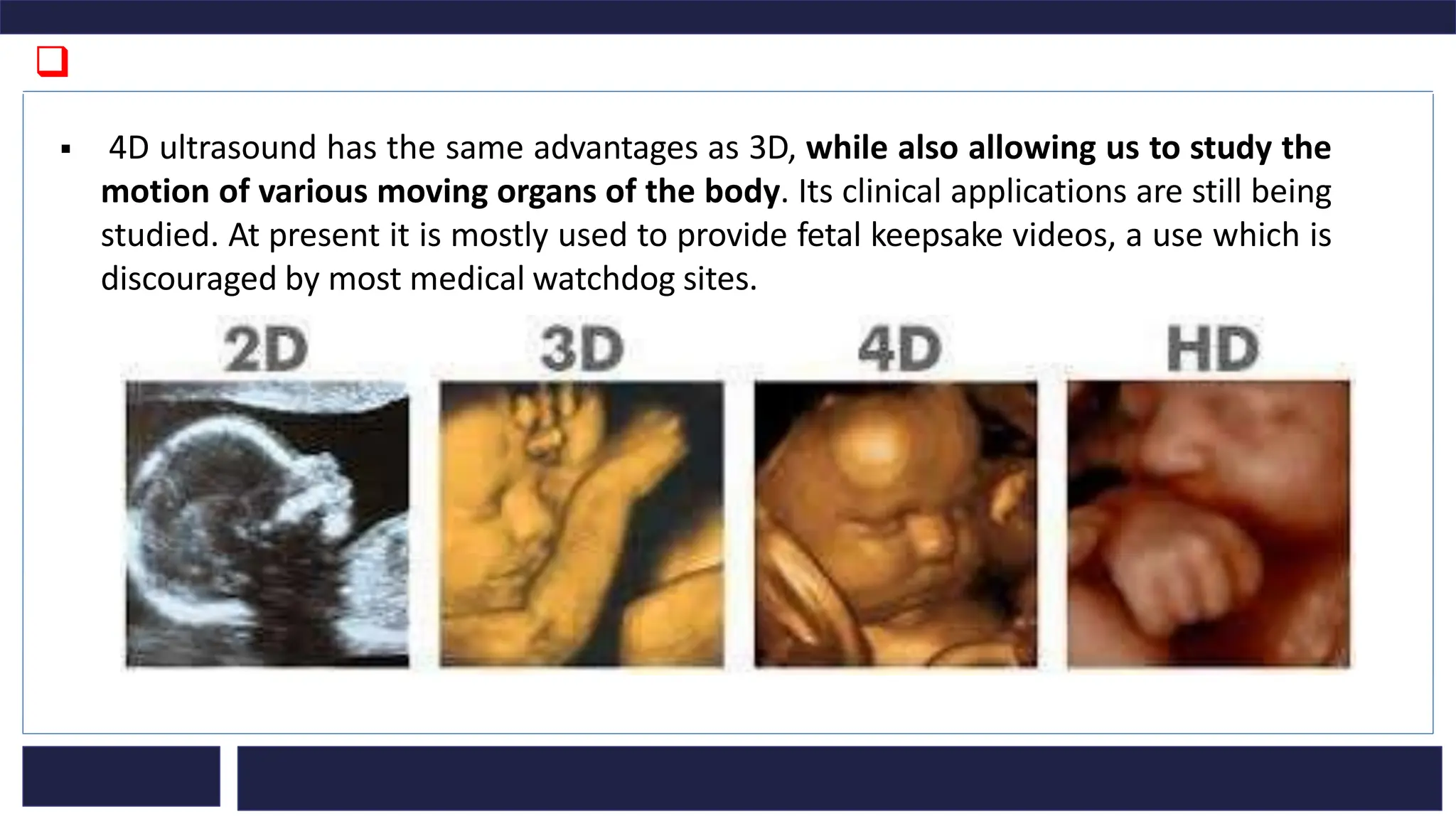
ULTRASOUND TRANSDUCER
❑ Introduction
➢ An ultrasound transducer, also called a probe, is a device that produces sound waves
that bounce off body tissues and make echoes.
➢ The transducer also receives the echoes and sends them to a computer that uses them
to create an image called sonogram.
➢ultrasound transducer is a very important part of the ultrasound machine and a core
part of the signal transmission chain for image reconstruction. Moreover, it is the
fundamental determinant of image quality.
➢ The essential element of each ultrasound transducer is a piezoelectric crystal. It serves
to generate as well as receive ultrasound waves.
➢ You can find ultrasound transducers in different shapes, sizes, and with diverse
features. That is because you need different specifications for maintaining image
quality across different parts of the body.
➢Transducers can be either passed over the surface of
the body – external transducers or can be inserted
into an orifice, such as the rectum or vagina
these are internal transducers.
❑ The ultrasound transducers differ in construction based on
1. Piezoelectric crystal arrangement
2. Aperture (footprint)
3. Frequency
❑ Types of ultrasound transducers
1. Linear Transducers
▪ the piezoelectric crystal arrangement is linear, the shape of the beam is
rectangular, and the near-field resolution is good
▪ The footprint, frequency, and applications of the linear transducer depend
on whether the product is for 2D or 3D imaging.
I. The linear transducer for 2D imaging has a
wide footprint and its central
frequency is 2.5Mhz – 12Mhz.
❑
➢ You can use this transducer for various applications, such as:
▪ Vascular examination
▪ Venipuncture, blood vessel visualization
▪ Breast
▪ Thyroid
▪ Tendon, arthrogenousimage of linear probe
▪ Rectangular Beam Shape
▪ Intraoperative, laparoscopy
▪ The thickness measurement of body fat and musculus for daily health care
check and locomotive syndrome check
▪ Photoacoustic imaging, ultrasonic velocity change imaging
❑
II. The linear transducer for 3D imaging has a wide footprint and a central frequency of
7.5Mhz – 11Mhz.
▪ You can use this transducer for various applications, such as:
▪ Breast
▪ Thyroid
▪ Arteria carotis of vascular application
❑
2. Convex Transducers
▪ The convex ultrasound transducer type is also called the curved transducer
because the piezoelectric crystal arrangement is curvilinear.
▪ the beam shape is convex and the transducer is good for in-depth examinations,
even though the
image resolution decreases when the depth increases.
▪ The footprint, frequency, and applications
also depend on whether
the product is for 2D or 3D imaging.
❑
I. The convex transducer for 2D imaging has a wide footprint and its central frequency is
5. MHz – 7.5MHz. You can use this transducer for various applications, such as:
▪ Abdominal examinations
▪ Transvaginal and trDiagnosis of organs
▪ ansrectal examinations
II. The convex transducer for 3D imaging has a wide field of view and a central
frequency of 3.5MHz – 6.5MHz. You can use it for abdominal examinations.
In addition to the convex transducers,
there is a subtype called micro convex. It has a much smaller footprint and typically,
physicians would use it in neonatal and paediatrics applications. .
❑
3. Phased Array Transducers
▪ This transducer is named after the piezoelectric crystal arrangement which is
called phased-array and it is the most commonly used crystal Phased Array
transducer has a small footprint and low frequency (its central frequency is
2Mhz – 7.5Mhz)
▪ The beam point is narrow but it expands depending on the applied frequency.
Furthermore, the beam shape is almost triangular and the near-field resolution
is poor.
▪ You can use this transducer for various applications,
such as:
▪ Cardiac examinations
▪ Abdominal examinations
▪ Brain examinations
❑
4. Pencil transducers
▪ also called CW Doppler probes, are utilized to measure blood flow and
speed of sound in blood. This probe has a small footprint and uses low
frequency (typically 2Mhz– 8Mhz).
❑
5. Endocavitary transducers
▪ These probes provide you with the opportunity to perform internal
examinations of the patient. Therefore, they are designed to fit in specific body
orifices. The endocavitary transducers include endovaginal, endorectal, and
endocavity transducers. Typically, they have small footprints and the frequency
varies in the range of 3.5Mhz – 11.5Mhz
❑
6. Transesophageal (TEE) transducers
▪ It has a small footprint and is used for internal examinations. It is often
employed in cardiology to obtain a better image of the heart through the
oesophagus. The frequency is middle, in the range of 3Mhz – 10Mhz.
❑
7. 4D transducer
▪ 3D imaging allows fetal structures and internal anatomy to be visualized as
static 3D images. However, 4D ultrasound allows us to add live streaming
video of the images, showing the motion of the fetal heart wall or valves, or
blood flow in various vessels. It is thus 3D ultrasound in live motion. It uses
either a 2D transducer which rapidly acquires
20-30 volumes or a matrix array 3D transducer is used.
❑
▪ 4D ultrasound has the same advantages as 3D, while also allowing us to study the
motion of various moving organs of the body. Its clinical applications are still being
studied. At present it is mostly used to provide fetal keepsake videos, a use which is
discouraged by most medical watchdog sites.

Ultrasound Transducer-biosensors and transducers

  • 1.
  • 2.
    ❑ Introduction ➢ Anultrasound transducer, also called a probe, is a device that produces sound waves that bounce off body tissues and make echoes. ➢ The transducer also receives the echoes and sends them to a computer that uses them to create an image called sonogram. ➢ultrasound transducer is a very important part of the ultrasound machine and a core part of the signal transmission chain for image reconstruction. Moreover, it is the fundamental determinant of image quality. ➢ The essential element of each ultrasound transducer is a piezoelectric crystal. It serves to generate as well as receive ultrasound waves.
  • 3.
    ➢ You canfind ultrasound transducers in different shapes, sizes, and with diverse features. That is because you need different specifications for maintaining image quality across different parts of the body. ➢Transducers can be either passed over the surface of the body – external transducers or can be inserted into an orifice, such as the rectum or vagina these are internal transducers.
  • 4.
    ❑ The ultrasoundtransducers differ in construction based on 1. Piezoelectric crystal arrangement 2. Aperture (footprint) 3. Frequency
  • 5.
    ❑ Types ofultrasound transducers 1. Linear Transducers ▪ the piezoelectric crystal arrangement is linear, the shape of the beam is rectangular, and the near-field resolution is good ▪ The footprint, frequency, and applications of the linear transducer depend on whether the product is for 2D or 3D imaging. I. The linear transducer for 2D imaging has a wide footprint and its central frequency is 2.5Mhz – 12Mhz.
  • 6.
    ❑ ➢ You canuse this transducer for various applications, such as: ▪ Vascular examination ▪ Venipuncture, blood vessel visualization ▪ Breast ▪ Thyroid ▪ Tendon, arthrogenousimage of linear probe ▪ Rectangular Beam Shape ▪ Intraoperative, laparoscopy ▪ The thickness measurement of body fat and musculus for daily health care check and locomotive syndrome check ▪ Photoacoustic imaging, ultrasonic velocity change imaging
  • 7.
    ❑ II. The lineartransducer for 3D imaging has a wide footprint and a central frequency of 7.5Mhz – 11Mhz. ▪ You can use this transducer for various applications, such as: ▪ Breast ▪ Thyroid ▪ Arteria carotis of vascular application
  • 8.
    ❑ 2. Convex Transducers ▪The convex ultrasound transducer type is also called the curved transducer because the piezoelectric crystal arrangement is curvilinear. ▪ the beam shape is convex and the transducer is good for in-depth examinations, even though the image resolution decreases when the depth increases. ▪ The footprint, frequency, and applications also depend on whether the product is for 2D or 3D imaging.
  • 9.
    ❑ I. The convextransducer for 2D imaging has a wide footprint and its central frequency is 5. MHz – 7.5MHz. You can use this transducer for various applications, such as: ▪ Abdominal examinations ▪ Transvaginal and trDiagnosis of organs ▪ ansrectal examinations II. The convex transducer for 3D imaging has a wide field of view and a central frequency of 3.5MHz – 6.5MHz. You can use it for abdominal examinations. In addition to the convex transducers, there is a subtype called micro convex. It has a much smaller footprint and typically, physicians would use it in neonatal and paediatrics applications. .
  • 10.
    ❑ 3. Phased ArrayTransducers ▪ This transducer is named after the piezoelectric crystal arrangement which is called phased-array and it is the most commonly used crystal Phased Array transducer has a small footprint and low frequency (its central frequency is 2Mhz – 7.5Mhz) ▪ The beam point is narrow but it expands depending on the applied frequency. Furthermore, the beam shape is almost triangular and the near-field resolution is poor. ▪ You can use this transducer for various applications, such as: ▪ Cardiac examinations ▪ Abdominal examinations ▪ Brain examinations
  • 11.
    ❑ 4. Pencil transducers ▪also called CW Doppler probes, are utilized to measure blood flow and speed of sound in blood. This probe has a small footprint and uses low frequency (typically 2Mhz– 8Mhz).
  • 12.
    ❑ 5. Endocavitary transducers ▪These probes provide you with the opportunity to perform internal examinations of the patient. Therefore, they are designed to fit in specific body orifices. The endocavitary transducers include endovaginal, endorectal, and endocavity transducers. Typically, they have small footprints and the frequency varies in the range of 3.5Mhz – 11.5Mhz
  • 13.
    ❑ 6. Transesophageal (TEE)transducers ▪ It has a small footprint and is used for internal examinations. It is often employed in cardiology to obtain a better image of the heart through the oesophagus. The frequency is middle, in the range of 3Mhz – 10Mhz.
  • 14.
    ❑ 7. 4D transducer ▪3D imaging allows fetal structures and internal anatomy to be visualized as static 3D images. However, 4D ultrasound allows us to add live streaming video of the images, showing the motion of the fetal heart wall or valves, or blood flow in various vessels. It is thus 3D ultrasound in live motion. It uses either a 2D transducer which rapidly acquires 20-30 volumes or a matrix array 3D transducer is used.
  • 15.
    ❑ ▪ 4D ultrasoundhas the same advantages as 3D, while also allowing us to study the motion of various moving organs of the body. Its clinical applications are still being studied. At present it is mostly used to provide fetal keepsake videos, a use which is discouraged by most medical watchdog sites.