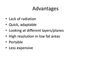
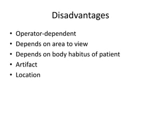
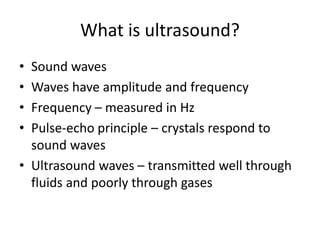
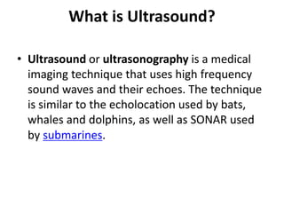
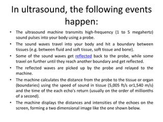
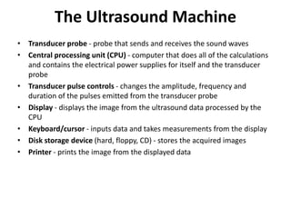
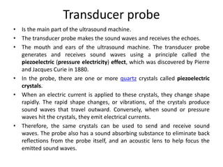
Ultrasound uses high-frequency sound waves to produce images of structures inside the body. It has several advantages over other imaging techniques, including lack of radiation exposure, low cost, portability, and ability to image different tissue planes. This document provides an introduction to ultrasound physics, terminology, machine components, and basic abdominal ultrasound imaging and interpretation for medical students. It discusses how ultrasound images are formed from sound wave reflections and demonstrates common ultrasound appearances of anatomical structures and pathologies.
Ultrasound Physics Made easy - By Dr Chandni WadhwaniChandni Wadhwani
History of ultrasound, Principle of Ultrasound.
Ultrasound wave and its interactions
Ultrasound machine and its parts, Image display, Artifacts and their clinical importance
what is Doppler ultrasound, Elastography and Recent advances in field of ultrasound.
Safety issues in ultrasound.
Learn from our Slideshare about the differences between ultrasound transducers. We also cover tips on how to treat your probes and how to select the right one.
• What is Ultrasound imaging?
• Why Ultrasound?
• Common Uses
• History
• Properties of Ultrasound
• Equipment types
• How does the procedure work?
• Physics
• Benefits and Risks etc.
Training Material inherited form Philips Basics of Ultrasonography. Covers the fundamentals of Ultrasound Waveform, Piezoelectric Effect, Phased Echo Concept, Goal of Ultrasound, Ultrasound Image Construction process, Types of Resolution, Probe Internals, The Doppler Effect, Spectrum Waveform and concept, Color Doppler, Components of Ultrasound.
Usg transducer and basic principles of ultrasound Doppler, this slide describe the basic physics of ultrasound transducer and Doppler , must know thing is given in this presentaion. Good review for radiology resident. Thanks.
Definition of Side lobes and the principle behind its production during ultrasound imaging. Side lobes artifact and its result on image. Explanation of harmonic imaging, its production and the techniques use to eliminate fundamental frequency to produce optimal harmonic images.
Francesca Gottschalk - How can education support child empowerment.pptxEduSkills OECD
Francesca Gottschalk from the OECD’s Centre for Educational Research and Innovation presents at the Ask an Expert Webinar: How can education support child empowerment?
Welcome to TechSoup New Member Orientation and Q&A (May 2024).pdfTechSoup
In this webinar you will learn how your organization can access TechSoup's wide variety of product discount and donation programs. From hardware to software, we'll give you a tour of the tools available to help your nonprofit with productivity, collaboration, financial management, donor tracking, security, and more.
The French Revolution, which began in 1789, was a period of radical social and political upheaval in France. It marked the decline of absolute monarchies, the rise of secular and democratic republics, and the eventual rise of Napoleon Bonaparte. This revolutionary period is crucial in understanding the transition from feudalism to modernity in Europe.
For more information, visit-www.vavaclasses.com
How to Make a Field invisible in Odoo 17Celine George
It is possible to hide or invisible some fields in odoo. Commonly using “invisible” attribute in the field definition to invisible the fields. This slide will show how to make a field invisible in odoo 17.
2024.06.01 Introducing a competency framework for languag learning materials ...Sandy Millin
http://sandymillin.wordpress.com/iateflwebinar2024
Published classroom materials form the basis of syllabuses, drive teacher professional development, and have a potentially huge influence on learners, teachers and education systems. All teachers also create their own materials, whether a few sentences on a blackboard, a highly-structured fully-realised online course, or anything in between. Despite this, the knowledge and skills needed to create effective language learning materials are rarely part of teacher training, and are mostly learnt by trial and error.
Knowledge and skills frameworks, generally called competency frameworks, for ELT teachers, trainers and managers have existed for a few years now. However, until I created one for my MA dissertation, there wasn’t one drawing together what we need to know and do to be able to effectively produce language learning materials.
This webinar will introduce you to my framework, highlighting the key competencies I identified from my research. It will also show how anybody involved in language teaching (any language, not just English!), teacher training, managing schools or developing language learning materials can benefit from using the framework.
Safalta Digital marketing institute in Noida, provide complete applications that encompass a huge range of virtual advertising and marketing additives, which includes search engine optimization, virtual communication advertising, pay-per-click on marketing, content material advertising, internet analytics, and greater. These university courses are designed for students who possess a comprehensive understanding of virtual marketing strategies and attributes.Safalta Digital Marketing Institute in Noida is a first choice for young individuals or students who are looking to start their careers in the field of digital advertising. The institute gives specialized courses designed and certification.
for beginners, providing thorough training in areas such as SEO, digital communication marketing, and PPC training in Noida. After finishing the program, students receive the certifications recognised by top different universitie, setting a strong foundation for a successful career in digital marketing.
Normal Labour/ Stages of Labour/ Mechanism of LabourWasim Ak
Normal labor is also termed spontaneous labor, defined as the natural physiological process through which the fetus, placenta, and membranes are expelled from the uterus through the birth canal at term (37 to 42 weeks
Unit 8 - Information and Communication Technology (Paper I).pdfThiyagu K
This slides describes the basic concepts of ICT, basics of Email, Emerging Technology and Digital Initiatives in Education. This presentations aligns with the UGC Paper I syllabus.
Read| The latest issue of The Challenger is here! We are thrilled to announce that our school paper has qualified for the NATIONAL SCHOOLS PRESS CONFERENCE (NSPC) 2024. Thank you for your unwavering support and trust. Dive into the stories that made us stand out!
Model Attribute Check Company Auto PropertyCeline George
In Odoo, the multi-company feature allows you to manage multiple companies within a single Odoo database instance. Each company can have its own configurations while still sharing common resources such as products, customers, and suppliers.
2. Learning objectives
• Present an introduction to medical
ultrasonography at the level of a third or
fourth year medical student.
• After the brief course, the student should be
able to:
– Understand basic physics and terminology of
ultrasound
– State the advantages and disadvantages of
ultrasound
– Understand how the images are obtained
– Begin to interpret simple ultrasound images.
3. Outline
• Brief history of ultrasound technology
• Advantages, disadvantages
• What is ultrasound?
• Physics
• Probes, controls, machine, Doppler
• Terminology
• What structures look like
• Abdominal ultrasound
• Example of liver/gallbladder
• Quick review questions
4. History
• Medical use – 1940s
• Developing technology – 1950s
• Advancements – 1960s/1970s
• Real time ultrasound – 1980s
• 3D and 4D images – 1990s
5. Advantages
• Lack of radiation
• Quick, adaptable
• Looking at different layers/planes
• High resolution in low fat areas
• Portable
• Less expensive
7. What is ultrasound?
• Sound waves
• Waves have amplitude and frequency
• Frequency – measured in Hz
• Pulse-echo principle – crystals respond to
sound waves
• Ultrasound waves – transmitted well through
fluids and poorly through gases
8. What is Ultrasound?
• Ultrasound or ultrasonography is a medical
imaging technique that uses high frequency
sound waves and their echoes. The technique
is similar to the echolocation used by bats,
whales and dolphins, as well as SONAR used
by submarines.
9. In ultrasound, the following events
happen:
• The ultrasound machine transmits high-frequency (1 to 5 megahertz)
sound pulses into your body using a probe.
• The sound waves travel into your body and hit a boundary between
tissues (e.g. between fluid and soft tissue, soft tissue and bone).
• Some of the sound waves get reflected back to the probe, while some
travel on further until they reach another boundary and get reflected.
• The reflected waves are picked up by the probe and relayed to the
machine.
• The machine calculates the distance from the probe to the tissue or organ
(boundaries) using the speed of sound in tissue (5,005 ft/s or1,540 m/s)
and the time of the each echo's return (usually on the order of millionths
of a second).
• The machine displays the distances and intensities of the echoes on the
screen, forming a two dimensional image like the one shown below.
10. The Ultrasound Machine
• Transducer probe - probe that sends and receives the sound waves
• Central processing unit (CPU) - computer that does all of the calculations
and contains the electrical power supplies for itself and the transducer
probe
• Transducer pulse controls - changes the amplitude, frequency and
duration of the pulses emitted from the transducer probe
• Display - displays the image from the ultrasound data processed by the
CPU
• Keyboard/cursor - inputs data and takes measurements from the display
• Disk storage device (hard, floppy, CD) - stores the acquired images
• Printer - prints the image from the displayed data
11. Transducer probe
• Is the main part of the ultrasound machine.
• The transducer probe makes the sound waves and receives the echoes.
• The mouth and ears of the ultrasound machine. The transducer probe
generates and receives sound waves using a principle called the
piezoelectric (pressure electricity) effect, which was discovered by Pierre
and Jacques Curie in 1880.
• In the probe, there are one or more quartz crystals called piezoelectric
crystals.
• When an electric current is applied to these crystals, they change shape
rapidly. The rapid shape changes, or vibrations, of the crystals produce
sound waves that travel outward. Conversely, when sound or pressure
waves hit the crystals, they emit electrical currents.
• Therefore, the same crystals can be used to send and receive sound
waves. The probe also has a sound absorbing substance to eliminate back
reflections from the probe itself, and an acoustic lens to help focus the
emitted sound waves.
12.
13.
14.
15. Physics
• Speed of ultrasound – average 1,540
m/second, but depends on tissue
• Acoustic impedance – density of tissue
multiplied by velocity of sound; harder
substances (like bone) usually have higher
acoustic impedance (than substances like
water or fat)
16. Terminology
• Echogenicity – measure of jumps in
impedance of an organ (NOT density)
• Hyperechoic – bright; many echos
• Hypoechoic – dark; few echos
• Anechoic – black; no echoes
• Shadowing
17. Frequencies
• Higher frequencies – better resolution but less
penetration [useful for thyroid, testes,
superficial blood vessels]
• Lower frequencies – better penetration but
worse resolution [useful for the abdomen,
aorta]
33. Doppler
Images from UCMC
Color Doppler – blue and red show
flow of blood
Spectral Doppler – waveform showing
flow (same as color, but quantitative)
• Tells you direction of flow, and its
magnitude
34. Doppler
Images from UCMC
Color Doppler – kidney , showing
segmental arteries and veins
Spectral Doppler – right testicle with
low impedance arterial waveform
• A little more detail
45. Gallstones – hyperechoic and
shadowing underneath
Image from UCMC
Kidney
Gallstone
Gallbladder
Shadowing
Liver
46. Liver/gallbladder example
• Now – try to think of an ultrasound from start
to finish; we will take the example of a
liver/gallbladder ultrasound
• Liver parenchyma
• Ducts
• Vascular system
• Gallbladder, stones
48. Liver/gallbladder example
• NPO 4-6 hours – gallbladder distended and
bowel gas minimal
• Longitudinal, transverse, and coronal scans in
supine and left posterior oblique positions
• Subcostal approach good for most of liver, but
may need intercostal approach for most
superior part
• Use Doppler to distinguish blood vessels and
ducts
49. Abdominal ultrasound
• Layperson description (what your patient gets
told):
http://www.nlm.nih.gov/medlineplus/ency/ar
ticle/003777.htm
• Example of how to do an abdominal
ultrasound:
http://www.youtube.com/watch?v=7Y6wFXf
muvg
51. Liver/gallbladder anatomy
IVC coursing
through liver
Aorta passing
anterior to IVC
and bifurcating
into right and left
common iliac
arteries
Portal vein
formed by
SMV and
splenic
vein, going
to liver
Branches of SMA
and SMV
Image from UCMC
53. Liver/gallbladder
CT – sagittal cut U/S – longitudinal orientation
Liver Gallbladder Right kidney Liver Right kidney
Images from UCMC
54. Liver/gallbladder
CT – axial cut
Right, middle, and left hepatic veins
Draining into the IVC
U/S – transverse orientation
Images from UCMC
56. Liver/gallbladder
U/S – transverse orientation
IVC
Pancreas
Left lobe of liver
Left kidney
Spleen
Splenic vein
CT – axial cut
Right portal vein
IVC
Aorta
Images from UCMC
57. Summary
• You should now understand ultrasound basics
• Physics, orientation, probes
• Terms, anatomy
• Much more to learn
58. Quick review questions
1. What is ultrasound? What types of tissues
does it travel well through? Poorly through?
59. Quick review questions
2. How does the frequency of the ultrasound
affect resolution and penetration?
60. Quick review questions
3. Name some types of hyperechoic,
hypoechoic, and anechoic tissues. Can you
picture them as they would appear on an
ultrasound image?
62. Quick review questions
5. What shapes do the probes come in? Can you
describe a few of the differences of the probe
types?
63. Quick review questions
6. What are the three main ways in which the
ultrasound probe can be held (i.e. the
orientations of the probes)?
64. Quick review questions
7. Name at least three indications for an
abdominal ultrasound.
What are some of the things you could tell a
patient when they are preparing for an
abdominal ultrasound and orders you might
need to write?