SlideShare a Scribd company logo
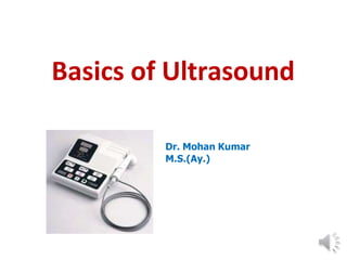
Basics of Ultrasound
Dr. Mohan Kumar
M.S.(Ay.)
History
The bat use Ultrasound for navigation
ULTRASOUND: BASIC
DEFINITION
 Ultrasound is acoustic(sound) energy in the form
of waves having a frequency above the human
hearing range(i.e. 20KHz)
 Ultrasound is a way of using sound waves to look
inside the human body.
Pan-Scanner - The transducer rotated in a semicircular arc
around the patient (1957)
Scan converter allowed for the first time to use the
upcoming computer technology to improve US
Ultrasound Parts
The Ultrasound Machine
A basic ultrasound machine has the followingparts:
1. Transducer probe - probe that sends and receives the sound
waves
2. Central processing unit (CPU) - computer that does all of the
calculations and contains the electrical power supplies for itself
and the transducer probe
3. Transducer pulse controls - changes the amplitude, frequency
and duration of the pulses emitted from the transducer probe
4. Display - displays the image from the ultrasound data processed
by the CPU
5. Keyboard/cursor - inputs data and takes measurements from
the display
6. Disk storage device (hard, floppy, CD) - stores the acquired
images
7. Printer - prints the image from the displayed data
ULTRASONOGRAPHY
• Ultrasonography or diagnostic sonography is an
ultrasound based diagnostic imaging technique used
for visualizing internal body structures.
MAIN IMAGING
MODES
GREY SCALE
IMAGING
A-Mode
B-Mode
M-Mode
DOPPLER IMAGING
Continuous wave Doppler
 Power Doppler
 Color Doppler
 Duplex Doppler
Pulsed wave Doppler
A MODE
Simplest form of ultrasound
imaging which is based on
the pulse-echo principle.
A scans can be used to
measure distances.
A scans only give one
dimensional information
 Not so useful for imaging
Used for echo-
encephalography and echo-
ophthalmoscopy
B MODE
 B stands for Brightness
B scans give two dimensional
information about the cross-
section.
Generally used to measure
cardiac chambers
dimensions, assess valvular
structure and function.
Development of the B-mode Ultrasound image quality
M MODE
 M stands for motion
This represents
movements of
structures over time.
M Mode is commonly
used for measuring
chamber dimensions.
This is analogous to
recording a video in
ultrasound.
DOPPLER IMAGING
It is a general term used to
visualize velocities of moving
tissues.
Doppler ultrasound evaluates
blood velocity as it flows
through a blood vessel.
Blood flow through the heart
and large vessels has certain
characteristics that can be
measured using Doppler
instruments.
BLOOD FLOW PATTERNS
LAMINAR FLOW
of flow
at vessel
• Layers
(normal)
• Slowest
wall
• Fastest within center
of vessel
TURBULENT
FLOW
• Obstructions
disrupt laminar flow
• Disordered
directions of flow
TYPES OF DOPPLER ULTRASOUND
1. CONTINUOUS WAVE DOPPLER (CW)
Uses different crystals to
send and receive the signal
One crystal constantly sends
a sound wave of a single
frequency,
constantly
the
receives
other
the
reflected signal
2. PULSED WAVE
DOPPLER
 Produces short
bursts/pulses of
sound
 Uses the same
crystals to send and
receive the signal
 This follows the same
pulse-echo technique
used in 2D image
formation.
3. COLOR
DOPPLER
Utilizes pulse-echo Doppler flow
principles to generate a color
image.
Image is superimposed on the
2D image.
The red and blue display
provides information regarding
DIRECTION and VELOCITY of
flow.
Used for general assessment of
flow in the region of interest
Gives only descriptive or semi
quantitative information on blood
flow.
4. POWER
DOPPLER
5 times more sensitive in
detecting blood flow than
color doppler.
It can get those images
that are impossible with
color doppler.
Used to evaluate blood
flow through vessels within
solid organs.
APPLICATIONS
 Obstetrics and Gynecology
1. Measuring the size of the fetus
2. Determining the sex of the baby
3. Monitoring the baby for various procedures
 Cardiology
1. Seeing the inside of the heart to identify abnormal functions
2. Measuring blood flow through the heart and major bloo
vessels
 Urology
1. Measuring blood flow through the kidney
2. Locating kidney stones
3. Detecting prostate cancer at early stage
RISKS
The two major risks involved with Ultrasound are:
 Development of heat:
Tissues or water absorb the ultrasound energy
which
increases their temperature locally.
 Formation of bubbles ( cavitation):
When dissolved gases come out of solution due
to
local heat caused by Ultrasound.
BENEFITS
 Images muscle, soft tissues very well
 Renders “live images” where most desirable
section is selected
 Shows structure of organs
 No long-term side-effects
 Widely available and comparatively flexible
 Highly portable
 Relatively inexpensive
 Spatial resolution is better in high frequency
ultrasound scanners
LIMITATIONS
 Sonographic devices have trouble penetrating bone
Sonography performs very poorly when there is a gas
between the transducer and organ of interest
Body habitus has large influence on image quality
Method is operator-dependent
No scout image as there is with CT and MRI

More Related Content

Similar to bams-4th-year-shalya-ultrasound-05-05-2020.pptx

Ultrasonography
UltrasonographyUltrasonography
UltrasonographySai Ram
 
Ultrasonography in Animals.pptxblba jhaha
Ultrasonography in Animals.pptxblba jhahaUltrasonography in Animals.pptxblba jhaha
Ultrasonography in Animals.pptxblba jhahaIzzatAftab
 
Ultrasonography
UltrasonographyUltrasonography
UltrasonographyAJAZ KHAN
 
Emergency us physics and instrumentation
Emergency us physics  and instrumentationEmergency us physics  and instrumentation
Emergency us physics and instrumentationdrnikahmad
 
Its is useful for studying the basic concepts of ultrasound for medical imagi...
Its is useful for studying the basic concepts of ultrasound for medical imagi...Its is useful for studying the basic concepts of ultrasound for medical imagi...
Its is useful for studying the basic concepts of ultrasound for medical imagi...DilshanDillu1
 
ultra sound.pptx
ultra sound.pptxultra sound.pptx
ultra sound.pptxAliMRiyath
 
Ultrasound Basics.ppt
Ultrasound Basics.pptUltrasound Basics.ppt
Ultrasound Basics.pptghadaElbanby1
 
Introduction To Radiology.pptx
Introduction To Radiology.pptxIntroduction To Radiology.pptx
Introduction To Radiology.pptxRT-Luqman Zarvaid
 
ULTRASOUND IMAGING.pptx
ULTRASOUND IMAGING.pptxULTRASOUND IMAGING.pptx
ULTRASOUND IMAGING.pptxJeyaDarshini1
 
3_2018_12_16!11_22_57_PM.pptx
3_2018_12_16!11_22_57_PM.pptx3_2018_12_16!11_22_57_PM.pptx
3_2018_12_16!11_22_57_PM.pptxKhaVuong2
 
USG THORAX [Autosaved].pptx
USG THORAX [Autosaved].pptxUSG THORAX [Autosaved].pptx
USG THORAX [Autosaved].pptxNarendra Tengli
 
ULTRASOUND MACHINE
ULTRASOUND  MACHINEULTRASOUND  MACHINE
ULTRASOUND MACHINEkaushikbaro
 

Similar to bams-4th-year-shalya-ultrasound-05-05-2020.pptx (20)

Ultrasonography
UltrasonographyUltrasonography
Ultrasonography
 
Us imaging
Us imagingUs imaging
Us imaging
 
Doppler ultrasonography
Doppler ultrasonographyDoppler ultrasonography
Doppler ultrasonography
 
Ultrasonography in Animals.pptxblba jhaha
Ultrasonography in Animals.pptxblba jhahaUltrasonography in Animals.pptxblba jhaha
Ultrasonography in Animals.pptxblba jhaha
 
Doppler effect
Doppler effectDoppler effect
Doppler effect
 
Doppler effect
Doppler effectDoppler effect
Doppler effect
 
Ultrasonography
UltrasonographyUltrasonography
Ultrasonography
 
Ultasound
UltasoundUltasound
Ultasound
 
Emergency us physics and instrumentation
Emergency us physics  and instrumentationEmergency us physics  and instrumentation
Emergency us physics and instrumentation
 
Its is useful for studying the basic concepts of ultrasound for medical imagi...
Its is useful for studying the basic concepts of ultrasound for medical imagi...Its is useful for studying the basic concepts of ultrasound for medical imagi...
Its is useful for studying the basic concepts of ultrasound for medical imagi...
 
ultra sound.pptx
ultra sound.pptxultra sound.pptx
ultra sound.pptx
 
Ultrasound Basics.ppt
Ultrasound Basics.pptUltrasound Basics.ppt
Ultrasound Basics.ppt
 
Introduction To Radiology.pptx
Introduction To Radiology.pptxIntroduction To Radiology.pptx
Introduction To Radiology.pptx
 
ULTRASOUND IMAGING.pptx
ULTRASOUND IMAGING.pptxULTRASOUND IMAGING.pptx
ULTRASOUND IMAGING.pptx
 
ULTRASOUND EQUIPMENT
ULTRASOUND EQUIPMENTULTRASOUND EQUIPMENT
ULTRASOUND EQUIPMENT
 
Ultra Sound Imaging
Ultra Sound ImagingUltra Sound Imaging
Ultra Sound Imaging
 
3_2018_12_16!11_22_57_PM.pptx
3_2018_12_16!11_22_57_PM.pptx3_2018_12_16!11_22_57_PM.pptx
3_2018_12_16!11_22_57_PM.pptx
 
USG THORAX [Autosaved].pptx
USG THORAX [Autosaved].pptxUSG THORAX [Autosaved].pptx
USG THORAX [Autosaved].pptx
 
ULTRASOUND MACHINE
ULTRASOUND  MACHINEULTRASOUND  MACHINE
ULTRASOUND MACHINE
 
Echocardiography an introduction
Echocardiography an introductionEchocardiography an introduction
Echocardiography an introduction
 

Recently uploaded

TEST BANK For Advanced Practice Nursing in the Care of Older Adults, 2nd Edit...
TEST BANK For Advanced Practice Nursing in the Care of Older Adults, 2nd Edit...TEST BANK For Advanced Practice Nursing in the Care of Older Adults, 2nd Edit...
TEST BANK For Advanced Practice Nursing in the Care of Older Adults, 2nd Edit...kevinkariuki227
 
ANATOMY OF THE LOWER URINARY TRACT AND MALE [Autosaved] [Autosaved].pptx
ANATOMY OF THE LOWER URINARY TRACT AND MALE [Autosaved] [Autosaved].pptxANATOMY OF THE LOWER URINARY TRACT AND MALE [Autosaved] [Autosaved].pptx
ANATOMY OF THE LOWER URINARY TRACT AND MALE [Autosaved] [Autosaved].pptxBright Chipili
 
Prix Galien International 2024 Forum Program
Prix Galien International 2024 Forum ProgramPrix Galien International 2024 Forum Program
Prix Galien International 2024 Forum ProgramLevi Shapiro
 
Lung Cancer: Artificial Intelligence, Synergetics, Complex System Analysis, S...
Lung Cancer: Artificial Intelligence, Synergetics, Complex System Analysis, S...Lung Cancer: Artificial Intelligence, Synergetics, Complex System Analysis, S...
Lung Cancer: Artificial Intelligence, Synergetics, Complex System Analysis, S...Oleg Kshivets
 
Compare home pulse pressure components collected directly from home
Compare home pulse pressure components collected directly from homeCompare home pulse pressure components collected directly from home
Compare home pulse pressure components collected directly from homeCatherine Liao
 
Scientificity and feasibility study of non-invasive central arterial pressure...
Scientificity and feasibility study of non-invasive central arterial pressure...Scientificity and feasibility study of non-invasive central arterial pressure...
Scientificity and feasibility study of non-invasive central arterial pressure...Catherine Liao
 
How to Give Better Lectures: Some Tips for Doctors
How to Give Better Lectures: Some Tips for DoctorsHow to Give Better Lectures: Some Tips for Doctors
How to Give Better Lectures: Some Tips for DoctorsLanceCatedral
 
Non-Invasive assessment of arterial stiffness in advanced heart failure patie...
Non-Invasive assessment of arterial stiffness in advanced heart failure patie...Non-Invasive assessment of arterial stiffness in advanced heart failure patie...
Non-Invasive assessment of arterial stiffness in advanced heart failure patie...Catherine Liao
 
Blue Printing in medical education by Dr.Mumtaz Ali.pptx
Blue Printing in medical education by Dr.Mumtaz Ali.pptxBlue Printing in medical education by Dr.Mumtaz Ali.pptx
Blue Printing in medical education by Dr.Mumtaz Ali.pptxNeurosurgeon Mumtaz Ali Narejo
 
Temporal, Infratemporal & Pterygopalatine BY Dr.RIG.pptx
Temporal, Infratemporal & Pterygopalatine BY Dr.RIG.pptxTemporal, Infratemporal & Pterygopalatine BY Dr.RIG.pptx
Temporal, Infratemporal & Pterygopalatine BY Dr.RIG.pptxDr. Rabia Inam Gandapore
 
Alcohol_Dr. Jeenal Mistry MD Pharmacology.pdf
Alcohol_Dr. Jeenal Mistry MD Pharmacology.pdfAlcohol_Dr. Jeenal Mistry MD Pharmacology.pdf
Alcohol_Dr. Jeenal Mistry MD Pharmacology.pdfDr Jeenal Mistry
 
TEST BANK For Williams' Essentials of Nutrition and Diet Therapy, 13th Editio...
TEST BANK For Williams' Essentials of Nutrition and Diet Therapy, 13th Editio...TEST BANK For Williams' Essentials of Nutrition and Diet Therapy, 13th Editio...
TEST BANK For Williams' Essentials of Nutrition and Diet Therapy, 13th Editio...kevinkariuki227
 
Antiplatelets in IHD, Dose Duration, DAPT vs SAPT
Antiplatelets in IHD, Dose Duration, DAPT vs SAPTAntiplatelets in IHD, Dose Duration, DAPT vs SAPT
Antiplatelets in IHD, Dose Duration, DAPT vs SAPTAkashGanganePatil1
 
Is preeclampsia and spontaneous preterm delivery associate with vascular and ...
Is preeclampsia and spontaneous preterm delivery associate with vascular and ...Is preeclampsia and spontaneous preterm delivery associate with vascular and ...
Is preeclampsia and spontaneous preterm delivery associate with vascular and ...Catherine Liao
 
1130525--家醫計畫2.0糖尿病照護研討會-社團法人高雄市醫師公會.pdf
1130525--家醫計畫2.0糖尿病照護研討會-社團法人高雄市醫師公會.pdf1130525--家醫計畫2.0糖尿病照護研討會-社團法人高雄市醫師公會.pdf
1130525--家醫計畫2.0糖尿病照護研討會-社團法人高雄市醫師公會.pdfKs doctor
 
Retinal consideration in cataract surgery
Retinal consideration in cataract surgeryRetinal consideration in cataract surgery
Retinal consideration in cataract surgeryKafrELShiekh University
 
Factors Affecting child behavior in Pediatric Dentistry
Factors Affecting child behavior in Pediatric DentistryFactors Affecting child behavior in Pediatric Dentistry
Factors Affecting child behavior in Pediatric DentistryDr Simran Deepak Vangani
 
Impact of cancers therapies on the loss in cardiac function, myocardial fffic...
Impact of cancers therapies on the loss in cardiac function, myocardial fffic...Impact of cancers therapies on the loss in cardiac function, myocardial fffic...
Impact of cancers therapies on the loss in cardiac function, myocardial fffic...Catherine Liao
 
Couples presenting to the infertility clinic- Do they really have infertility...
Couples presenting to the infertility clinic- Do they really have infertility...Couples presenting to the infertility clinic- Do they really have infertility...
Couples presenting to the infertility clinic- Do they really have infertility...Sujoy Dasgupta
 
Presentació "Advancing Emergency Medicine Education through Virtual Reality"
Presentació "Advancing Emergency Medicine Education through Virtual Reality"Presentació "Advancing Emergency Medicine Education through Virtual Reality"
Presentació "Advancing Emergency Medicine Education through Virtual Reality"Badalona Serveis Assistencials
 

Recently uploaded (20)

TEST BANK For Advanced Practice Nursing in the Care of Older Adults, 2nd Edit...
TEST BANK For Advanced Practice Nursing in the Care of Older Adults, 2nd Edit...TEST BANK For Advanced Practice Nursing in the Care of Older Adults, 2nd Edit...
TEST BANK For Advanced Practice Nursing in the Care of Older Adults, 2nd Edit...
 
ANATOMY OF THE LOWER URINARY TRACT AND MALE [Autosaved] [Autosaved].pptx
ANATOMY OF THE LOWER URINARY TRACT AND MALE [Autosaved] [Autosaved].pptxANATOMY OF THE LOWER URINARY TRACT AND MALE [Autosaved] [Autosaved].pptx
ANATOMY OF THE LOWER URINARY TRACT AND MALE [Autosaved] [Autosaved].pptx
 
Prix Galien International 2024 Forum Program
Prix Galien International 2024 Forum ProgramPrix Galien International 2024 Forum Program
Prix Galien International 2024 Forum Program
 
Lung Cancer: Artificial Intelligence, Synergetics, Complex System Analysis, S...
Lung Cancer: Artificial Intelligence, Synergetics, Complex System Analysis, S...Lung Cancer: Artificial Intelligence, Synergetics, Complex System Analysis, S...
Lung Cancer: Artificial Intelligence, Synergetics, Complex System Analysis, S...
 
Compare home pulse pressure components collected directly from home
Compare home pulse pressure components collected directly from homeCompare home pulse pressure components collected directly from home
Compare home pulse pressure components collected directly from home
 
Scientificity and feasibility study of non-invasive central arterial pressure...
Scientificity and feasibility study of non-invasive central arterial pressure...Scientificity and feasibility study of non-invasive central arterial pressure...
Scientificity and feasibility study of non-invasive central arterial pressure...
 
How to Give Better Lectures: Some Tips for Doctors
How to Give Better Lectures: Some Tips for DoctorsHow to Give Better Lectures: Some Tips for Doctors
How to Give Better Lectures: Some Tips for Doctors
 
Non-Invasive assessment of arterial stiffness in advanced heart failure patie...
Non-Invasive assessment of arterial stiffness in advanced heart failure patie...Non-Invasive assessment of arterial stiffness in advanced heart failure patie...
Non-Invasive assessment of arterial stiffness in advanced heart failure patie...
 
Blue Printing in medical education by Dr.Mumtaz Ali.pptx
Blue Printing in medical education by Dr.Mumtaz Ali.pptxBlue Printing in medical education by Dr.Mumtaz Ali.pptx
Blue Printing in medical education by Dr.Mumtaz Ali.pptx
 
Temporal, Infratemporal & Pterygopalatine BY Dr.RIG.pptx
Temporal, Infratemporal & Pterygopalatine BY Dr.RIG.pptxTemporal, Infratemporal & Pterygopalatine BY Dr.RIG.pptx
Temporal, Infratemporal & Pterygopalatine BY Dr.RIG.pptx
 
Alcohol_Dr. Jeenal Mistry MD Pharmacology.pdf
Alcohol_Dr. Jeenal Mistry MD Pharmacology.pdfAlcohol_Dr. Jeenal Mistry MD Pharmacology.pdf
Alcohol_Dr. Jeenal Mistry MD Pharmacology.pdf
 
TEST BANK For Williams' Essentials of Nutrition and Diet Therapy, 13th Editio...
TEST BANK For Williams' Essentials of Nutrition and Diet Therapy, 13th Editio...TEST BANK For Williams' Essentials of Nutrition and Diet Therapy, 13th Editio...
TEST BANK For Williams' Essentials of Nutrition and Diet Therapy, 13th Editio...
 
Antiplatelets in IHD, Dose Duration, DAPT vs SAPT
Antiplatelets in IHD, Dose Duration, DAPT vs SAPTAntiplatelets in IHD, Dose Duration, DAPT vs SAPT
Antiplatelets in IHD, Dose Duration, DAPT vs SAPT
 
Is preeclampsia and spontaneous preterm delivery associate with vascular and ...
Is preeclampsia and spontaneous preterm delivery associate with vascular and ...Is preeclampsia and spontaneous preterm delivery associate with vascular and ...
Is preeclampsia and spontaneous preterm delivery associate with vascular and ...
 
1130525--家醫計畫2.0糖尿病照護研討會-社團法人高雄市醫師公會.pdf
1130525--家醫計畫2.0糖尿病照護研討會-社團法人高雄市醫師公會.pdf1130525--家醫計畫2.0糖尿病照護研討會-社團法人高雄市醫師公會.pdf
1130525--家醫計畫2.0糖尿病照護研討會-社團法人高雄市醫師公會.pdf
 
Retinal consideration in cataract surgery
Retinal consideration in cataract surgeryRetinal consideration in cataract surgery
Retinal consideration in cataract surgery
 
Factors Affecting child behavior in Pediatric Dentistry
Factors Affecting child behavior in Pediatric DentistryFactors Affecting child behavior in Pediatric Dentistry
Factors Affecting child behavior in Pediatric Dentistry
 
Impact of cancers therapies on the loss in cardiac function, myocardial fffic...
Impact of cancers therapies on the loss in cardiac function, myocardial fffic...Impact of cancers therapies on the loss in cardiac function, myocardial fffic...
Impact of cancers therapies on the loss in cardiac function, myocardial fffic...
 
Couples presenting to the infertility clinic- Do they really have infertility...
Couples presenting to the infertility clinic- Do they really have infertility...Couples presenting to the infertility clinic- Do they really have infertility...
Couples presenting to the infertility clinic- Do they really have infertility...
 
Presentació "Advancing Emergency Medicine Education through Virtual Reality"
Presentació "Advancing Emergency Medicine Education through Virtual Reality"Presentació "Advancing Emergency Medicine Education through Virtual Reality"
Presentació "Advancing Emergency Medicine Education through Virtual Reality"
 

bams-4th-year-shalya-ultrasound-05-05-2020.pptx

  • 1. Basics of Ultrasound Dr. Mohan Kumar M.S.(Ay.)
  • 2. History The bat use Ultrasound for navigation
  • 3. ULTRASOUND: BASIC DEFINITION  Ultrasound is acoustic(sound) energy in the form of waves having a frequency above the human hearing range(i.e. 20KHz)  Ultrasound is a way of using sound waves to look inside the human body.
  • 4. Pan-Scanner - The transducer rotated in a semicircular arc around the patient (1957)
  • 5. Scan converter allowed for the first time to use the upcoming computer technology to improve US
  • 7. The Ultrasound Machine A basic ultrasound machine has the followingparts: 1. Transducer probe - probe that sends and receives the sound waves 2. Central processing unit (CPU) - computer that does all of the calculations and contains the electrical power supplies for itself and the transducer probe 3. Transducer pulse controls - changes the amplitude, frequency and duration of the pulses emitted from the transducer probe 4. Display - displays the image from the ultrasound data processed by the CPU 5. Keyboard/cursor - inputs data and takes measurements from the display 6. Disk storage device (hard, floppy, CD) - stores the acquired images 7. Printer - prints the image from the displayed data
  • 8. ULTRASONOGRAPHY • Ultrasonography or diagnostic sonography is an ultrasound based diagnostic imaging technique used for visualizing internal body structures.
  • 9. MAIN IMAGING MODES GREY SCALE IMAGING A-Mode B-Mode M-Mode DOPPLER IMAGING Continuous wave Doppler  Power Doppler  Color Doppler  Duplex Doppler Pulsed wave Doppler
  • 10. A MODE Simplest form of ultrasound imaging which is based on the pulse-echo principle. A scans can be used to measure distances. A scans only give one dimensional information  Not so useful for imaging Used for echo- encephalography and echo- ophthalmoscopy
  • 11. B MODE  B stands for Brightness B scans give two dimensional information about the cross- section. Generally used to measure cardiac chambers dimensions, assess valvular structure and function.
  • 12. Development of the B-mode Ultrasound image quality
  • 13. M MODE  M stands for motion This represents movements of structures over time. M Mode is commonly used for measuring chamber dimensions. This is analogous to recording a video in ultrasound.
  • 14. DOPPLER IMAGING It is a general term used to visualize velocities of moving tissues. Doppler ultrasound evaluates blood velocity as it flows through a blood vessel. Blood flow through the heart and large vessels has certain characteristics that can be measured using Doppler instruments.
  • 15. BLOOD FLOW PATTERNS LAMINAR FLOW of flow at vessel • Layers (normal) • Slowest wall • Fastest within center of vessel TURBULENT FLOW • Obstructions disrupt laminar flow • Disordered directions of flow
  • 16. TYPES OF DOPPLER ULTRASOUND 1. CONTINUOUS WAVE DOPPLER (CW) Uses different crystals to send and receive the signal One crystal constantly sends a sound wave of a single frequency, constantly the receives other the reflected signal
  • 17. 2. PULSED WAVE DOPPLER  Produces short bursts/pulses of sound  Uses the same crystals to send and receive the signal  This follows the same pulse-echo technique used in 2D image formation.
  • 18. 3. COLOR DOPPLER Utilizes pulse-echo Doppler flow principles to generate a color image. Image is superimposed on the 2D image. The red and blue display provides information regarding DIRECTION and VELOCITY of flow. Used for general assessment of flow in the region of interest Gives only descriptive or semi quantitative information on blood flow.
  • 19. 4. POWER DOPPLER 5 times more sensitive in detecting blood flow than color doppler. It can get those images that are impossible with color doppler. Used to evaluate blood flow through vessels within solid organs.
  • 20. APPLICATIONS  Obstetrics and Gynecology 1. Measuring the size of the fetus 2. Determining the sex of the baby 3. Monitoring the baby for various procedures  Cardiology 1. Seeing the inside of the heart to identify abnormal functions 2. Measuring blood flow through the heart and major bloo vessels  Urology 1. Measuring blood flow through the kidney 2. Locating kidney stones 3. Detecting prostate cancer at early stage
  • 21. RISKS The two major risks involved with Ultrasound are:  Development of heat: Tissues or water absorb the ultrasound energy which increases their temperature locally.  Formation of bubbles ( cavitation): When dissolved gases come out of solution due to local heat caused by Ultrasound.
  • 22. BENEFITS  Images muscle, soft tissues very well  Renders “live images” where most desirable section is selected  Shows structure of organs  No long-term side-effects  Widely available and comparatively flexible  Highly portable  Relatively inexpensive  Spatial resolution is better in high frequency ultrasound scanners
  • 23. LIMITATIONS  Sonographic devices have trouble penetrating bone Sonography performs very poorly when there is a gas between the transducer and organ of interest Body habitus has large influence on image quality Method is operator-dependent No scout image as there is with CT and MRI