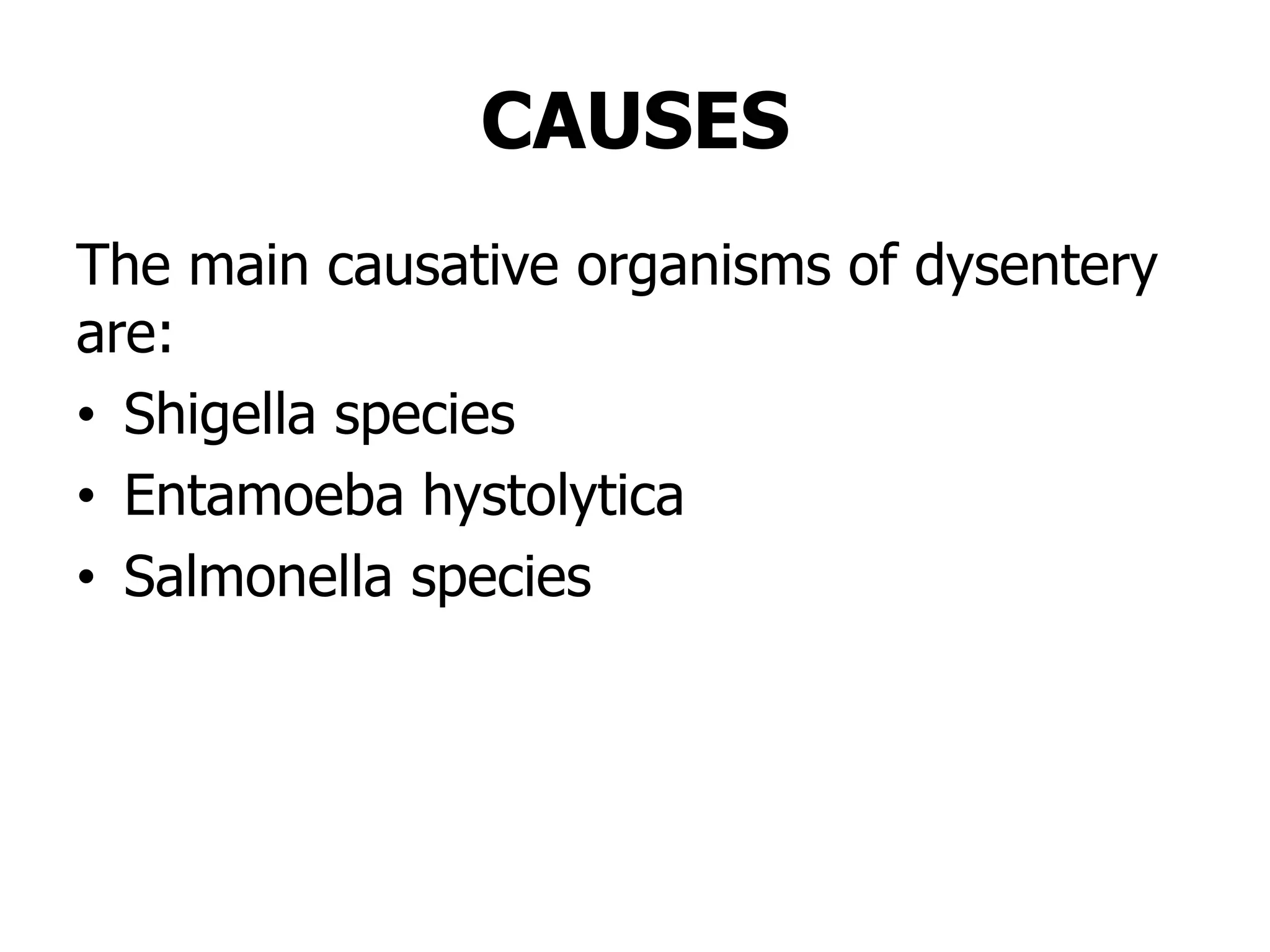
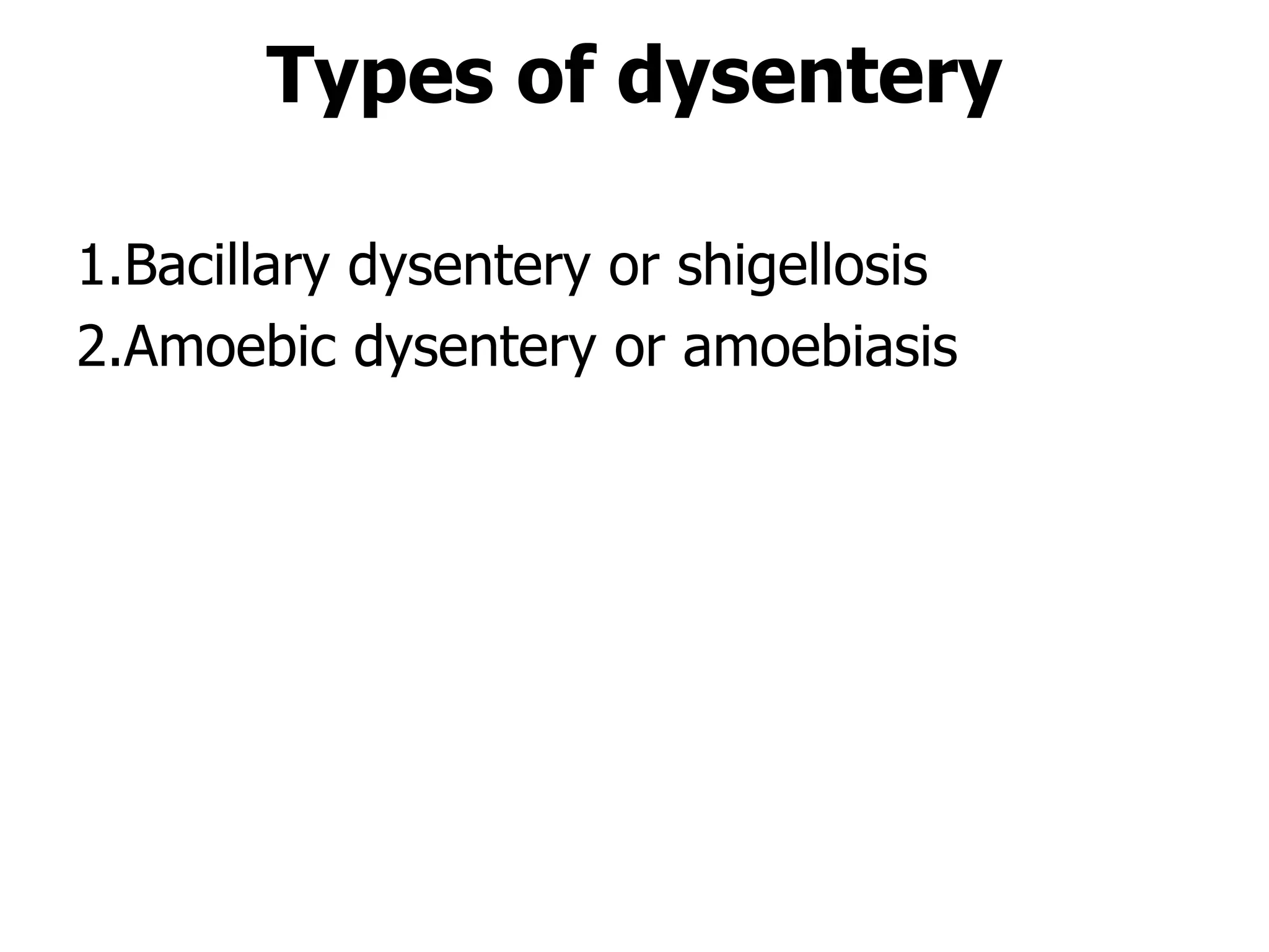
DYSENTERY
BY NORAH NTABO
BSC NRS-UNZA
INTRODUCTION
• Dysentery is a blood diarrheal disease;
• It is a notifiable infectious disease
commonly caused by bacillary or amoebic
dysentery,
• Transmitted through oral feacal root and
• Most prevalent in unhygienic areas like
nursing homes.
• In Zambia the incidence of dysentery is 7
per 1000
GENERAL OBJECTIVE
• At the end of the lecture/ discussion
students should be able to acquire
knowledge in dysentery, so that they can
use the knowledge to identify, treat and
prevent the occurrences of dysentery in
the communities.
SPECIFIC OBJECTIVES
At the end of this lecture/discussion students
should be able to:
1.Define dysentery
2.List the causes of Dysentery
3.Mention the two main types of dysentery
4.Describe of management of bacillary and
amoebic dysentery
DEFINITION
• Dysentery is an inflammatory disorder of
the intestine, especially of the colon, that
results in severe diarrhoea containing
blood and mucus in the faeces
characterised by fever, abdominal pain,
and rectal tenesmus caused by any kind of
infection (Basavanthappa B.T. 2005) .
CAUSES
The main causative organisms of dysentery
are:
• Shigella species
• Entamoeba hystolytica
• Salmonella species
Types of dysentery
1.Bacillary dysentery or shigellosis
2.Amoebic dysentery or amoebiasis
Bacillary Dysentery
/Shigellosis
DEFINITION
• Bacillary dysentery is an acute
inflammation and ulceration of large
intestines characterized by small frequent
bowel movements consisting of blood and
mucous in stool caused by shigella. (Beer
M.H etal, 2006).
CAUSES
• Bacillary dysentery is caused by non-
motile gram negative bacteria of the
genus Shigella.
Shigella is in 4 strains:
• Shigella flexneri
• Shigella boydii
• Shigella dysenteriae
• Shigella sonnei
Predisposing factors
• These are best summarized by the 6 F’s
which are Formites, Food, Faeces,
Fingers, Fluids and Flies.
• If these are well taken care then the
problem is solved.
• The predisposing factors include;
• Poor feeding methods, example, use of
dirty feeding bottles for infants, eating
unboiled and improperly prepared foods.
Predisposing factors cont…
• Poor personal hygiene especially hand
hygiene(hand washing, long unkempt
finger nails and so forth)
• Poor source, treatment and storage
facilities for water to drink
• Poor sanitation - Rubbish pits or dumping
sites; Sewerage lines.
• Overcrowding
EPIDEMIOLOGY
• It is most prevalent in unhygienic areas of
the tropics,
• But because it is easily spread, sporadic
outbreaks are common in all parts of the
world.
• Occurs among confined populations, such
as those in nursing homes, large
institutions subject to overcrowding
MODE OF TRANSMISSION
• The route of transmission of shigella is fecal
oral route.
• The bacilli are excreted in faeces and through
poor sanitation and bad hygiene, food and
water can then become contaminated.
• Flies also frequently cause contamination of
food and are prevalent mode of spread of
dysentery.
INCUBATION PERIOD
• The incubation period of shigella is 1 - 7 days.
PATHOPHYSIOGY
• When the bacillus enters the GIT, it
invades the large intestine causing
inflammation of the mucosa leading to
ulceration and bleeding of the mucosa
• Stool would be blood stained and mucoid.
• In the later stage, pus forms due to
infection.
• Adjacent lymph nodes may be affected
resulting into fever
SIGNS AND
SYMPTOMS
SIGNS AND SYMPTOMS
• Sudden onset of s/s
• Fever which results from infection and
inflammatory reaction.
• S/s of dehydration such as loss of skin turgor, as
a result of diarrhoea.
• Dehydration may or may not be present in this
condition because patient passes small amount
of stool, but it’s the frequency which is increased
• Abdominal discomfort: This may be due to
irritation of the mucosal lining of the GIT by the
bacteria and usually it is an early symptom of
S/S CONT….
• Nausea and Vomiting: This may be due to
irritation of mucosal lining of the GIT
(stomach).
• Colic abdominal pains: May be due to
inflammatory reaction in the mucosal lining
of the large intestines.
• Bloody diarrhoea - This may be due to
damage of the mucosal lining of the large
intestines during inflammation. Damage to
the mucosal lining may also cause damage
to the capillaries.
S/S CONT….
• The passage of bloody diarrhoea is usually
accompanied by Urgency and tenesmus.
(Urgency is the urge to open bowels at
very frequent intervals even if small
amounts of stool are passed and
tenesmus is a painful ineffective straining
to empty the bowels
MANAGEMENT
Aims
1.To correct electrolyte and fluid imbalance
3.To identify and eliminate the causative
organism
3.To prevent and manage complications
INVESTIGATIONS
• Microscopic examination of a fresh stool
specimen
• Rectal swab for culture and sensitivity.
• Stool should be cultured within a few hours of
collection.
• Detection of the organism in stool confirms
diagnosis.
• Immunofluorescent techniques to detect
organism in stool.
• Sigmoidoscopy reveals a red, bleeding mucosa
with patches of necrotic membrane which may
separate to leave ulcerated areas.
FLUID THERAPY
• Fluid and electrolyte replacement
• Oral rehydration is usually required to
restore fluid and electrolyte imbalances.
• However, each patient should be assessed
for the degree of dehydration and the
appropriate fluid replacement therapy
given.
DRUGS
• Antibiotics are administered to shorten the
duration of illness and prevent relapse.
• Any of the following are given while
waiting for result of culture and sensitivity:
• Nalidixic acid 1g PO qid for 7 to 14 days
• Ciprofloxacin 500mg PO BD for 5 days
• Co-otrimoxazole (Septrin ;Trimethoprime-
Sulfamethoxazole ) 960mg PO BD for 5/7
• Ampicillin 500mg qid for 5 days
AMOEBIC
DYSENTERY/AMOEBIASIS
DEFINITIONS
• Amoebiasis is an infection of the large
intestines caused by Entamoeba
hystolytica, a single celled parasite
(Berkow et al, 1997).
CAUSE
• The cause of amoebic dysentery is
entamoeba hystolytica
PREDISPOSING FACTORS
• Refer to bacillary dysentery
EPIDEMIOLOGY
• Entamoeba histolytica has a worldwide
distribution and is endemic in most
countries with poor sanitation and low
socioeconomic conditions.
• Use of night soil for agricultural purposes
favours the spread of the disease.
• The organism is acquired when cysts are
ingested.
MODE OF TRANSMISSION
• Faecal-oral route; vectors such as flies,
cockroaches and rodents are capable of
carrying cysts and contaminate food and
water
INCUBATION PERIOD
• It may take 2 weeks or years.
• Human beings are the principal
reservoirs/carriers.
PATHOPHYSIOLOGY
• Ingested cysts enter the alimentary tract
through the mouth to the stomach where
they eccyst during digestion.
• Motile trophozoites are released which
multiply, invade and ulcerate the intestinal
mucosa of the large bowels, forming flask
like ulcers.
• Some of the amoeba goes through the
mesenteric artery and reach the liver causing
total destruction of the liver resulting in
amoebic hepatocellular necrosis and then
liver abscess.
SIGNS AND SYMPTOMS
• On set is gradual and associated with
abdominal discomfort.
• Mildly loose stools (diarrhoea) with or without
blood and mucus.
• Diarrhoea may alternate with constipation.
• Tenderness may develop over the caecum,
transverse colon or sigmoid
• Fever may be present
• Abdominal pains that may be on and off.
• If there is hepatic amoebiasis, there would be
body malaise, swinging temperature,
sweating, and enlarged tender liver.
• Foul-smelly stool.
• Weight loss in chronic cases.
INVESTIGATIONS
• Stool for m/c/s
• History of blood stained stool.
• Physical inspection will reveal
dehydration.
• Rectal swab culture.
• Blood for Hb.
• Sigmoidoscopy will review ulcers.
• Liver scan will review Liver abscess.
TREATMENT
• Flagyl 200-400mg tids
• Septrin 960mg bd x 5-7 days
• Furamide[diloxanidefuroate] 500mg tds
for 10 days
• For Hepatic Amoebiasis give Flagyl and
Chloroquine 600mg od for 2 days and
then 300mg od for 21 days.
• Panadol 1 gram tds x3/7
• Intravenous fluids [Ringers Lactate)
NURSING CARE
Aims
1.To prevent further spread of infection
2.To replace lost fluids and electrolytes
3.To prevent complications such as shock
4.To identify any contacts
ENVIRONMENT
• Admit patient in an isolation room away
from other patients to prevent spread of
infection to other people.
• The room should be well lit for easy
observation and ventilated environment to
promote air circulation.
• Patient should be nursed near the toilet
for convenience.
• Equipment such as drip stands,
intravenous set and observations tray
should be within patient’s environment.
OBSERVATION
• Observe general condition of patient.
• Monitor vital signs such as temperature, pulse,
respirations and blood pressure frequently.
• The frequency of vital sign observations depends
on patient’s condition.
• Monitor the intake and output and record on the
fluid balance charts.
• Monitor stool for amount, consistency and color
and report.
• Observe for any signs of dehydration such as
loss of skin elasticity, sunken eyes, and thirsty
and dry mucus membranes of the mouth.
INFECTION PREVENTION
• Isolate patient away from other patients to
prevent spread of infection.
• People who come in contact with this
patient should observe isolation
techniques such, putting on gowns and
masks whenever they enter the room,
washing hands before and after attending
to the patient.
• Restrict visitors because they can also get
the infection.
IP cont…
• The linen which is used by the patient
should be disinfected with JIK 1:6 and
should be labeled “infectious” before
sending it to the laundry.
• It should not be mixed with other linen
from the wards.
• All utensils used by patient should be
disinfected
• Administer prescribed medication to treat
the causative organism.
NUTRITION
• Give some copious drinks
• Light mixed diet free from irritants.
• If patient is unable to take food and fluids
orally commence him/her on intravenous
fluids.
• Maintain strict intake and output.
• Record intake and output, time
commenced IVF, type of fluid and date
started.
HYGIENE
• Assisted /bed bath can be given depending on
the condition of the patient to promote
comfort, self esteem and to remove dirty.
• Assist the patient with oral care to prevent
complications of a dirty mouth such as mouth
infections and also promote salivation as the
patient’s mouth can be dry due to excessive
loss of fluids.
HYGIENE cont…
• Change linen whenever soiled and
disinfect the linen with Jik 1:6 before
sending to the laundry. Ensure perineal
area is cleaned
PSYCHOLOGICAL CARE
• Patients with dysentery may feel as if they
have been neglected.
• The nurse needs to give proper
psychological care to allay anxiety.
• Explain the disease process to patient which
should include the cause, mode of
transmission, signs and symptoms,
treatment and complications.
PSYCHOLOGICAL CARE
cont…
• Explain to the patient the reason for isolation
which is prevention of spread of infection.
• Explain also to the significant others on why
they are not allowed to visit the patient.
• Any procedure which is done to patient should
be explained to gain his/her cooperation.
• Allow patients to ask questions and answer
them truthfully.
MEDICATION
• Administer prescribed drugs as prescribed
and observe for side effects.
• Administer fluids according to patient’s
condition.
ELIMINATION
• Observe intake and output and record.
• Observe stool for amount, contents and
odor.
• Provide bed pan in the initial stage but as
condition improves, encourage patient to
go to the toilet.
PREVENTION AND CONTROL
OF DYSENTERY
• We have discussed the mode of
transmission and predisposing factors of
dysentery.
• From our discussion, how can dysentery
be prevented?
Dysentery can be prevented by
doing the following measures:
Improved Environmental Sanitation:
measures include:
• Provision of safe and adequate water supply.
• Safe and adequate disposal of human excreta
through use of pit latrines or toilets
• Food safety against faecal contamination
• Provision of information, education and
communication about dysentery.
• Discourage use of untreated human excreta
for manure.
Prevention cont…
Early Diagnosis and Treatment of
Cases and Carriers
• Prompt detection and appropriate and
adequate treatment of both cases and
carriers
• Regular screening of food handlers
Prevention cont…
Improved Personal and Communal Hygiene
• Adequate hand washing with soap under running
water after using the toilet and before handling
and eating food.
• Use of pit latrines or toilets for defaecation.
• Children should not be allowed to defaecate on
the ground.
• Toilet training pots should be used and disinfected
after use.
• Children’s stools should be disposed off in the
toilet or pit latrine
• Boil water for drinking and for washing vegetables
and fruits.
• Avoid eating raw vegetable and fruit salads.
COMPLICATIONS
• Perforation of the colon
• Peritonitis
• Rectal prolapse
• Haematogenous dissemination of the shigellas
(rare) causing abscesses and meningitis
• Toxic megacolon
• Hemiplegia
• Encephalopathy
• Septicaemia
• Hyponatraemia
• Reiter’s syndrome
• Liver abscess
SUMMARY
• Dysentery is an inflammatory disorder of the
intestine, especially of the colon, that results
in severe diarrhoea containing blood and
mucus in the faeces with fever, abdominal
pain, and rectal tenesmus caused by
infection.
• The main types of dysentery are Bacillary
dysentery or shigellosis and Amoebic
dysentery or amoebiasis.
• They are both treated using antibiotics like
flagyl or Nalidixic and nursed in isolation/
revise barrier.
ASSINGMENT
• Read and write notes on Reiter’s
syndrome and Toxic megacolon
REFRRRENCES
• Basavanthappa B.T, (2005), Medical Surgical
Nursing, New Delhi, Jaypee Brothers, India.
• Beer M.H etal, (2006).The Merck manual of
diagnosis and therapy, 18th Edition, Merck &
Co.Inc, USA.
• Berkow.R, et al (1997), The Merck Manual of
Medical Information, Merck Research
Laboratories, New Jersey. Smeltzer etal, (2010).
• Bruuner and Suddarth Medical-Surgical Nursing,
10th Edition, Elsevier, Missouri
• Cahill, M. (1998), Diseases, Springhouse
Corporation, Pennsylvania.
THANK YOU

DYSENTERY ppt.pptx

  • 1.
  • 2.
    INTRODUCTION • Dysentery isa blood diarrheal disease; • It is a notifiable infectious disease commonly caused by bacillary or amoebic dysentery, • Transmitted through oral feacal root and • Most prevalent in unhygienic areas like nursing homes. • In Zambia the incidence of dysentery is 7 per 1000
  • 3.
    GENERAL OBJECTIVE • Atthe end of the lecture/ discussion students should be able to acquire knowledge in dysentery, so that they can use the knowledge to identify, treat and prevent the occurrences of dysentery in the communities.
  • 4.
    SPECIFIC OBJECTIVES At theend of this lecture/discussion students should be able to: 1.Define dysentery 2.List the causes of Dysentery 3.Mention the two main types of dysentery 4.Describe of management of bacillary and amoebic dysentery
  • 5.
    DEFINITION • Dysentery isan inflammatory disorder of the intestine, especially of the colon, that results in severe diarrhoea containing blood and mucus in the faeces characterised by fever, abdominal pain, and rectal tenesmus caused by any kind of infection (Basavanthappa B.T. 2005) .
  • 6.
    CAUSES The main causativeorganisms of dysentery are: • Shigella species • Entamoeba hystolytica • Salmonella species
  • 7.
    Types of dysentery 1.Bacillarydysentery or shigellosis 2.Amoebic dysentery or amoebiasis
  • 8.
    Bacillary Dysentery /Shigellosis DEFINITION • Bacillarydysentery is an acute inflammation and ulceration of large intestines characterized by small frequent bowel movements consisting of blood and mucous in stool caused by shigella. (Beer M.H etal, 2006).
  • 9.
    CAUSES • Bacillary dysenteryis caused by non- motile gram negative bacteria of the genus Shigella. Shigella is in 4 strains: • Shigella flexneri • Shigella boydii • Shigella dysenteriae • Shigella sonnei
  • 10.
    Predisposing factors • Theseare best summarized by the 6 F’s which are Formites, Food, Faeces, Fingers, Fluids and Flies. • If these are well taken care then the problem is solved. • The predisposing factors include; • Poor feeding methods, example, use of dirty feeding bottles for infants, eating unboiled and improperly prepared foods.
  • 11.
    Predisposing factors cont… •Poor personal hygiene especially hand hygiene(hand washing, long unkempt finger nails and so forth) • Poor source, treatment and storage facilities for water to drink • Poor sanitation - Rubbish pits or dumping sites; Sewerage lines. • Overcrowding
  • 12.
    EPIDEMIOLOGY • It ismost prevalent in unhygienic areas of the tropics, • But because it is easily spread, sporadic outbreaks are common in all parts of the world. • Occurs among confined populations, such as those in nursing homes, large institutions subject to overcrowding
  • 13.
    MODE OF TRANSMISSION •The route of transmission of shigella is fecal oral route. • The bacilli are excreted in faeces and through poor sanitation and bad hygiene, food and water can then become contaminated. • Flies also frequently cause contamination of food and are prevalent mode of spread of dysentery. INCUBATION PERIOD • The incubation period of shigella is 1 - 7 days.
  • 14.
    PATHOPHYSIOGY • When thebacillus enters the GIT, it invades the large intestine causing inflammation of the mucosa leading to ulceration and bleeding of the mucosa • Stool would be blood stained and mucoid. • In the later stage, pus forms due to infection. • Adjacent lymph nodes may be affected resulting into fever
  • 15.
  • 16.
    SIGNS AND SYMPTOMS •Sudden onset of s/s • Fever which results from infection and inflammatory reaction. • S/s of dehydration such as loss of skin turgor, as a result of diarrhoea. • Dehydration may or may not be present in this condition because patient passes small amount of stool, but it’s the frequency which is increased • Abdominal discomfort: This may be due to irritation of the mucosal lining of the GIT by the bacteria and usually it is an early symptom of
  • 17.
    S/S CONT…. • Nauseaand Vomiting: This may be due to irritation of mucosal lining of the GIT (stomach). • Colic abdominal pains: May be due to inflammatory reaction in the mucosal lining of the large intestines. • Bloody diarrhoea - This may be due to damage of the mucosal lining of the large intestines during inflammation. Damage to the mucosal lining may also cause damage to the capillaries.
  • 18.
    S/S CONT…. • Thepassage of bloody diarrhoea is usually accompanied by Urgency and tenesmus. (Urgency is the urge to open bowels at very frequent intervals even if small amounts of stool are passed and tenesmus is a painful ineffective straining to empty the bowels
  • 19.
    MANAGEMENT Aims 1.To correct electrolyteand fluid imbalance 3.To identify and eliminate the causative organism 3.To prevent and manage complications
  • 20.
    INVESTIGATIONS • Microscopic examinationof a fresh stool specimen • Rectal swab for culture and sensitivity. • Stool should be cultured within a few hours of collection. • Detection of the organism in stool confirms diagnosis. • Immunofluorescent techniques to detect organism in stool. • Sigmoidoscopy reveals a red, bleeding mucosa with patches of necrotic membrane which may separate to leave ulcerated areas.
  • 21.
    FLUID THERAPY • Fluidand electrolyte replacement • Oral rehydration is usually required to restore fluid and electrolyte imbalances. • However, each patient should be assessed for the degree of dehydration and the appropriate fluid replacement therapy given.
  • 22.
    DRUGS • Antibiotics areadministered to shorten the duration of illness and prevent relapse. • Any of the following are given while waiting for result of culture and sensitivity: • Nalidixic acid 1g PO qid for 7 to 14 days • Ciprofloxacin 500mg PO BD for 5 days • Co-otrimoxazole (Septrin ;Trimethoprime- Sulfamethoxazole ) 960mg PO BD for 5/7 • Ampicillin 500mg qid for 5 days
  • 23.
    AMOEBIC DYSENTERY/AMOEBIASIS DEFINITIONS • Amoebiasis isan infection of the large intestines caused by Entamoeba hystolytica, a single celled parasite (Berkow et al, 1997).
  • 24.
    CAUSE • The causeof amoebic dysentery is entamoeba hystolytica PREDISPOSING FACTORS • Refer to bacillary dysentery EPIDEMIOLOGY • Entamoeba histolytica has a worldwide distribution and is endemic in most countries with poor sanitation and low socioeconomic conditions. • Use of night soil for agricultural purposes favours the spread of the disease. • The organism is acquired when cysts are ingested.
  • 25.
    MODE OF TRANSMISSION •Faecal-oral route; vectors such as flies, cockroaches and rodents are capable of carrying cysts and contaminate food and water INCUBATION PERIOD • It may take 2 weeks or years. • Human beings are the principal reservoirs/carriers.
  • 26.
    PATHOPHYSIOLOGY • Ingested cystsenter the alimentary tract through the mouth to the stomach where they eccyst during digestion. • Motile trophozoites are released which multiply, invade and ulcerate the intestinal mucosa of the large bowels, forming flask like ulcers. • Some of the amoeba goes through the mesenteric artery and reach the liver causing total destruction of the liver resulting in amoebic hepatocellular necrosis and then liver abscess.
  • 27.
    SIGNS AND SYMPTOMS •On set is gradual and associated with abdominal discomfort. • Mildly loose stools (diarrhoea) with or without blood and mucus. • Diarrhoea may alternate with constipation. • Tenderness may develop over the caecum, transverse colon or sigmoid • Fever may be present • Abdominal pains that may be on and off. • If there is hepatic amoebiasis, there would be body malaise, swinging temperature, sweating, and enlarged tender liver. • Foul-smelly stool. • Weight loss in chronic cases.
  • 28.
    INVESTIGATIONS • Stool form/c/s • History of blood stained stool. • Physical inspection will reveal dehydration. • Rectal swab culture. • Blood for Hb. • Sigmoidoscopy will review ulcers. • Liver scan will review Liver abscess.
  • 29.
    TREATMENT • Flagyl 200-400mgtids • Septrin 960mg bd x 5-7 days • Furamide[diloxanidefuroate] 500mg tds for 10 days • For Hepatic Amoebiasis give Flagyl and Chloroquine 600mg od for 2 days and then 300mg od for 21 days. • Panadol 1 gram tds x3/7 • Intravenous fluids [Ringers Lactate)
  • 30.
    NURSING CARE Aims 1.To preventfurther spread of infection 2.To replace lost fluids and electrolytes 3.To prevent complications such as shock 4.To identify any contacts
  • 31.
    ENVIRONMENT • Admit patientin an isolation room away from other patients to prevent spread of infection to other people. • The room should be well lit for easy observation and ventilated environment to promote air circulation. • Patient should be nursed near the toilet for convenience. • Equipment such as drip stands, intravenous set and observations tray should be within patient’s environment.
  • 32.
    OBSERVATION • Observe generalcondition of patient. • Monitor vital signs such as temperature, pulse, respirations and blood pressure frequently. • The frequency of vital sign observations depends on patient’s condition. • Monitor the intake and output and record on the fluid balance charts. • Monitor stool for amount, consistency and color and report. • Observe for any signs of dehydration such as loss of skin elasticity, sunken eyes, and thirsty and dry mucus membranes of the mouth.
  • 33.
    INFECTION PREVENTION • Isolatepatient away from other patients to prevent spread of infection. • People who come in contact with this patient should observe isolation techniques such, putting on gowns and masks whenever they enter the room, washing hands before and after attending to the patient. • Restrict visitors because they can also get the infection.
  • 34.
    IP cont… • Thelinen which is used by the patient should be disinfected with JIK 1:6 and should be labeled “infectious” before sending it to the laundry. • It should not be mixed with other linen from the wards. • All utensils used by patient should be disinfected • Administer prescribed medication to treat the causative organism.
  • 35.
    NUTRITION • Give somecopious drinks • Light mixed diet free from irritants. • If patient is unable to take food and fluids orally commence him/her on intravenous fluids. • Maintain strict intake and output. • Record intake and output, time commenced IVF, type of fluid and date started.
  • 36.
    HYGIENE • Assisted /bedbath can be given depending on the condition of the patient to promote comfort, self esteem and to remove dirty. • Assist the patient with oral care to prevent complications of a dirty mouth such as mouth infections and also promote salivation as the patient’s mouth can be dry due to excessive loss of fluids.
  • 37.
    HYGIENE cont… • Changelinen whenever soiled and disinfect the linen with Jik 1:6 before sending to the laundry. Ensure perineal area is cleaned
  • 38.
    PSYCHOLOGICAL CARE • Patientswith dysentery may feel as if they have been neglected. • The nurse needs to give proper psychological care to allay anxiety. • Explain the disease process to patient which should include the cause, mode of transmission, signs and symptoms, treatment and complications.
  • 39.
    PSYCHOLOGICAL CARE cont… • Explainto the patient the reason for isolation which is prevention of spread of infection. • Explain also to the significant others on why they are not allowed to visit the patient. • Any procedure which is done to patient should be explained to gain his/her cooperation. • Allow patients to ask questions and answer them truthfully.
  • 40.
    MEDICATION • Administer prescribeddrugs as prescribed and observe for side effects. • Administer fluids according to patient’s condition.
  • 41.
    ELIMINATION • Observe intakeand output and record. • Observe stool for amount, contents and odor. • Provide bed pan in the initial stage but as condition improves, encourage patient to go to the toilet.
  • 42.
    PREVENTION AND CONTROL OFDYSENTERY • We have discussed the mode of transmission and predisposing factors of dysentery. • From our discussion, how can dysentery be prevented?
  • 43.
    Dysentery can beprevented by doing the following measures: Improved Environmental Sanitation: measures include: • Provision of safe and adequate water supply. • Safe and adequate disposal of human excreta through use of pit latrines or toilets • Food safety against faecal contamination • Provision of information, education and communication about dysentery. • Discourage use of untreated human excreta for manure.
  • 44.
    Prevention cont… Early Diagnosisand Treatment of Cases and Carriers • Prompt detection and appropriate and adequate treatment of both cases and carriers • Regular screening of food handlers
  • 45.
    Prevention cont… Improved Personaland Communal Hygiene • Adequate hand washing with soap under running water after using the toilet and before handling and eating food. • Use of pit latrines or toilets for defaecation. • Children should not be allowed to defaecate on the ground. • Toilet training pots should be used and disinfected after use. • Children’s stools should be disposed off in the toilet or pit latrine • Boil water for drinking and for washing vegetables and fruits. • Avoid eating raw vegetable and fruit salads.
  • 46.
    COMPLICATIONS • Perforation ofthe colon • Peritonitis • Rectal prolapse • Haematogenous dissemination of the shigellas (rare) causing abscesses and meningitis • Toxic megacolon • Hemiplegia • Encephalopathy • Septicaemia • Hyponatraemia • Reiter’s syndrome • Liver abscess
  • 47.
    SUMMARY • Dysentery isan inflammatory disorder of the intestine, especially of the colon, that results in severe diarrhoea containing blood and mucus in the faeces with fever, abdominal pain, and rectal tenesmus caused by infection. • The main types of dysentery are Bacillary dysentery or shigellosis and Amoebic dysentery or amoebiasis. • They are both treated using antibiotics like flagyl or Nalidixic and nursed in isolation/ revise barrier.
  • 48.
    ASSINGMENT • Read andwrite notes on Reiter’s syndrome and Toxic megacolon
  • 49.
    REFRRRENCES • Basavanthappa B.T,(2005), Medical Surgical Nursing, New Delhi, Jaypee Brothers, India. • Beer M.H etal, (2006).The Merck manual of diagnosis and therapy, 18th Edition, Merck & Co.Inc, USA. • Berkow.R, et al (1997), The Merck Manual of Medical Information, Merck Research Laboratories, New Jersey. Smeltzer etal, (2010). • Bruuner and Suddarth Medical-Surgical Nursing, 10th Edition, Elsevier, Missouri • Cahill, M. (1998), Diseases, Springhouse Corporation, Pennsylvania.
  • 50.