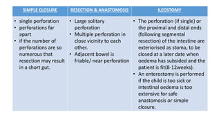
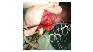
TYPHOID ILEAL
PERFORATION
Dr. Prince T. Ankrah
Snr. House Officer
(GENERAL SURGERY DEPARTMENT)
ENRH, December 2018.
OUTLINE
• INTRODUCTION
• EPIDEMIOLOGY
• AETIOLOGY
• PATHOPHYSIOLOGY
• CLINICAL FEATURES
• INVESTIGATIONS
• MANAGEMENT
• POST OP COMPLICATIONS
• TAKE-HOME POINTS
INTRODUCTION
The most common surgical complication in cases of
typhoid fever.
Mainly a problem in developing countries due to poor
sanitation and late diagnosis/proper management.
Diagnosis is mainly clinical
Surgery remains the gold standard of treatment
EPIDEMIOLOGY
• Global incidence of typhoid fever is 21million cases annually
with 1-4% mortality
• Children account for >50% of all cases of typhoid ileal
perforation with peak age of 5-9years.
• Has higher incidence in rainy season.
Aetiology
• Salmonella Typhii
• And rarely by Salmonella Paratyphii A, B,C.
• Transmission is mainly via feco-oral route.
Pathogensis
• S. typhi and paratyphi enter the host’s system primarily via
the distal ileum
• Attachment to the epithelium over clusters of lymphoid
tissue in the ileum (Peyer patches).
1.
• Ingestion of contaminated food
2
• Intestinal mucosa invasion of ingested bacilli
3
• Taken up & transported to regional lymph node
4
• Multiplication in the intestinal lymphoid tissue
5
• Interaction with enterocytes & M cells (ileal Peyer’s patches)
6
• Entry of bacilli into the bloodstream
7
• Invasion of the gallbladder, biliary system and lymphatic tissues of the bowel
8
• Passage into the intestinal tract (stool)
CLINICAL FEATURES
• History of fever, 2-3weeks preceding the onset of abdominal
pain.
• Abdominal pain
• ± hematochezia prior to onset of pain
• Diarrhea or constipation.
• ± jaundice may be a complaint.
ON GENERAL EXAMINATION
Depending on the stage and severirty of the illness one could notice
• A very ill-looking patient
• Dehydrated
• Pale
• Febrile
• Wasted
CVS- there could be signs of shock
CHEST- usually clinically clear but crepitation could indicate pneumonia
co-morbidity
ABDOMEN
• Generalized tenderness
• Rebound tenderness
• Guarding
• Rigidity
• Diminished or absent bowel sounds
• Tenderness and fullness in the recto-vesical or recto-uterine pouch,
suggesting a pelvic collection of pus.
• Blood may be seen on the examining finger in patients with bleeding.
INVESTIGATIONS
Diagnosis is usually clinical, based on a good Hx + PE findings.
Investigations done are usually to:
Support the diagnosis
Identify deficits & likely complications
Ascertain fitness of the patient for surgery
FBC, BUE & Cr, BF for MP’s,GXM, plain CXR, ABD xray, blood & urine
cultures
INITIAL MANAGEMENT
• Correction of fluid and electrolyte deficits
• Gastric decompression (NG tube)
• Urethral catheter to monitor fluid output
• Blood transfusion if warranted
• IV antibiotic therapy
• Antipyrexics
DEFINITIVE MANAGEMENT
SURGERY IS MAINSTAY to get perforation corrected. Could be achieved
by laparotomy + options:
1. Simple closure of perforation
2. Segmental resection and anastomosis
3. Enterostomy
SIMPLE CLOSURE RESECTION & ANASTOMOSIS ILEOSTOMY
• single perforation
• perforations far
apart
• if the number of
perforations are so
numerous that
resection may result
in a short gut.
• Large solitary
perforation
• Multiple perforation in
close vicinity to each
other.
• Adjacent bowel is
friable/ near perforation
• The perforation (if single) or
the proximal and distal ends
(following segmental
resection) of the intestine are
exteriorised as stoma, to be
closed at a later date when
oedema has subsided and the
patient is fit(8-12weeks).
• An enterostomy is performed
if the child is too sick or
intestinal oedema is too
extensive for safe
anastomosis or simple
closure.
POST OP MANAGEMENT
• Strict fluid and electrolyte mgt
• Antibiotic cover
• Correction of anemia
• Close monitoring of vital signs
• Daily monitoring for intra-abdominal collection
• Wound dressing
• Adequate nutritional support
Post OP complications
• Prolonged ileus
• Surgical site infection
• Abdominal wound dehiscence
• Anastomotic leakage or complete breakdown of the anastomosis
• Enterocutaneous fistula
• Intraperitoneal abscess
• Adhesion intestinal obstruction
• Transient psychosis
• Electrolyte imbalance
TAKE HOME POINTS
• Ileal perforation is the most common and serious surgical
complication of typhoid infection especially in developing
countries.
• Early diagnosis, adequate initial resuscitation, and proper
choice of surgical procedure go a long way to reduce its
morbidity and mortality.
References

Typhoid ileal perforation

  • 1.
    TYPHOID ILEAL PERFORATION Dr. PrinceT. Ankrah Snr. House Officer (GENERAL SURGERY DEPARTMENT) ENRH, December 2018.
  • 2.
    OUTLINE • INTRODUCTION • EPIDEMIOLOGY •AETIOLOGY • PATHOPHYSIOLOGY • CLINICAL FEATURES • INVESTIGATIONS • MANAGEMENT • POST OP COMPLICATIONS • TAKE-HOME POINTS
  • 3.
    INTRODUCTION The most commonsurgical complication in cases of typhoid fever. Mainly a problem in developing countries due to poor sanitation and late diagnosis/proper management. Diagnosis is mainly clinical Surgery remains the gold standard of treatment
  • 4.
    EPIDEMIOLOGY • Global incidenceof typhoid fever is 21million cases annually with 1-4% mortality • Children account for >50% of all cases of typhoid ileal perforation with peak age of 5-9years. • Has higher incidence in rainy season.
  • 5.
    Aetiology • Salmonella Typhii •And rarely by Salmonella Paratyphii A, B,C. • Transmission is mainly via feco-oral route.
  • 6.
    Pathogensis • S. typhiand paratyphi enter the host’s system primarily via the distal ileum • Attachment to the epithelium over clusters of lymphoid tissue in the ileum (Peyer patches).
  • 7.
    1. • Ingestion ofcontaminated food 2 • Intestinal mucosa invasion of ingested bacilli 3 • Taken up & transported to regional lymph node 4 • Multiplication in the intestinal lymphoid tissue 5 • Interaction with enterocytes & M cells (ileal Peyer’s patches) 6 • Entry of bacilli into the bloodstream 7 • Invasion of the gallbladder, biliary system and lymphatic tissues of the bowel 8 • Passage into the intestinal tract (stool)
  • 8.
    CLINICAL FEATURES • Historyof fever, 2-3weeks preceding the onset of abdominal pain. • Abdominal pain • ± hematochezia prior to onset of pain • Diarrhea or constipation. • ± jaundice may be a complaint.
  • 9.
    ON GENERAL EXAMINATION Dependingon the stage and severirty of the illness one could notice • A very ill-looking patient • Dehydrated • Pale • Febrile • Wasted CVS- there could be signs of shock CHEST- usually clinically clear but crepitation could indicate pneumonia co-morbidity
  • 10.
    ABDOMEN • Generalized tenderness •Rebound tenderness • Guarding • Rigidity • Diminished or absent bowel sounds • Tenderness and fullness in the recto-vesical or recto-uterine pouch, suggesting a pelvic collection of pus. • Blood may be seen on the examining finger in patients with bleeding.
  • 11.
    INVESTIGATIONS Diagnosis is usuallyclinical, based on a good Hx + PE findings. Investigations done are usually to: Support the diagnosis Identify deficits & likely complications Ascertain fitness of the patient for surgery FBC, BUE & Cr, BF for MP’s,GXM, plain CXR, ABD xray, blood & urine cultures
  • 12.
    INITIAL MANAGEMENT • Correctionof fluid and electrolyte deficits • Gastric decompression (NG tube) • Urethral catheter to monitor fluid output • Blood transfusion if warranted • IV antibiotic therapy • Antipyrexics
  • 13.
    DEFINITIVE MANAGEMENT SURGERY ISMAINSTAY to get perforation corrected. Could be achieved by laparotomy + options: 1. Simple closure of perforation 2. Segmental resection and anastomosis 3. Enterostomy
  • 14.
    SIMPLE CLOSURE RESECTION& ANASTOMOSIS ILEOSTOMY • single perforation • perforations far apart • if the number of perforations are so numerous that resection may result in a short gut. • Large solitary perforation • Multiple perforation in close vicinity to each other. • Adjacent bowel is friable/ near perforation • The perforation (if single) or the proximal and distal ends (following segmental resection) of the intestine are exteriorised as stoma, to be closed at a later date when oedema has subsided and the patient is fit(8-12weeks). • An enterostomy is performed if the child is too sick or intestinal oedema is too extensive for safe anastomosis or simple closure.
  • 17.
    POST OP MANAGEMENT •Strict fluid and electrolyte mgt • Antibiotic cover • Correction of anemia • Close monitoring of vital signs • Daily monitoring for intra-abdominal collection • Wound dressing • Adequate nutritional support
  • 18.
    Post OP complications •Prolonged ileus • Surgical site infection • Abdominal wound dehiscence • Anastomotic leakage or complete breakdown of the anastomosis • Enterocutaneous fistula • Intraperitoneal abscess • Adhesion intestinal obstruction • Transient psychosis • Electrolyte imbalance
  • 19.
    TAKE HOME POINTS •Ileal perforation is the most common and serious surgical complication of typhoid infection especially in developing countries. • Early diagnosis, adequate initial resuscitation, and proper choice of surgical procedure go a long way to reduce its morbidity and mortality.
  • 20.