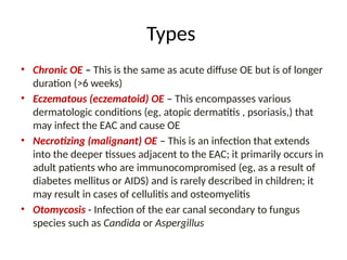
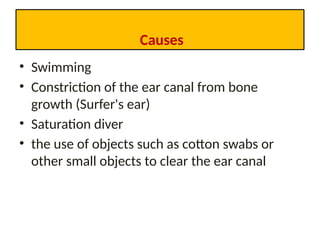
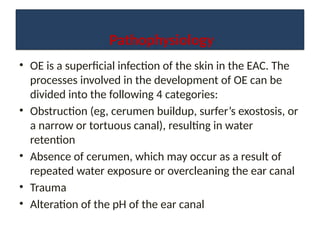
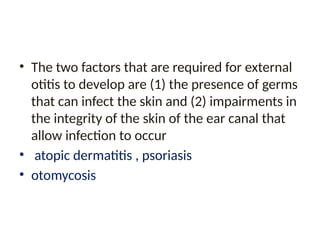
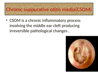
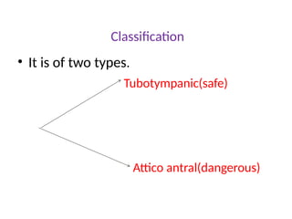
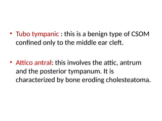
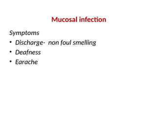
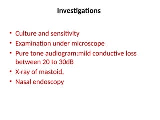
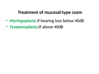
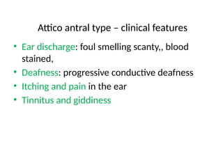
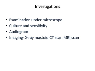
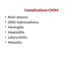
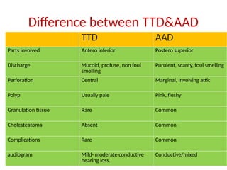
EAR DISORDERS
Review of anatomy
• The ear has external, middle, and inner portions. The
outer ear is called the pinna and is made of ridged
cartilage covered by skin. Sound funnels through the
pinna into the external auditory canal, a short tube that
ends at the eardrum (tympanic membrane).
• Sound causes the vibration of eardrum and its tiny
attached bones in the middle portion of the ear, and the
vibrations are conducted to the nearby cochlea. The
spiral-shaped cochlea is part of the inner ear; it
transforms sound into nerve impulses that travel to the
brain.
Infections of the External Ear
• Otitis Externa is an infection of the external
auditory canal (EAC) that can be divided
according to the time course of the infection:
acute, subacute, or chronic
• Acute: less than 6 weeks of duration.
Types
• Chronic OE – This is the same as acute diffuse OE but is of longer
duration (>6 weeks)
• Eczematous (eczematoid) OE – This encompasses various
dermatologic conditions (eg, atopic dermatitis , psoriasis,) that
may infect the EAC and cause OE
• Necrotizing (malignant) OE – This is an infection that extends
into the deeper tissues adjacent to the EAC; it primarily occurs in
adult patients who are immunocompromised (eg, as a result of
diabetes mellitus or AIDS) and is rarely described in children; it
may result in cases of cellulitis and osteomyelitis
• Otomycosis - Infection of the ear canal secondary to fungus
species such as Candida or Aspergillus
Causes
• Swimming
• Constriction of the ear canal from bone
growth (Surfer's ear)
• Saturation diver
• the use of objects such as cotton swabs or
other small objects to clear the ear canal
Pathophysiology
• OE is a superficial infection of the skin in the EAC. The
processes involved in the development of OE can be
divided into the following 4 categories:
• Obstruction (eg, cerumen buildup, surfer’s exostosis, or
a narrow or tortuous canal), resulting in water
retention
• Absence of cerumen, which may occur as a result of
repeated water exposure or overcleaning the ear canal
• Trauma
• Alteration of the pH of the ear canal
• The two factors that are required for external
otitis to develop are (1) the presence of germs
that can infect the skin and (2) impairments in
the integrity of the skin of the ear canal that
allow infection to occur
• atopic dermatitis , psoriasis
• otomycosis
Symptoms
• Drainage from the ear - yellow, yellow-green, foul smelling,
persistent
• Ear pain - felt deep inside the ear and may get worse when
moving head
• Hearing loss
• Itching of the ear or ear canal
• Fever
• Trouble swallowing
• Weakness in the face
• Voice loss
Diagnosis
• When the ear is inspected, the canal appears
red and swollen in well-developed cases.
• physical examination
• Otoscope :narrowing of the ear canal from
inflammation and the presence of drainage
and debris.
• Culture of the drainage
Treatment
• Aural toilet
• Aural toilet must be performed and can be
done most conveniently by dry mopping. The
ear is cleaned with a gentle rotatory action.
Once the cotton wool is soiled it is replaced.
• Dressings
If the otitis externa is severe, a length of 1 cm
ribbon gauze, impregnated with appropriate
medication, should be inserted gently into the
meatus, and renewed daily until the meatus
has returned to normal
• The following medications are of value on
the dressing:
• 8% aluminium acetate;
• 10% ichthammol in glycerine;
• ointment of gramicidin, neomycin, nystatin
and triamcinolone
• (Tri-Adcortyl);
• other medication may be used as dictated by
the result of culture.
• If fungal otitis externa is present, dressings of
3% amphotericin, miconazole
• or nystatin may be used.
Otitis media
• Inflamation of middle ear.
Types
Acute
suppurative
non suppurative
Chronic
suppurative
Acute Otitis Media
• Acute otitis media, i.e. acute inflammation of the
middle-ear cavity, is a common condition and is
frequently bilateral. It occurs most commonly in
children and it is important that it is managed with
care to prevent subsequent complications. It most
commonly follows an acute upper respiratory tract
infection and may be viral or bacterial.
Pathology
• Acute otitis media is an infection of the
mucous membrane of the whole of the
middle-ear cleft Eustachian tube, tympanic
cavity, mastoid antrum and air cells.
• The bacteria responsible for acute otitis media
are: Streptococcus pneumonia 35%,
Haemophilus influenzae 25%, Moraxella
catarrhalis 15%. Group A streptococci and
Staphylococcus aureus may also be
responsible.
• The sequence of events in acute otitis media is
as follows:
• organisms invade the mucous membrane
causing inflammation, oedema, exudate and
later, pus;
• oedema closes the Eustachian tube,
preventing aeration and drainage;
• pressure from the pus rises, causing the drum
to bulge;
• necrosis of the tympanic membrane results in
perforation;
• the ear continues to drain until the infection
resolves
Causes
• Common cold
• Acute tonsillitis
• Influenza
• whooping cough
Symptoms, signs
• Earache
• Deafness
It is conductive in nature and may be
accompanied by tinnitus
• Pyrexia
• Tenderness
There is usually some tenderness to pressure on
the mastoid antrum.
contd…….
• The tympanic membrane varies in appearance
• Redness and fullness of the drum;.
• Bulging, with loss of landmarks. Purple
colour..
• Perforation with otorrhoea.
Treatment
• Antibiotics:, amoxycillin will be more effective.
Co-amoxiclav is useful in Moraxella infections.
• Analgesics
• Nasal vasoconstrictors
• Ear drops (steroides or antibiotics)
• Myringotomy is necessary when bulging of
the tympanic membrane persists, despite
adequate antibiotic therapy
Chronic suppurative otitis media(CSOM)
• CSOM is a chronic inflammatory process
involving the middle ear cleft producing
irreversible pathological changes .
causes
• Late treatment of acute otitis media.
• Inadequate or inappropriate antibiotic
therapy.
• Upper airway infection.
• Lowered resistance, e.g. malnutrition,
anaemia,immunological impairment.
• Particularly virulent infection, e.g. measles.
Classification
• It is of two types.
Tubotympanic(safe)
Attico antral(dangerous)
Types of CSOM
Mucosal disease with tympanic membrane
perforation (tubo-tympanic disease, relatively
safe).
Bony:
cholesteatoma—dangerous (attico-antral
disease).
• Tubo tympanic : this is a benign type of CSOM
confined only to the middle ear cleft.
• Attico antral: this involves the attic, antrum
and the posterior tympanum. It is
characterized by bone eroding cholesteatoma.
Mucosal infection
Symptoms
• Discharge- non foul smelling
• Deafness
• Earache
• Signs: discharge, tympanic membrane
perforation,
• Tuning fork test: rinne-negative
• Weber-lateralised to one side
Investigations
• Culture and sensitivity
• Examination under microscope
• Pure tone audiogram:mild conductive loss
between 20 to 30dB
• X-ray of mastoid,
• Nasal endoscopy
Treatment of mucosal-type csom
• Myringoplasty if hearing loss below 40dB
• Tympanoplasty:if above 40dB
Attico antral type – clinical features
• Ear discharge: foul smelling scanty,, blood
stained,
• Deafness: progressive conductive deafness
• Itching and pain in the ear
• Tinnitus and giddiness
Sign
• In Otoscopic examination: foul smelling discharge in
the ext. Auditory canal
• Granulation tissue in the meatus
• Attic or marginal perforation of tymanic membrane
• Cholesteatoma
• Mastoid tenderness
• Tuning fork test-Rinne negative, weber localised to
lateral side,
Investigations
• Examination under microscope
• Culture and sensitivity
• Audiogram
• Imaging- X-ray mastoid,CT scan,MRI scan
Management
• Goal – to make the ear safe and dry
• To restore and improve hearing
• Surgical management
• Main line treatment.
• 1. canal wall down mastoidectomy:consists of
radical and modified radical mastoidectomy.
These procedures ensures safety and dry ear but
functional improvement may not be achieved.
• 2. Canal wall up mastoidectomy: or combined
approach tympanoplasty, where functional
improvement can be achieved but not the
safety.
Medical management
• It is used only for patient who are unfit for
surgery. Topical antibiotic and steroid are
used.
• In some cases 5- flurouracil used.
Complications-CSOM
• Brain abscess
• Otitic hydrocephalus
• Meningitis
• Mastoiditis
• Labyrynthitis
• Petrositis
Difference between TTD&AAD
TTD AAD
Parts involved Antero inferior Postero superior
Discharge Mucoid, profuse, non foul
smelling
Purulent, scanty, foul smelling
Perforation Central Marginal, Involving attic
Polyp Usually pale Pink, fleshy
Granulation tissue Rare Common
Cholesteatoma Absent Common
Complications Rare Common
audiogram Mild- moderate conductive
hearing loss.
Conductive/mixed

ear disorders otitis media , causes , symptoms

  • 1.
  • 3.
    Review of anatomy •The ear has external, middle, and inner portions. The outer ear is called the pinna and is made of ridged cartilage covered by skin. Sound funnels through the pinna into the external auditory canal, a short tube that ends at the eardrum (tympanic membrane). • Sound causes the vibration of eardrum and its tiny attached bones in the middle portion of the ear, and the vibrations are conducted to the nearby cochlea. The spiral-shaped cochlea is part of the inner ear; it transforms sound into nerve impulses that travel to the brain.
  • 4.
    Infections of theExternal Ear • Otitis Externa is an infection of the external auditory canal (EAC) that can be divided according to the time course of the infection: acute, subacute, or chronic • Acute: less than 6 weeks of duration.
  • 5.
    Types • Chronic OE– This is the same as acute diffuse OE but is of longer duration (>6 weeks) • Eczematous (eczematoid) OE – This encompasses various dermatologic conditions (eg, atopic dermatitis , psoriasis,) that may infect the EAC and cause OE • Necrotizing (malignant) OE – This is an infection that extends into the deeper tissues adjacent to the EAC; it primarily occurs in adult patients who are immunocompromised (eg, as a result of diabetes mellitus or AIDS) and is rarely described in children; it may result in cases of cellulitis and osteomyelitis • Otomycosis - Infection of the ear canal secondary to fungus species such as Candida or Aspergillus
  • 6.
    Causes • Swimming • Constrictionof the ear canal from bone growth (Surfer's ear) • Saturation diver • the use of objects such as cotton swabs or other small objects to clear the ear canal
  • 7.
    Pathophysiology • OE isa superficial infection of the skin in the EAC. The processes involved in the development of OE can be divided into the following 4 categories: • Obstruction (eg, cerumen buildup, surfer’s exostosis, or a narrow or tortuous canal), resulting in water retention • Absence of cerumen, which may occur as a result of repeated water exposure or overcleaning the ear canal • Trauma • Alteration of the pH of the ear canal
  • 8.
    • The twofactors that are required for external otitis to develop are (1) the presence of germs that can infect the skin and (2) impairments in the integrity of the skin of the ear canal that allow infection to occur • atopic dermatitis , psoriasis • otomycosis
  • 9.
    Symptoms • Drainage fromthe ear - yellow, yellow-green, foul smelling, persistent • Ear pain - felt deep inside the ear and may get worse when moving head • Hearing loss • Itching of the ear or ear canal • Fever • Trouble swallowing • Weakness in the face • Voice loss
  • 10.
    Diagnosis • When theear is inspected, the canal appears red and swollen in well-developed cases. • physical examination • Otoscope :narrowing of the ear canal from inflammation and the presence of drainage and debris. • Culture of the drainage
  • 11.
    Treatment • Aural toilet •Aural toilet must be performed and can be done most conveniently by dry mopping. The ear is cleaned with a gentle rotatory action. Once the cotton wool is soiled it is replaced.
  • 12.
    • Dressings If theotitis externa is severe, a length of 1 cm ribbon gauze, impregnated with appropriate medication, should be inserted gently into the meatus, and renewed daily until the meatus has returned to normal
  • 13.
    • The followingmedications are of value on the dressing: • 8% aluminium acetate; • 10% ichthammol in glycerine; • ointment of gramicidin, neomycin, nystatin and triamcinolone • (Tri-Adcortyl);
  • 14.
    • other medicationmay be used as dictated by the result of culture. • If fungal otitis externa is present, dressings of 3% amphotericin, miconazole • or nystatin may be used.
  • 15.
    Otitis media • Inflamationof middle ear. Types Acute suppurative non suppurative Chronic suppurative
  • 16.
    Acute Otitis Media •Acute otitis media, i.e. acute inflammation of the middle-ear cavity, is a common condition and is frequently bilateral. It occurs most commonly in children and it is important that it is managed with care to prevent subsequent complications. It most commonly follows an acute upper respiratory tract infection and may be viral or bacterial.
  • 17.
    Pathology • Acute otitismedia is an infection of the mucous membrane of the whole of the middle-ear cleft Eustachian tube, tympanic cavity, mastoid antrum and air cells.
  • 18.
    • The bacteriaresponsible for acute otitis media are: Streptococcus pneumonia 35%, Haemophilus influenzae 25%, Moraxella catarrhalis 15%. Group A streptococci and Staphylococcus aureus may also be responsible.
  • 19.
    • The sequenceof events in acute otitis media is as follows: • organisms invade the mucous membrane causing inflammation, oedema, exudate and later, pus; • oedema closes the Eustachian tube, preventing aeration and drainage;
  • 20.
    • pressure fromthe pus rises, causing the drum to bulge; • necrosis of the tympanic membrane results in perforation; • the ear continues to drain until the infection resolves
  • 21.
    Causes • Common cold •Acute tonsillitis • Influenza • whooping cough
  • 22.
    Symptoms, signs • Earache •Deafness It is conductive in nature and may be accompanied by tinnitus • Pyrexia • Tenderness There is usually some tenderness to pressure on the mastoid antrum.
  • 23.
    contd……. • The tympanicmembrane varies in appearance • Redness and fullness of the drum;. • Bulging, with loss of landmarks. Purple colour.. • Perforation with otorrhoea.
  • 24.
    Treatment • Antibiotics:, amoxycillinwill be more effective. Co-amoxiclav is useful in Moraxella infections.
  • 25.
    • Analgesics • Nasalvasoconstrictors • Ear drops (steroides or antibiotics) • Myringotomy is necessary when bulging of the tympanic membrane persists, despite adequate antibiotic therapy
  • 26.
    Chronic suppurative otitismedia(CSOM) • CSOM is a chronic inflammatory process involving the middle ear cleft producing irreversible pathological changes .
  • 27.
    causes • Late treatmentof acute otitis media. • Inadequate or inappropriate antibiotic therapy. • Upper airway infection. • Lowered resistance, e.g. malnutrition, anaemia,immunological impairment. • Particularly virulent infection, e.g. measles.
  • 28.
    Classification • It isof two types. Tubotympanic(safe) Attico antral(dangerous)
  • 29.
    Types of CSOM Mucosaldisease with tympanic membrane perforation (tubo-tympanic disease, relatively safe). Bony: cholesteatoma—dangerous (attico-antral disease).
  • 30.
    • Tubo tympanic: this is a benign type of CSOM confined only to the middle ear cleft. • Attico antral: this involves the attic, antrum and the posterior tympanum. It is characterized by bone eroding cholesteatoma.
  • 31.
    Mucosal infection Symptoms • Discharge-non foul smelling • Deafness • Earache
  • 32.
    • Signs: discharge,tympanic membrane perforation, • Tuning fork test: rinne-negative • Weber-lateralised to one side
  • 33.
    Investigations • Culture andsensitivity • Examination under microscope • Pure tone audiogram:mild conductive loss between 20 to 30dB • X-ray of mastoid, • Nasal endoscopy
  • 34.
    Treatment of mucosal-typecsom • Myringoplasty if hearing loss below 40dB • Tympanoplasty:if above 40dB
  • 35.
    Attico antral type– clinical features • Ear discharge: foul smelling scanty,, blood stained, • Deafness: progressive conductive deafness • Itching and pain in the ear • Tinnitus and giddiness
  • 36.
    Sign • In Otoscopicexamination: foul smelling discharge in the ext. Auditory canal • Granulation tissue in the meatus • Attic or marginal perforation of tymanic membrane • Cholesteatoma • Mastoid tenderness • Tuning fork test-Rinne negative, weber localised to lateral side,
  • 37.
    Investigations • Examination undermicroscope • Culture and sensitivity • Audiogram • Imaging- X-ray mastoid,CT scan,MRI scan
  • 38.
    Management • Goal –to make the ear safe and dry • To restore and improve hearing • Surgical management • Main line treatment. • 1. canal wall down mastoidectomy:consists of radical and modified radical mastoidectomy. These procedures ensures safety and dry ear but functional improvement may not be achieved.
  • 39.
    • 2. Canalwall up mastoidectomy: or combined approach tympanoplasty, where functional improvement can be achieved but not the safety.
  • 40.
    Medical management • Itis used only for patient who are unfit for surgery. Topical antibiotic and steroid are used. • In some cases 5- flurouracil used.
  • 41.
    Complications-CSOM • Brain abscess •Otitic hydrocephalus • Meningitis • Mastoiditis • Labyrynthitis • Petrositis
  • 42.
    Difference between TTD&AAD TTDAAD Parts involved Antero inferior Postero superior Discharge Mucoid, profuse, non foul smelling Purulent, scanty, foul smelling Perforation Central Marginal, Involving attic Polyp Usually pale Pink, fleshy Granulation tissue Rare Common Cholesteatoma Absent Common Complications Rare Common audiogram Mild- moderate conductive hearing loss. Conductive/mixed